User login
Case 1
A 65-year-old woman presents to the urgent care center with several growths inside a tattoo on her left leg that developed several weeks before presentation. Patient states she has had the tattoo for approximately 15 years, but had a revision done to the original artwork at a local tattoo parlor 4 months ago; she noted that the skin lesions appeared one month after this revision, have rapidly increased in size, and are occasionally pruritic.
Patient has a medical history of breast cancer, for which she was diagnosed and treated at age 50 years, and she is also a cigarette smoker. She denies a prior history of skin cancer. Physical examination reveals scattered exophytic nodules, with the largest nodule measuring 1.2 cm in diameter.
What is your diagnosis?
Case 2
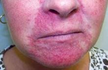
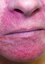
A 48-year-old woman presents to the urgent care center with dermatitis around her nose and mouth, which she states has been progressing in severity over the past several months and is at times pruritic. She had been treating the site twice daily with topical betamethasone diproprionate cream and had also been on intermittent doses of oral corticosteroids. Physical examination reveals a pronounced erythematous papulopustular eruption of the affected areas. The rash did not involve her neck, forehead, or scalp.
What is your diagnosis?
Mr Himmelsbach is a nurse practitioner at berks Plastic Surgery in Wyomissing, Pennsylvania. Dr Schleicher, editor of “diagnosis at a Glance,” is director of the dermdOX center in Hazleton, Pennsylvania; a clinical instructor of dermatology at King’s college in Wilkes-barre, Pennsylvania; an associate professor of medicine at the commonwealth medical college in Scranton, Pennsylvania; and an adjunct assistant professor of dermatology at the University of Pennsylvania in Philadelphia. He is also a member of the emerGeNcY medIcINe editorial board. Ms Remaley is a physician assistant at reading dermatology Associates in reading, Pennsylvania.
Answer
Case 1
Biopsy of the two largest lesions revealed keratoacanthoma (KA); excisional surgeries were subsequently performed on the other lesions. KAs originate within pilosebaceous glands and are classified as a variant of invasive squamous cell carcinoma. The lesions are characterized by rapid growth, potential for spontaneous involution, and low incidence of metastatic spread. Although KAs have been linked to chronic tar exposure in industrial workers, they more commonly occur in cigarette smokers and in a significant percentage of metastatic melanoma patients treated with BRAF inhibitors. KA developing in a tattoo is a rare occurrence, and the association in this case with recent tattoo ink application is an intriguing one.
Case 2
Steroid-induced facial dermatitis manifests as an eruption of papules and pustules on an erythematous scaling base classically involving the nasolabial folds and perioral area. A clear zone may be present around the vermillion border. This rash is caused by prolonged treatment of blemishes or rashes with mid-to-high potency topical corticosteroids. During treatment, the complexion initially improves but then gradually worsens. Upon discontinuation of corticosteroid therapy, a rebound flare ensues, often triggering resumption of the precipitating medication. Management is difficult, though most cases respond to substitution with a low-potency corticosteroid followed by application of either pimecrolimus or a sulfur-containing lotion. 32
Case 1
A 65-year-old woman presents to the urgent care center with several growths inside a tattoo on her left leg that developed several weeks before presentation. Patient states she has had the tattoo for approximately 15 years, but had a revision done to the original artwork at a local tattoo parlor 4 months ago; she noted that the skin lesions appeared one month after this revision, have rapidly increased in size, and are occasionally pruritic.
Patient has a medical history of breast cancer, for which she was diagnosed and treated at age 50 years, and she is also a cigarette smoker. She denies a prior history of skin cancer. Physical examination reveals scattered exophytic nodules, with the largest nodule measuring 1.2 cm in diameter.
What is your diagnosis?
Case 2
A 48-year-old woman presents to the urgent care center with dermatitis around her nose and mouth, which she states has been progressing in severity over the past several months and is at times pruritic. She had been treating the site twice daily with topical betamethasone diproprionate cream and had also been on intermittent doses of oral corticosteroids. Physical examination reveals a pronounced erythematous papulopustular eruption of the affected areas. The rash did not involve her neck, forehead, or scalp.
What is your diagnosis?
Mr Himmelsbach is a nurse practitioner at berks Plastic Surgery in Wyomissing, Pennsylvania. Dr Schleicher, editor of “diagnosis at a Glance,” is director of the dermdOX center in Hazleton, Pennsylvania; a clinical instructor of dermatology at King’s college in Wilkes-barre, Pennsylvania; an associate professor of medicine at the commonwealth medical college in Scranton, Pennsylvania; and an adjunct assistant professor of dermatology at the University of Pennsylvania in Philadelphia. He is also a member of the emerGeNcY medIcINe editorial board. Ms Remaley is a physician assistant at reading dermatology Associates in reading, Pennsylvania.
Answer
Case 1
Biopsy of the two largest lesions revealed keratoacanthoma (KA); excisional surgeries were subsequently performed on the other lesions. KAs originate within pilosebaceous glands and are classified as a variant of invasive squamous cell carcinoma. The lesions are characterized by rapid growth, potential for spontaneous involution, and low incidence of metastatic spread. Although KAs have been linked to chronic tar exposure in industrial workers, they more commonly occur in cigarette smokers and in a significant percentage of metastatic melanoma patients treated with BRAF inhibitors. KA developing in a tattoo is a rare occurrence, and the association in this case with recent tattoo ink application is an intriguing one.
Case 2
Steroid-induced facial dermatitis manifests as an eruption of papules and pustules on an erythematous scaling base classically involving the nasolabial folds and perioral area. A clear zone may be present around the vermillion border. This rash is caused by prolonged treatment of blemishes or rashes with mid-to-high potency topical corticosteroids. During treatment, the complexion initially improves but then gradually worsens. Upon discontinuation of corticosteroid therapy, a rebound flare ensues, often triggering resumption of the precipitating medication. Management is difficult, though most cases respond to substitution with a low-potency corticosteroid followed by application of either pimecrolimus or a sulfur-containing lotion. 32


