User login
Give guidance, not just Viagra, to men with diabetes and related impotence
Diabetic men with erectile dysfunction might consider a prescription for a drug like Viagra to be a permanent cure for a temporary issue: Take a pill, problem solved. But the truth, a leading urologist says, is entirely different.
“If a diabetic patient has erectile dysfunction, it’s not enough to provide Viagra [sildenafil] or Cialis [tadalafil] and then send him on his merry way,” J. Francois Eid, MD, a New York City urologist, said at the annual meeting of the American Association of Diabetes Educators. “It’s important to let individuals know the drug has not cured the erectile dysfunction. If patients don’t take care of the diabetes, the erectile dysfunction progresses.”
In an interview, Dr. Eid shared several messages for medical professionals who treat men with diabetes and related erectile dysfunction:
• Diabetes has “devastating” effects on the penis, and may even cause it to shrink.
• Long-term uncontrolled diabetes can make ED permanent.
• While ED drugs often fail in men with diabetes, several other options exist; and penis implants may provide significant relief.
How big is the problem? An estimated 50%-75% of men with diabetes experience from some degree of ED, which is thought to be three times more common in diabetic men than other men, according to Dr. Eid.
“Diabetes has a devastating effect on the muscle tissue inside the penis,” he said. “All the tiny little arteries that feed blood to the muscle get occluded. Little by little, the muscle inside the penis shrinks.” Indeed, some diabetics with ED complain that their penises have shrunk, he said.
Diabetic damage doesn’t stop with these small vessels, he said. “You really have two parallel situations: You need blood flow that feeds the muscle of the penis, and you need an artery dedicated to bringing blood rapidly when a man becomes aroused and wants to be sexually active,” he said. “That artery is also affected by diabetes. They’ll say ‘I can get a partial erection, but I can’t maintain it.’ ”
What comes after an ED diagnosis in diabetic patients? Often, Dr. Eid will instantly refer these men to a cardiologist. “If a patient has diabetes and is newly diagnosed, a significant portion of these men are going to develop coronary artery disease in the next 2-3 years,” he said. “One of the things we do is recommend is that they see a cardiologist and perhaps have a stress test or some sort of evaluation.”
Dr. Eid also urges these patients to treat their diabetes in order to avoid developing ED for life. “They need to manage their diabetes and make sure they control it so the ED will not progress and will stabilize, as a result,” he said. “If the diabetes is controlled after the patient is first diagnosed, then the erections will come back. But if the patient has diabetes for many years, and suddenly decides it’s time to control it, they cannot prevent the damage that’s already been done.”
As for treatments, patients with diabetes and related ED should begin with medications like sildenafil and tadalafil, he recommended. But research findings suggest that the drugs will fail in half of men with type 2 diabetes, he said.
Other options include penile self-injections, vacuum devices, and penile implants.
The injections “can work well and are painless, but men detest having to inject themselves before sexual activity,” Dr. Eid said. And he said men rarely have success using vacuum devices, which are available over the counter.
By contrast, penis implants can successfully treat erectile dysfunction in many cases, Dr. Eid said. Men trigger erections by squeezing a pump that is implanted into the scrotum. Fluid then flows from an implanted reservoir into a cylinder implanted in the penis.
Men are often pleased by penis implants because they can have sex spontaneously without having to plan for it ahead of time, as required by medications. “They’re completely transformed,” Dr. Eid said. “They feel that they’re cured.”
Dr. Eid receives or has received research support/grants from American Medical Systems, Coloplast, Lilly ICOS, Bayer, Vivus, Pharmacia-Upjohn, and Pfizer. He is or was a consultant and on the speakers bureau for Coloplast, American Medical Systems, Lilly ICOS, Bayer and Pfizer.
Diabetic men with erectile dysfunction might consider a prescription for a drug like Viagra to be a permanent cure for a temporary issue: Take a pill, problem solved. But the truth, a leading urologist says, is entirely different.
“If a diabetic patient has erectile dysfunction, it’s not enough to provide Viagra [sildenafil] or Cialis [tadalafil] and then send him on his merry way,” J. Francois Eid, MD, a New York City urologist, said at the annual meeting of the American Association of Diabetes Educators. “It’s important to let individuals know the drug has not cured the erectile dysfunction. If patients don’t take care of the diabetes, the erectile dysfunction progresses.”
In an interview, Dr. Eid shared several messages for medical professionals who treat men with diabetes and related erectile dysfunction:
• Diabetes has “devastating” effects on the penis, and may even cause it to shrink.
• Long-term uncontrolled diabetes can make ED permanent.
• While ED drugs often fail in men with diabetes, several other options exist; and penis implants may provide significant relief.
How big is the problem? An estimated 50%-75% of men with diabetes experience from some degree of ED, which is thought to be three times more common in diabetic men than other men, according to Dr. Eid.
“Diabetes has a devastating effect on the muscle tissue inside the penis,” he said. “All the tiny little arteries that feed blood to the muscle get occluded. Little by little, the muscle inside the penis shrinks.” Indeed, some diabetics with ED complain that their penises have shrunk, he said.
Diabetic damage doesn’t stop with these small vessels, he said. “You really have two parallel situations: You need blood flow that feeds the muscle of the penis, and you need an artery dedicated to bringing blood rapidly when a man becomes aroused and wants to be sexually active,” he said. “That artery is also affected by diabetes. They’ll say ‘I can get a partial erection, but I can’t maintain it.’ ”
What comes after an ED diagnosis in diabetic patients? Often, Dr. Eid will instantly refer these men to a cardiologist. “If a patient has diabetes and is newly diagnosed, a significant portion of these men are going to develop coronary artery disease in the next 2-3 years,” he said. “One of the things we do is recommend is that they see a cardiologist and perhaps have a stress test or some sort of evaluation.”
Dr. Eid also urges these patients to treat their diabetes in order to avoid developing ED for life. “They need to manage their diabetes and make sure they control it so the ED will not progress and will stabilize, as a result,” he said. “If the diabetes is controlled after the patient is first diagnosed, then the erections will come back. But if the patient has diabetes for many years, and suddenly decides it’s time to control it, they cannot prevent the damage that’s already been done.”
As for treatments, patients with diabetes and related ED should begin with medications like sildenafil and tadalafil, he recommended. But research findings suggest that the drugs will fail in half of men with type 2 diabetes, he said.
Other options include penile self-injections, vacuum devices, and penile implants.
The injections “can work well and are painless, but men detest having to inject themselves before sexual activity,” Dr. Eid said. And he said men rarely have success using vacuum devices, which are available over the counter.
By contrast, penis implants can successfully treat erectile dysfunction in many cases, Dr. Eid said. Men trigger erections by squeezing a pump that is implanted into the scrotum. Fluid then flows from an implanted reservoir into a cylinder implanted in the penis.
Men are often pleased by penis implants because they can have sex spontaneously without having to plan for it ahead of time, as required by medications. “They’re completely transformed,” Dr. Eid said. “They feel that they’re cured.”
Dr. Eid receives or has received research support/grants from American Medical Systems, Coloplast, Lilly ICOS, Bayer, Vivus, Pharmacia-Upjohn, and Pfizer. He is or was a consultant and on the speakers bureau for Coloplast, American Medical Systems, Lilly ICOS, Bayer and Pfizer.
Diabetic men with erectile dysfunction might consider a prescription for a drug like Viagra to be a permanent cure for a temporary issue: Take a pill, problem solved. But the truth, a leading urologist says, is entirely different.
“If a diabetic patient has erectile dysfunction, it’s not enough to provide Viagra [sildenafil] or Cialis [tadalafil] and then send him on his merry way,” J. Francois Eid, MD, a New York City urologist, said at the annual meeting of the American Association of Diabetes Educators. “It’s important to let individuals know the drug has not cured the erectile dysfunction. If patients don’t take care of the diabetes, the erectile dysfunction progresses.”
In an interview, Dr. Eid shared several messages for medical professionals who treat men with diabetes and related erectile dysfunction:
• Diabetes has “devastating” effects on the penis, and may even cause it to shrink.
• Long-term uncontrolled diabetes can make ED permanent.
• While ED drugs often fail in men with diabetes, several other options exist; and penis implants may provide significant relief.
How big is the problem? An estimated 50%-75% of men with diabetes experience from some degree of ED, which is thought to be three times more common in diabetic men than other men, according to Dr. Eid.
“Diabetes has a devastating effect on the muscle tissue inside the penis,” he said. “All the tiny little arteries that feed blood to the muscle get occluded. Little by little, the muscle inside the penis shrinks.” Indeed, some diabetics with ED complain that their penises have shrunk, he said.
Diabetic damage doesn’t stop with these small vessels, he said. “You really have two parallel situations: You need blood flow that feeds the muscle of the penis, and you need an artery dedicated to bringing blood rapidly when a man becomes aroused and wants to be sexually active,” he said. “That artery is also affected by diabetes. They’ll say ‘I can get a partial erection, but I can’t maintain it.’ ”
What comes after an ED diagnosis in diabetic patients? Often, Dr. Eid will instantly refer these men to a cardiologist. “If a patient has diabetes and is newly diagnosed, a significant portion of these men are going to develop coronary artery disease in the next 2-3 years,” he said. “One of the things we do is recommend is that they see a cardiologist and perhaps have a stress test or some sort of evaluation.”
Dr. Eid also urges these patients to treat their diabetes in order to avoid developing ED for life. “They need to manage their diabetes and make sure they control it so the ED will not progress and will stabilize, as a result,” he said. “If the diabetes is controlled after the patient is first diagnosed, then the erections will come back. But if the patient has diabetes for many years, and suddenly decides it’s time to control it, they cannot prevent the damage that’s already been done.”
As for treatments, patients with diabetes and related ED should begin with medications like sildenafil and tadalafil, he recommended. But research findings suggest that the drugs will fail in half of men with type 2 diabetes, he said.
Other options include penile self-injections, vacuum devices, and penile implants.
The injections “can work well and are painless, but men detest having to inject themselves before sexual activity,” Dr. Eid said. And he said men rarely have success using vacuum devices, which are available over the counter.
By contrast, penis implants can successfully treat erectile dysfunction in many cases, Dr. Eid said. Men trigger erections by squeezing a pump that is implanted into the scrotum. Fluid then flows from an implanted reservoir into a cylinder implanted in the penis.
Men are often pleased by penis implants because they can have sex spontaneously without having to plan for it ahead of time, as required by medications. “They’re completely transformed,” Dr. Eid said. “They feel that they’re cured.”
Dr. Eid receives or has received research support/grants from American Medical Systems, Coloplast, Lilly ICOS, Bayer, Vivus, Pharmacia-Upjohn, and Pfizer. He is or was a consultant and on the speakers bureau for Coloplast, American Medical Systems, Lilly ICOS, Bayer and Pfizer.
EXPERT ANALYSIS FROM AADE 16
Close monitoring of psoriasis patients can delay PsA onset
NEWPORT BEACH, CALIF. – A patient with psoriasis can develop crippling psoriatic arthritis (PsA) within 5 to 10 years of diagnosis, but monitoring patients for signs of trouble can help prevent the onset of PsA, according to Alan Menter, MD.
Even a simple foot examination can make a huge difference, noted Dr. Menter, chief of the division of dermatology and director of the Psoriasis Research Institute at Baylor University Medical Center, Dallas. “At every visit, you and I should be looking for early signs of joint disease,” he said at the Skin Disease Education Foundation’s Women’s & Pediatric Dermatology Seminar. “We should not let these patients develop any joint disease because we have drugs that can prevent joint destruction.”

Dr. Menter pointed out that PsA is a disease that is distinct from psoriasis. “It’s linked to psoriasis, but genetically, there are differences,” he said, “and immunologically, what goes on in skin is not identical.”
He provided the following pearls regarding diagnosing PsA:
• Be on the lookout for “sausage fingers” and “sausage toes,” both signs of PsA. “You and I are very visual people, and we can see a swollen toe or finger very easily,” Dr. Menter said. “I take the shoes off every psoriasis patient at every visit and run my thumb and index finger down the Achilles. I look for a swollen Achilles – classic enthesitis.” In some cases, swollen big toes in psoriasis patients may be misdiagnosed as gout instead of PsA, he noted.
• Ask patients about how their joints feel when they wake up in the morning: Do they have swelling and tenderness? “That’s an early marker of psoriatic arthritis disease,” Dr. Menter said. In contrast, in a patient with osteoarthritis, “the more they use their joints, the worse it gets.”
• The severity of psoriasis has nothing to do with the severity of PsA. “You can have 50% of the body covered with psoriasis but no arthritis,” he said. “Or you can have someone with one patch of psoriasis on the scalp with devastating joint disease.”
• Be aware that there are five PsA subtypes that can occur in combination with each other:
1. Dactylitis. This is the form that causes the “sausage digit.”
2. Asymmetric oligoarthritis. This is the type most commonly seen on presentation, when there are few joints affected.
3. Symmetric arthritis. This form is more common in females and difficult to differentiate from rheumatoid arthritis.
4. Distal interphalangeal joint arthritis. This type is often linked to dactylitis and nail dystrophy.
5. Arthritis mutilans. This is more common in females, linked to long disease duration, and present in an estimated 5% of cases.
Dr. Menter suggested that dermatologists refer suspected cases of PsA to a rheumatologist. Since patients may have to wait 6-10 weeks for an appointment, he recommended that dermatologists consider NSAIDs, such as the over-the-counter naproxen and prescription meloxicam and celecoxib in the meantime. Dermatologists may also consider bringing up the use of methotrexate and biologics, he said.
Dr. Menter disclosed relationships with multiple pharmaceutical companies, including AbbVie, Allergan, Amgen, Boehringer Ingelheim, Eli Lilly, Merck, Novartis, and Pfizer.
SDEF and this news organization are owned by Frontline Medical Communications.
NEWPORT BEACH, CALIF. – A patient with psoriasis can develop crippling psoriatic arthritis (PsA) within 5 to 10 years of diagnosis, but monitoring patients for signs of trouble can help prevent the onset of PsA, according to Alan Menter, MD.
Even a simple foot examination can make a huge difference, noted Dr. Menter, chief of the division of dermatology and director of the Psoriasis Research Institute at Baylor University Medical Center, Dallas. “At every visit, you and I should be looking for early signs of joint disease,” he said at the Skin Disease Education Foundation’s Women’s & Pediatric Dermatology Seminar. “We should not let these patients develop any joint disease because we have drugs that can prevent joint destruction.”
Dr. Menter pointed out that PsA is a disease that is distinct from psoriasis. “It’s linked to psoriasis, but genetically, there are differences,” he said, “and immunologically, what goes on in skin is not identical.”
He provided the following pearls regarding diagnosing PsA:
• Be on the lookout for “sausage fingers” and “sausage toes,” both signs of PsA. “You and I are very visual people, and we can see a swollen toe or finger very easily,” Dr. Menter said. “I take the shoes off every psoriasis patient at every visit and run my thumb and index finger down the Achilles. I look for a swollen Achilles – classic enthesitis.” In some cases, swollen big toes in psoriasis patients may be misdiagnosed as gout instead of PsA, he noted.
• Ask patients about how their joints feel when they wake up in the morning: Do they have swelling and tenderness? “That’s an early marker of psoriatic arthritis disease,” Dr. Menter said. In contrast, in a patient with osteoarthritis, “the more they use their joints, the worse it gets.”
• The severity of psoriasis has nothing to do with the severity of PsA. “You can have 50% of the body covered with psoriasis but no arthritis,” he said. “Or you can have someone with one patch of psoriasis on the scalp with devastating joint disease.”
• Be aware that there are five PsA subtypes that can occur in combination with each other:
1. Dactylitis. This is the form that causes the “sausage digit.”
2. Asymmetric oligoarthritis. This is the type most commonly seen on presentation, when there are few joints affected.
3. Symmetric arthritis. This form is more common in females and difficult to differentiate from rheumatoid arthritis.
4. Distal interphalangeal joint arthritis. This type is often linked to dactylitis and nail dystrophy.
5. Arthritis mutilans. This is more common in females, linked to long disease duration, and present in an estimated 5% of cases.
Dr. Menter suggested that dermatologists refer suspected cases of PsA to a rheumatologist. Since patients may have to wait 6-10 weeks for an appointment, he recommended that dermatologists consider NSAIDs, such as the over-the-counter naproxen and prescription meloxicam and celecoxib in the meantime. Dermatologists may also consider bringing up the use of methotrexate and biologics, he said.
Dr. Menter disclosed relationships with multiple pharmaceutical companies, including AbbVie, Allergan, Amgen, Boehringer Ingelheim, Eli Lilly, Merck, Novartis, and Pfizer.
SDEF and this news organization are owned by Frontline Medical Communications.
NEWPORT BEACH, CALIF. – A patient with psoriasis can develop crippling psoriatic arthritis (PsA) within 5 to 10 years of diagnosis, but monitoring patients for signs of trouble can help prevent the onset of PsA, according to Alan Menter, MD.
Even a simple foot examination can make a huge difference, noted Dr. Menter, chief of the division of dermatology and director of the Psoriasis Research Institute at Baylor University Medical Center, Dallas. “At every visit, you and I should be looking for early signs of joint disease,” he said at the Skin Disease Education Foundation’s Women’s & Pediatric Dermatology Seminar. “We should not let these patients develop any joint disease because we have drugs that can prevent joint destruction.”
Dr. Menter pointed out that PsA is a disease that is distinct from psoriasis. “It’s linked to psoriasis, but genetically, there are differences,” he said, “and immunologically, what goes on in skin is not identical.”
He provided the following pearls regarding diagnosing PsA:
• Be on the lookout for “sausage fingers” and “sausage toes,” both signs of PsA. “You and I are very visual people, and we can see a swollen toe or finger very easily,” Dr. Menter said. “I take the shoes off every psoriasis patient at every visit and run my thumb and index finger down the Achilles. I look for a swollen Achilles – classic enthesitis.” In some cases, swollen big toes in psoriasis patients may be misdiagnosed as gout instead of PsA, he noted.
• Ask patients about how their joints feel when they wake up in the morning: Do they have swelling and tenderness? “That’s an early marker of psoriatic arthritis disease,” Dr. Menter said. In contrast, in a patient with osteoarthritis, “the more they use their joints, the worse it gets.”
• The severity of psoriasis has nothing to do with the severity of PsA. “You can have 50% of the body covered with psoriasis but no arthritis,” he said. “Or you can have someone with one patch of psoriasis on the scalp with devastating joint disease.”
• Be aware that there are five PsA subtypes that can occur in combination with each other:
1. Dactylitis. This is the form that causes the “sausage digit.”
2. Asymmetric oligoarthritis. This is the type most commonly seen on presentation, when there are few joints affected.
3. Symmetric arthritis. This form is more common in females and difficult to differentiate from rheumatoid arthritis.
4. Distal interphalangeal joint arthritis. This type is often linked to dactylitis and nail dystrophy.
5. Arthritis mutilans. This is more common in females, linked to long disease duration, and present in an estimated 5% of cases.
Dr. Menter suggested that dermatologists refer suspected cases of PsA to a rheumatologist. Since patients may have to wait 6-10 weeks for an appointment, he recommended that dermatologists consider NSAIDs, such as the over-the-counter naproxen and prescription meloxicam and celecoxib in the meantime. Dermatologists may also consider bringing up the use of methotrexate and biologics, he said.
Dr. Menter disclosed relationships with multiple pharmaceutical companies, including AbbVie, Allergan, Amgen, Boehringer Ingelheim, Eli Lilly, Merck, Novartis, and Pfizer.
SDEF and this news organization are owned by Frontline Medical Communications.
EXPERT ANALYSIS FROM SDEF WOMEN'S & PEDIATRIC DERMATOLOGY SEMINAR
Study provides best support to date for using ultrasound to detect gout
Ultrasound examination of patients with at least one swollen joint or a subcutaneous nodule detected gout with a sensitivity of 76.9% and specificity of 84.3% in an international, multicenter study.
“Ultrasound is a good test for gout,” said study lead author Alexis R. Ogdie-Beatty, MD, of the University of Pennsylvania, Philadelphia. “It has high specificity, even among patients with early disease. The sensitivity was lower so it doesn’t pick up all cases of gout, although it was higher in people with a longer duration of disease.”

But gout remains difficult to diagnose because of the many similar types of inflammatory arthritis. A 2016 report from the Agency for Healthcare Research and Quality says proper diagnosis is a “major challenge,” especially in primary care and urgent/emergency settings.
Monosodium urate (MSU) crystal analysis via joint aspiration is considered the “gold standard” of gout diagnostic tools, but the agency report notes that it “can be technically difficult to perform and painful to the patient.”
There are other challenges. “Sometimes we see people between flares, or it’s already started to improve so there’s not enough fluid to perform an arthrocentesis,” Dr. Ogdie-Beatty said in an interview. “Additionally, while rheumatologists are generally very good at joint aspirations, many primary care physicians are not specifically trained for this. Thus, an imaging study can be helpful in these cases.”
The new study (Arthritis Rheumatol. 2016 Oct 16. doi: 10.1002/art.39959) follows up on findings from the multicenter, cross-sectional Study for Updated Gout Classification Criteria, which independently linked ultrasound analysis to diagnosis of gout with an odds ratio of 7.2 (Arthritis Care Res. 2015 Sep;67[9]:1304-15). One goal of the new study is to determine specificity and sensitivity of ultrasound exams.
The researchers examined data from the previous study, which enrolled consecutive patients in clinical practice settings across 25 countries who had at least one swollen joint or a subcutaneous nodule in whom gout was on the differential diagnosis. They underwent ultrasound examinations (most commonly of the knees, metatarsophalangeal joints, and ankles) and were deemed to have a “positive test” if at least one of three signs appeared: a “double contour” sign, tophus, or a “snowstorm” appearance. However, the study did not require a specific ultrasound scanning protocol or training.
MSU crystal examinations confirmed which of the subjects actually had gout. In total, ultrasound examinations were performed on 824 patients, of whom 416 were confirmed to have gout (mean age, 60, and 87% male), and the other 408 did not have gout (mean age, 60, and 54% male).
The researchers found that the sensitivity of the ultrasound exams was 76.9%, meaning they correctly identified patients with gout just over three-quarters of the time. Dr. Ogdie-Beatty referred to this as “moderate” sensitivity. The researchers found that the sensitivity was highest in patients who’d had disease for 2 or more years and in those who didn’t show clinical signs of tophi.
The specificity for these signs – the percentage of the time that ultrasound exams correctly identified patients without gout – was 84.3%. The positive and negative predictive values were 83.3% and 78.1%, respectively.
Dr. Ogdie-Beatty said the cost of ultrasound examinations is variable, although insurance covers it for trained providers. It’s unclear how commonly ultrasound examinations are used to detect gout, she said, but “many rheumatologists now have ultrasound as a part of their practice, and ultrasound training during fellowship has become important.”
Dr. Ogdie-Beatty reported receiving consulting fees from Novartis, and her coauthors reported various financial relationships with industry. The study was supported by various National Institutes of Health grants to several authors, as well as the American College of Rheumatology, the European League Against Rheumatism, and various arthritis-related organizations.
The new study provides evidence that both answers and raises questions about the technical standards by which musculoskeletal ultrasound examination is used to detect gout and about what we mean when we make a clinical diagnosis of gout.
The study’s results showed that ultrasound provided excellent specificity for gout in the setting of acute calcium pyrophosphate deposition disease (CPPD), also known as pseudogout, particularly in distinguishing the deposition of monosodium urate on the surface of cartilage (double contour sign), and calcium pyrophosphate within the matrix of the cartilage (interface sign). Ultrasound’s specificity for gout apart from CPPD was particularly high when more than one feature of gout was present because one feature of gout was more often present in patients with CPPD than in non-CPPD controls, but this was not the case when two features of gout were present. However, this leaves open the possibility that some patients may concurrently have both CPPD and gout, which the study does not fully address. Apart from the level of operator experience in using ultrasound, diagnostic bias did not seem to play a role in the diagnostic utility of ultrasound because clinical appearance of scanned joints or presence of tophi did not greatly affect its sensitivity when considering that patients with tophi have more advanced disease and probably have more obvious ultrasound findings.
The study’s demonstration of ultrasound’s ability to identify tophi is important because rheumatologists consider the presence of tophi as marking the transition from hyperuricemia to gout, which often involves of inflammation and tissue damage. Many other studies have noted how people with hyperuricemia can have the double contour sign without meeting clinical criteria for gout.
It still remains important to note that, even though the test characteristics of ultrasound in the study should be generalizable, its ability to support a diagnosis of acute gout does not rule out infection in the same joint.
Eugene Y. Kissin, MD, is with the division of rheumatology at Boston University, and Michael H. Pillinger, MD, is with the division of rheumatology at New York University, New York. Their comments are derived from their editorial accompanying the study by Dr. Ogdie-Beatty and her colleagues (Arthritis Rheumatol. 2016 Oct 16. doi: 10.1002/art.39958). Dr. Kissin reported no disclosures. Dr. Pillinger reported consulting for AstraZeneca, Crealta/Horizon, and Sobi, and he has served as an investigator for a trial sponsored by Takeda.
The new study provides evidence that both answers and raises questions about the technical standards by which musculoskeletal ultrasound examination is used to detect gout and about what we mean when we make a clinical diagnosis of gout.
The study’s results showed that ultrasound provided excellent specificity for gout in the setting of acute calcium pyrophosphate deposition disease (CPPD), also known as pseudogout, particularly in distinguishing the deposition of monosodium urate on the surface of cartilage (double contour sign), and calcium pyrophosphate within the matrix of the cartilage (interface sign). Ultrasound’s specificity for gout apart from CPPD was particularly high when more than one feature of gout was present because one feature of gout was more often present in patients with CPPD than in non-CPPD controls, but this was not the case when two features of gout were present. However, this leaves open the possibility that some patients may concurrently have both CPPD and gout, which the study does not fully address. Apart from the level of operator experience in using ultrasound, diagnostic bias did not seem to play a role in the diagnostic utility of ultrasound because clinical appearance of scanned joints or presence of tophi did not greatly affect its sensitivity when considering that patients with tophi have more advanced disease and probably have more obvious ultrasound findings.
The study’s demonstration of ultrasound’s ability to identify tophi is important because rheumatologists consider the presence of tophi as marking the transition from hyperuricemia to gout, which often involves of inflammation and tissue damage. Many other studies have noted how people with hyperuricemia can have the double contour sign without meeting clinical criteria for gout.
It still remains important to note that, even though the test characteristics of ultrasound in the study should be generalizable, its ability to support a diagnosis of acute gout does not rule out infection in the same joint.
Eugene Y. Kissin, MD, is with the division of rheumatology at Boston University, and Michael H. Pillinger, MD, is with the division of rheumatology at New York University, New York. Their comments are derived from their editorial accompanying the study by Dr. Ogdie-Beatty and her colleagues (Arthritis Rheumatol. 2016 Oct 16. doi: 10.1002/art.39958). Dr. Kissin reported no disclosures. Dr. Pillinger reported consulting for AstraZeneca, Crealta/Horizon, and Sobi, and he has served as an investigator for a trial sponsored by Takeda.
The new study provides evidence that both answers and raises questions about the technical standards by which musculoskeletal ultrasound examination is used to detect gout and about what we mean when we make a clinical diagnosis of gout.
The study’s results showed that ultrasound provided excellent specificity for gout in the setting of acute calcium pyrophosphate deposition disease (CPPD), also known as pseudogout, particularly in distinguishing the deposition of monosodium urate on the surface of cartilage (double contour sign), and calcium pyrophosphate within the matrix of the cartilage (interface sign). Ultrasound’s specificity for gout apart from CPPD was particularly high when more than one feature of gout was present because one feature of gout was more often present in patients with CPPD than in non-CPPD controls, but this was not the case when two features of gout were present. However, this leaves open the possibility that some patients may concurrently have both CPPD and gout, which the study does not fully address. Apart from the level of operator experience in using ultrasound, diagnostic bias did not seem to play a role in the diagnostic utility of ultrasound because clinical appearance of scanned joints or presence of tophi did not greatly affect its sensitivity when considering that patients with tophi have more advanced disease and probably have more obvious ultrasound findings.
The study’s demonstration of ultrasound’s ability to identify tophi is important because rheumatologists consider the presence of tophi as marking the transition from hyperuricemia to gout, which often involves of inflammation and tissue damage. Many other studies have noted how people with hyperuricemia can have the double contour sign without meeting clinical criteria for gout.
It still remains important to note that, even though the test characteristics of ultrasound in the study should be generalizable, its ability to support a diagnosis of acute gout does not rule out infection in the same joint.
Eugene Y. Kissin, MD, is with the division of rheumatology at Boston University, and Michael H. Pillinger, MD, is with the division of rheumatology at New York University, New York. Their comments are derived from their editorial accompanying the study by Dr. Ogdie-Beatty and her colleagues (Arthritis Rheumatol. 2016 Oct 16. doi: 10.1002/art.39958). Dr. Kissin reported no disclosures. Dr. Pillinger reported consulting for AstraZeneca, Crealta/Horizon, and Sobi, and he has served as an investigator for a trial sponsored by Takeda.
Ultrasound examination of patients with at least one swollen joint or a subcutaneous nodule detected gout with a sensitivity of 76.9% and specificity of 84.3% in an international, multicenter study.
“Ultrasound is a good test for gout,” said study lead author Alexis R. Ogdie-Beatty, MD, of the University of Pennsylvania, Philadelphia. “It has high specificity, even among patients with early disease. The sensitivity was lower so it doesn’t pick up all cases of gout, although it was higher in people with a longer duration of disease.”
But gout remains difficult to diagnose because of the many similar types of inflammatory arthritis. A 2016 report from the Agency for Healthcare Research and Quality says proper diagnosis is a “major challenge,” especially in primary care and urgent/emergency settings.
Monosodium urate (MSU) crystal analysis via joint aspiration is considered the “gold standard” of gout diagnostic tools, but the agency report notes that it “can be technically difficult to perform and painful to the patient.”
There are other challenges. “Sometimes we see people between flares, or it’s already started to improve so there’s not enough fluid to perform an arthrocentesis,” Dr. Ogdie-Beatty said in an interview. “Additionally, while rheumatologists are generally very good at joint aspirations, many primary care physicians are not specifically trained for this. Thus, an imaging study can be helpful in these cases.”
The new study (Arthritis Rheumatol. 2016 Oct 16. doi: 10.1002/art.39959) follows up on findings from the multicenter, cross-sectional Study for Updated Gout Classification Criteria, which independently linked ultrasound analysis to diagnosis of gout with an odds ratio of 7.2 (Arthritis Care Res. 2015 Sep;67[9]:1304-15). One goal of the new study is to determine specificity and sensitivity of ultrasound exams.
The researchers examined data from the previous study, which enrolled consecutive patients in clinical practice settings across 25 countries who had at least one swollen joint or a subcutaneous nodule in whom gout was on the differential diagnosis. They underwent ultrasound examinations (most commonly of the knees, metatarsophalangeal joints, and ankles) and were deemed to have a “positive test” if at least one of three signs appeared: a “double contour” sign, tophus, or a “snowstorm” appearance. However, the study did not require a specific ultrasound scanning protocol or training.
MSU crystal examinations confirmed which of the subjects actually had gout. In total, ultrasound examinations were performed on 824 patients, of whom 416 were confirmed to have gout (mean age, 60, and 87% male), and the other 408 did not have gout (mean age, 60, and 54% male).
The researchers found that the sensitivity of the ultrasound exams was 76.9%, meaning they correctly identified patients with gout just over three-quarters of the time. Dr. Ogdie-Beatty referred to this as “moderate” sensitivity. The researchers found that the sensitivity was highest in patients who’d had disease for 2 or more years and in those who didn’t show clinical signs of tophi.
The specificity for these signs – the percentage of the time that ultrasound exams correctly identified patients without gout – was 84.3%. The positive and negative predictive values were 83.3% and 78.1%, respectively.
Dr. Ogdie-Beatty said the cost of ultrasound examinations is variable, although insurance covers it for trained providers. It’s unclear how commonly ultrasound examinations are used to detect gout, she said, but “many rheumatologists now have ultrasound as a part of their practice, and ultrasound training during fellowship has become important.”
Dr. Ogdie-Beatty reported receiving consulting fees from Novartis, and her coauthors reported various financial relationships with industry. The study was supported by various National Institutes of Health grants to several authors, as well as the American College of Rheumatology, the European League Against Rheumatism, and various arthritis-related organizations.
Ultrasound examination of patients with at least one swollen joint or a subcutaneous nodule detected gout with a sensitivity of 76.9% and specificity of 84.3% in an international, multicenter study.
“Ultrasound is a good test for gout,” said study lead author Alexis R. Ogdie-Beatty, MD, of the University of Pennsylvania, Philadelphia. “It has high specificity, even among patients with early disease. The sensitivity was lower so it doesn’t pick up all cases of gout, although it was higher in people with a longer duration of disease.”
But gout remains difficult to diagnose because of the many similar types of inflammatory arthritis. A 2016 report from the Agency for Healthcare Research and Quality says proper diagnosis is a “major challenge,” especially in primary care and urgent/emergency settings.
Monosodium urate (MSU) crystal analysis via joint aspiration is considered the “gold standard” of gout diagnostic tools, but the agency report notes that it “can be technically difficult to perform and painful to the patient.”
There are other challenges. “Sometimes we see people between flares, or it’s already started to improve so there’s not enough fluid to perform an arthrocentesis,” Dr. Ogdie-Beatty said in an interview. “Additionally, while rheumatologists are generally very good at joint aspirations, many primary care physicians are not specifically trained for this. Thus, an imaging study can be helpful in these cases.”
The new study (Arthritis Rheumatol. 2016 Oct 16. doi: 10.1002/art.39959) follows up on findings from the multicenter, cross-sectional Study for Updated Gout Classification Criteria, which independently linked ultrasound analysis to diagnosis of gout with an odds ratio of 7.2 (Arthritis Care Res. 2015 Sep;67[9]:1304-15). One goal of the new study is to determine specificity and sensitivity of ultrasound exams.
The researchers examined data from the previous study, which enrolled consecutive patients in clinical practice settings across 25 countries who had at least one swollen joint or a subcutaneous nodule in whom gout was on the differential diagnosis. They underwent ultrasound examinations (most commonly of the knees, metatarsophalangeal joints, and ankles) and were deemed to have a “positive test” if at least one of three signs appeared: a “double contour” sign, tophus, or a “snowstorm” appearance. However, the study did not require a specific ultrasound scanning protocol or training.
MSU crystal examinations confirmed which of the subjects actually had gout. In total, ultrasound examinations were performed on 824 patients, of whom 416 were confirmed to have gout (mean age, 60, and 87% male), and the other 408 did not have gout (mean age, 60, and 54% male).
The researchers found that the sensitivity of the ultrasound exams was 76.9%, meaning they correctly identified patients with gout just over three-quarters of the time. Dr. Ogdie-Beatty referred to this as “moderate” sensitivity. The researchers found that the sensitivity was highest in patients who’d had disease for 2 or more years and in those who didn’t show clinical signs of tophi.
The specificity for these signs – the percentage of the time that ultrasound exams correctly identified patients without gout – was 84.3%. The positive and negative predictive values were 83.3% and 78.1%, respectively.
Dr. Ogdie-Beatty said the cost of ultrasound examinations is variable, although insurance covers it for trained providers. It’s unclear how commonly ultrasound examinations are used to detect gout, she said, but “many rheumatologists now have ultrasound as a part of their practice, and ultrasound training during fellowship has become important.”
Dr. Ogdie-Beatty reported receiving consulting fees from Novartis, and her coauthors reported various financial relationships with industry. The study was supported by various National Institutes of Health grants to several authors, as well as the American College of Rheumatology, the European League Against Rheumatism, and various arthritis-related organizations.
FROM ARTHRITIS & RHEUMATOLOGY
Key clinical point:
Major finding: Examiners looked for at least one of three potential signs of gout. The sensitivity for gout was 76.9%, and the specificity was 84.3%.
Data source: 824 patients with potential signs of gout who underwent ultrasound examination in an international, multicenter, observational cross-sectional study
Disclosures: Dr. Ogdie-Beatty reported receiving consulting fees from Novartis, and her coauthors reported various financial relationships with industry. The study was supported by various National Institutes of Health grants to several authors, as well as the American College of Rheumatology, the European League Against Rheumatism, and various arthritis-related organizations.
UC patients who failed infliximab gain from vedolizumab
Vedolizumab (Entyvio) was much more effective than placebo as a treatment for ulcerative colitis (UC) in a range of patients, researchers found in a follow-up analysis of trial data.
Patients who’d previously failed TNF-antagonist therapy with infliximab, however, fared worse at 6 and 52 weeks than those who hadn’t tried the treatment. Still, Brian G. Feagan, MD, of Western University, London, Ont., and fellow researchers report that vedolizumab had a “consistent benefit” in patients with moderate to severe active UC, regardless of whether their experience with TNF-antagonist therapy was poor or nonexistent. Remission rates at 52 weeks reached as high as 46.9%.
According to background information in the study, tumor necrosis factor–alpha (TNF) antagonist therapy has significantly improved treatment for ulcerative colitis, but many patients fail to respond. There is also a risk of infection.
The new study, published online Sept. 14 in Clinical Gastroenterology and Hepatology (2016. doi: 10.1016/j.cgh.2016.08.044), offers prespecified exploratory and post hoc analyses of vedolizumab vs. placebo in sets of UC patients from a phase III trial.
In a blinded group, 374 patients were randomly assigned to receive intravenous vedolizumab or placebo on days 1 and 15; 521 other patients in an open-label group received intravenous vedolizumab in the same fashion.
Patients in both groups who responded to vedolizumab at 6 weeks were randomly assigned to one of three groups: They continued vedolizumab every 8 weeks or every 4 weeks, or they received a placebo at week 6 for as many as 46 weeks. Patients who initially received the placebo either continued taking it over the maintenance period or stopped; those who didn’t respond to vedolizumab continued to get it every 4 weeks through week 52.
The new study focuses on 464 patients had not taken a TNF antagonist previously and 367 who’d failed TNF-antagonist therapy.
At week 6, 53.1% of the vedolizumab patients who hadn’t tried a TNF antagonist saw a clinical response, compared with 26.3% of counterparts who took a placebo (absolute difference, 26.4%; 95% confidence interval, 12.4-40.4; relative risk, 2.0; 95% CI, 1.3-3.0).
Among patients who had failed TNF antagonists, 39.0% saw a clinical response on vedolizumab at 6 weeks, and 20.6% saw one on placebo (absolute difference, 18.1%; 95% CI, 2.8-33.5; RR, 1.9; 95% CI, 1.1-3.2.). All those who’d failed TNF antagonists had taken infliximab because it was the only drug of its type available at the time of enrollment.
At week 52, the percentage of TNF antagonist–naive patients who were in remission was 46.9% in those on vedolizumab and 19.0% in those on placebo (absolute difference: 28.0%; 95% CI, 14.9-41.1; RR, 2.5; 95% CI, 1.5-4.0.) Among those who’d failed TNF-antagonist therapy, 36.1% of those on vedolizumab reached remission, and just 5.3% of those on placebo did (absolute difference, 29.5%; 95% CI, 12.8-46.1; RR, 6.6; 95% CI, 1.7-26.5.)
“These analyses show that vedolizumab had a consistent benefit for inducing and maintaining clinical response and remission in both TNF-naive and TNF-failure patients with moderately to severely active UC,” the authors write. “Further, no increased rates of serious infections were observed with vedolizumab treatment relative to placebo in either subgroup.”
The study authors caution against comparing the efficacy statistics in this study to the results of trials of TNF antagonists. While it’s “alluring” to make the comparison, they write, “We would caution against using such an approach to determine relative efficacy, given the confounding effects of differences in patient populations, outcome definitions, and, notably, trial design.”
They note that a study comparing the safety and efficacy of vedolizumab to adalimumab (Humira) in ulcerative colitis is underway (NCT02497469) – it’s now recruiting patients – and “the results of such studies will be critical in informing payers regarding the relative value of these agents.”
What’s next? The researchers note that TNF-antagonist alternatives to infliximab now exist. “Randomized trials should be performed comparing vedolizumab to the use of a second TNF antagonist in TNF -antagonist failure patients with adequate serum drug concentrations,” they write.
Millennium Pharmaceuticals (Takeda Pharmaceuticals) funded the clinical studies. Some of the study authors are employees of Takeda. Other study authors have received support from Takeda, Millennium, or both.
Vedolizumab (Entyvio) was much more effective than placebo as a treatment for ulcerative colitis (UC) in a range of patients, researchers found in a follow-up analysis of trial data.
Patients who’d previously failed TNF-antagonist therapy with infliximab, however, fared worse at 6 and 52 weeks than those who hadn’t tried the treatment. Still, Brian G. Feagan, MD, of Western University, London, Ont., and fellow researchers report that vedolizumab had a “consistent benefit” in patients with moderate to severe active UC, regardless of whether their experience with TNF-antagonist therapy was poor or nonexistent. Remission rates at 52 weeks reached as high as 46.9%.
According to background information in the study, tumor necrosis factor–alpha (TNF) antagonist therapy has significantly improved treatment for ulcerative colitis, but many patients fail to respond. There is also a risk of infection.
The new study, published online Sept. 14 in Clinical Gastroenterology and Hepatology (2016. doi: 10.1016/j.cgh.2016.08.044), offers prespecified exploratory and post hoc analyses of vedolizumab vs. placebo in sets of UC patients from a phase III trial.
In a blinded group, 374 patients were randomly assigned to receive intravenous vedolizumab or placebo on days 1 and 15; 521 other patients in an open-label group received intravenous vedolizumab in the same fashion.
Patients in both groups who responded to vedolizumab at 6 weeks were randomly assigned to one of three groups: They continued vedolizumab every 8 weeks or every 4 weeks, or they received a placebo at week 6 for as many as 46 weeks. Patients who initially received the placebo either continued taking it over the maintenance period or stopped; those who didn’t respond to vedolizumab continued to get it every 4 weeks through week 52.
The new study focuses on 464 patients had not taken a TNF antagonist previously and 367 who’d failed TNF-antagonist therapy.
At week 6, 53.1% of the vedolizumab patients who hadn’t tried a TNF antagonist saw a clinical response, compared with 26.3% of counterparts who took a placebo (absolute difference, 26.4%; 95% confidence interval, 12.4-40.4; relative risk, 2.0; 95% CI, 1.3-3.0).
Among patients who had failed TNF antagonists, 39.0% saw a clinical response on vedolizumab at 6 weeks, and 20.6% saw one on placebo (absolute difference, 18.1%; 95% CI, 2.8-33.5; RR, 1.9; 95% CI, 1.1-3.2.). All those who’d failed TNF antagonists had taken infliximab because it was the only drug of its type available at the time of enrollment.
At week 52, the percentage of TNF antagonist–naive patients who were in remission was 46.9% in those on vedolizumab and 19.0% in those on placebo (absolute difference: 28.0%; 95% CI, 14.9-41.1; RR, 2.5; 95% CI, 1.5-4.0.) Among those who’d failed TNF-antagonist therapy, 36.1% of those on vedolizumab reached remission, and just 5.3% of those on placebo did (absolute difference, 29.5%; 95% CI, 12.8-46.1; RR, 6.6; 95% CI, 1.7-26.5.)
“These analyses show that vedolizumab had a consistent benefit for inducing and maintaining clinical response and remission in both TNF-naive and TNF-failure patients with moderately to severely active UC,” the authors write. “Further, no increased rates of serious infections were observed with vedolizumab treatment relative to placebo in either subgroup.”
The study authors caution against comparing the efficacy statistics in this study to the results of trials of TNF antagonists. While it’s “alluring” to make the comparison, they write, “We would caution against using such an approach to determine relative efficacy, given the confounding effects of differences in patient populations, outcome definitions, and, notably, trial design.”
They note that a study comparing the safety and efficacy of vedolizumab to adalimumab (Humira) in ulcerative colitis is underway (NCT02497469) – it’s now recruiting patients – and “the results of such studies will be critical in informing payers regarding the relative value of these agents.”
What’s next? The researchers note that TNF-antagonist alternatives to infliximab now exist. “Randomized trials should be performed comparing vedolizumab to the use of a second TNF antagonist in TNF -antagonist failure patients with adequate serum drug concentrations,” they write.
Millennium Pharmaceuticals (Takeda Pharmaceuticals) funded the clinical studies. Some of the study authors are employees of Takeda. Other study authors have received support from Takeda, Millennium, or both.
Vedolizumab (Entyvio) was much more effective than placebo as a treatment for ulcerative colitis (UC) in a range of patients, researchers found in a follow-up analysis of trial data.
Patients who’d previously failed TNF-antagonist therapy with infliximab, however, fared worse at 6 and 52 weeks than those who hadn’t tried the treatment. Still, Brian G. Feagan, MD, of Western University, London, Ont., and fellow researchers report that vedolizumab had a “consistent benefit” in patients with moderate to severe active UC, regardless of whether their experience with TNF-antagonist therapy was poor or nonexistent. Remission rates at 52 weeks reached as high as 46.9%.
According to background information in the study, tumor necrosis factor–alpha (TNF) antagonist therapy has significantly improved treatment for ulcerative colitis, but many patients fail to respond. There is also a risk of infection.
The new study, published online Sept. 14 in Clinical Gastroenterology and Hepatology (2016. doi: 10.1016/j.cgh.2016.08.044), offers prespecified exploratory and post hoc analyses of vedolizumab vs. placebo in sets of UC patients from a phase III trial.
In a blinded group, 374 patients were randomly assigned to receive intravenous vedolizumab or placebo on days 1 and 15; 521 other patients in an open-label group received intravenous vedolizumab in the same fashion.
Patients in both groups who responded to vedolizumab at 6 weeks were randomly assigned to one of three groups: They continued vedolizumab every 8 weeks or every 4 weeks, or they received a placebo at week 6 for as many as 46 weeks. Patients who initially received the placebo either continued taking it over the maintenance period or stopped; those who didn’t respond to vedolizumab continued to get it every 4 weeks through week 52.
The new study focuses on 464 patients had not taken a TNF antagonist previously and 367 who’d failed TNF-antagonist therapy.
At week 6, 53.1% of the vedolizumab patients who hadn’t tried a TNF antagonist saw a clinical response, compared with 26.3% of counterparts who took a placebo (absolute difference, 26.4%; 95% confidence interval, 12.4-40.4; relative risk, 2.0; 95% CI, 1.3-3.0).
Among patients who had failed TNF antagonists, 39.0% saw a clinical response on vedolizumab at 6 weeks, and 20.6% saw one on placebo (absolute difference, 18.1%; 95% CI, 2.8-33.5; RR, 1.9; 95% CI, 1.1-3.2.). All those who’d failed TNF antagonists had taken infliximab because it was the only drug of its type available at the time of enrollment.
At week 52, the percentage of TNF antagonist–naive patients who were in remission was 46.9% in those on vedolizumab and 19.0% in those on placebo (absolute difference: 28.0%; 95% CI, 14.9-41.1; RR, 2.5; 95% CI, 1.5-4.0.) Among those who’d failed TNF-antagonist therapy, 36.1% of those on vedolizumab reached remission, and just 5.3% of those on placebo did (absolute difference, 29.5%; 95% CI, 12.8-46.1; RR, 6.6; 95% CI, 1.7-26.5.)
“These analyses show that vedolizumab had a consistent benefit for inducing and maintaining clinical response and remission in both TNF-naive and TNF-failure patients with moderately to severely active UC,” the authors write. “Further, no increased rates of serious infections were observed with vedolizumab treatment relative to placebo in either subgroup.”
The study authors caution against comparing the efficacy statistics in this study to the results of trials of TNF antagonists. While it’s “alluring” to make the comparison, they write, “We would caution against using such an approach to determine relative efficacy, given the confounding effects of differences in patient populations, outcome definitions, and, notably, trial design.”
They note that a study comparing the safety and efficacy of vedolizumab to adalimumab (Humira) in ulcerative colitis is underway (NCT02497469) – it’s now recruiting patients – and “the results of such studies will be critical in informing payers regarding the relative value of these agents.”
What’s next? The researchers note that TNF-antagonist alternatives to infliximab now exist. “Randomized trials should be performed comparing vedolizumab to the use of a second TNF antagonist in TNF -antagonist failure patients with adequate serum drug concentrations,” they write.
Millennium Pharmaceuticals (Takeda Pharmaceuticals) funded the clinical studies. Some of the study authors are employees of Takeda. Other study authors have received support from Takeda, Millennium, or both.
Key clinical point: , but they didn’t fare as well as those who never tried TNF-antagonist therapy.
Major finding: Of UC patients who took vedolizumab for 52 weeks, 46.9% of those who’d never tried TNF-antagonist therapy were in remission, compared with 36.1% of those who failed infliximab.
Data source: Post hoc analysis of 831 patients with moderate to severe UC in a multicenter, randomized, phase III trial of vedolizumab vs. placebo.
Disclosures: Millennium Pharmaceuticals (Takeda Pharmaceuticals) funded the clinical studies. Some of the study authors are employees of Takeda. Other study authors have received support from Takeda, Millennium, or both.
Pharmacists urge more focus on kidney disease indicator
SAN DIEGO – Assessing the risk of kidney disease in diabetes requires estimating the glomerular filtration rate.
“One of the big campaigns is Know Your Numbers, and we think of ABC,” also known as hemoglobin A1c, blood pressure, and cholesterol levels, noted Andrew Bzowyckyj, PharmD, BCPS, CDE. “Now we have eGFR, which measures kidney function. It’s something quantifiable that patients can follow.”
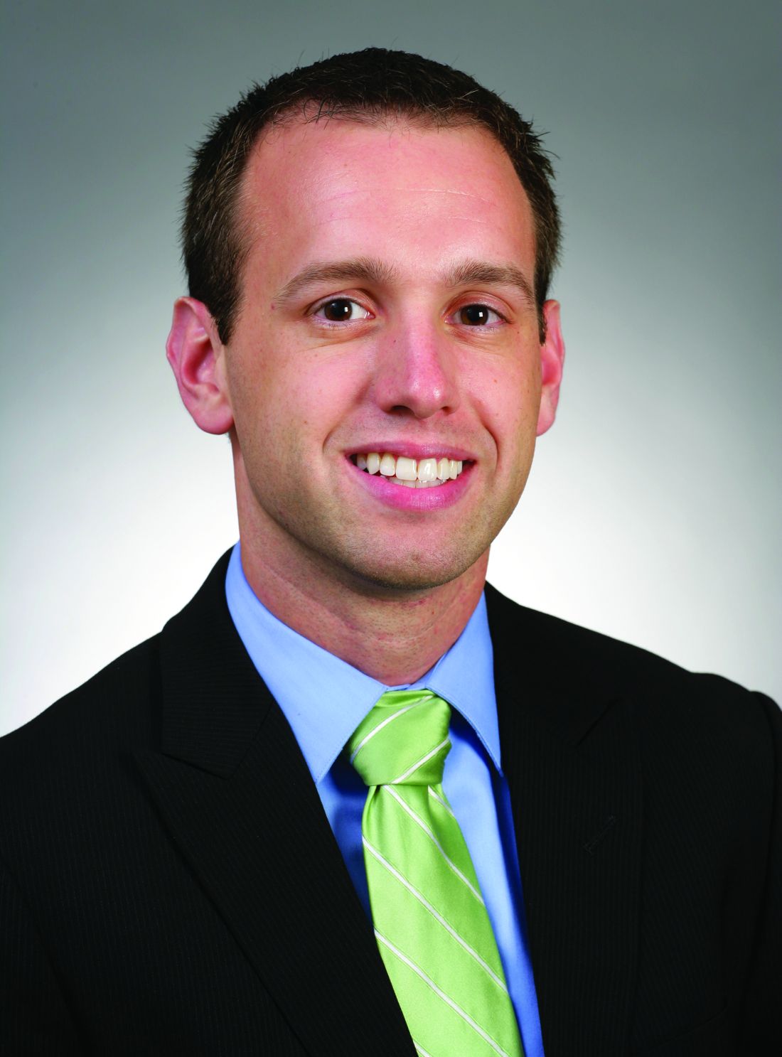
Dr. Bzowyckyj of the University of Missouri-Kansas City School of Pharmacy, and Ashley Crowl, PharmD, BCACP, of the School of Pharmacy at the University of Kansas, spoke about the importance of eGFR at the annual meeting of the American Association of Diabetes Educators.
At issue has been the high rate of chronic kidney disease in diabetics. According to the Centers for Disease Control and Prevention, in 2011, about 50,000 people in the United States began treatment for kidney failure caused by diabetes. And almost 230,000 people were on dialysis or had gotten kidney transplants necessitated by kidney failure that year.
“Educators need to realize their role is not only to manage diabetes but also prevent and educate about concurrent kidney disease,” according to Dr. Bzowyckyj. “We want to empower educators to know they can make a difference.”
It’s important to understand that the serum creatinine level (SCr) itself shouldn’t be used alone to determine kidney function, the pharmacists noted. These tests provide some insight into kidney function by measuring the level of the waste product creatinine; higher levels are a sign that the kidneys aren’t removing waste properly.
But the levels can be misleading. “Some health care professionals see an elevated SCr and quickly label a patient with chronic kidney disease. But other factors need to be considered before providing this diagnosis. You cannot look at one SCr level, which can be increased or decreased by many factors.”
For example, “if you’re a body builder, you’ll have a high creatinine, but your kidneys could be just fine,” Dr. Bzowyckyj said. Dehydration, rhabdomyolysis, and medical problems related to pregnancy can also boost levels, while lower muscle mass and malnutrition can lower them.

According to the National Kidney Foundation, eGFR in most healthy people is 90 mL/min per 1.73 m2 or above. An eGFR of less than 90 mL/min per 1.73 m2 for 3 months or more is a sign of kidney disease.
The two pharmacists said that it’s important for diabetes educators to monitor eGFR because it helps them get a better idea about whether they should urge a physician to adjust the medications taken by their patients. With the help of eGFR, “we can get more people more appropriately on metformin,” Dr. Crowl says.
The FDA is on board. In April, the agency announced labeling changes for metformin drugs “to expand metformin’s use in certain patients with reduced kidney function” and recommended that measurements such as eGFR be used instead of SCr to determine whether a patient with kidney disease should take metformin.
The FDA now believes that “metformin can be used safely in patients with mild impairment in kidney function and in some patients with moderate impairment in kidney function.” It is not recommended for those with severe cases, the agency has said.
Dr. Bzowyckyj and Dr. Crowl reported having no relevant financial disclosures.
SAN DIEGO – Assessing the risk of kidney disease in diabetes requires estimating the glomerular filtration rate.
“One of the big campaigns is Know Your Numbers, and we think of ABC,” also known as hemoglobin A1c, blood pressure, and cholesterol levels, noted Andrew Bzowyckyj, PharmD, BCPS, CDE. “Now we have eGFR, which measures kidney function. It’s something quantifiable that patients can follow.”
Dr. Bzowyckyj of the University of Missouri-Kansas City School of Pharmacy, and Ashley Crowl, PharmD, BCACP, of the School of Pharmacy at the University of Kansas, spoke about the importance of eGFR at the annual meeting of the American Association of Diabetes Educators.
At issue has been the high rate of chronic kidney disease in diabetics. According to the Centers for Disease Control and Prevention, in 2011, about 50,000 people in the United States began treatment for kidney failure caused by diabetes. And almost 230,000 people were on dialysis or had gotten kidney transplants necessitated by kidney failure that year.
“Educators need to realize their role is not only to manage diabetes but also prevent and educate about concurrent kidney disease,” according to Dr. Bzowyckyj. “We want to empower educators to know they can make a difference.”
It’s important to understand that the serum creatinine level (SCr) itself shouldn’t be used alone to determine kidney function, the pharmacists noted. These tests provide some insight into kidney function by measuring the level of the waste product creatinine; higher levels are a sign that the kidneys aren’t removing waste properly.
But the levels can be misleading. “Some health care professionals see an elevated SCr and quickly label a patient with chronic kidney disease. But other factors need to be considered before providing this diagnosis. You cannot look at one SCr level, which can be increased or decreased by many factors.”
For example, “if you’re a body builder, you’ll have a high creatinine, but your kidneys could be just fine,” Dr. Bzowyckyj said. Dehydration, rhabdomyolysis, and medical problems related to pregnancy can also boost levels, while lower muscle mass and malnutrition can lower them.
According to the National Kidney Foundation, eGFR in most healthy people is 90 mL/min per 1.73 m2 or above. An eGFR of less than 90 mL/min per 1.73 m2 for 3 months or more is a sign of kidney disease.
The two pharmacists said that it’s important for diabetes educators to monitor eGFR because it helps them get a better idea about whether they should urge a physician to adjust the medications taken by their patients. With the help of eGFR, “we can get more people more appropriately on metformin,” Dr. Crowl says.
The FDA is on board. In April, the agency announced labeling changes for metformin drugs “to expand metformin’s use in certain patients with reduced kidney function” and recommended that measurements such as eGFR be used instead of SCr to determine whether a patient with kidney disease should take metformin.
The FDA now believes that “metformin can be used safely in patients with mild impairment in kidney function and in some patients with moderate impairment in kidney function.” It is not recommended for those with severe cases, the agency has said.
Dr. Bzowyckyj and Dr. Crowl reported having no relevant financial disclosures.
SAN DIEGO – Assessing the risk of kidney disease in diabetes requires estimating the glomerular filtration rate.
“One of the big campaigns is Know Your Numbers, and we think of ABC,” also known as hemoglobin A1c, blood pressure, and cholesterol levels, noted Andrew Bzowyckyj, PharmD, BCPS, CDE. “Now we have eGFR, which measures kidney function. It’s something quantifiable that patients can follow.”
Dr. Bzowyckyj of the University of Missouri-Kansas City School of Pharmacy, and Ashley Crowl, PharmD, BCACP, of the School of Pharmacy at the University of Kansas, spoke about the importance of eGFR at the annual meeting of the American Association of Diabetes Educators.
At issue has been the high rate of chronic kidney disease in diabetics. According to the Centers for Disease Control and Prevention, in 2011, about 50,000 people in the United States began treatment for kidney failure caused by diabetes. And almost 230,000 people were on dialysis or had gotten kidney transplants necessitated by kidney failure that year.
“Educators need to realize their role is not only to manage diabetes but also prevent and educate about concurrent kidney disease,” according to Dr. Bzowyckyj. “We want to empower educators to know they can make a difference.”
It’s important to understand that the serum creatinine level (SCr) itself shouldn’t be used alone to determine kidney function, the pharmacists noted. These tests provide some insight into kidney function by measuring the level of the waste product creatinine; higher levels are a sign that the kidneys aren’t removing waste properly.
But the levels can be misleading. “Some health care professionals see an elevated SCr and quickly label a patient with chronic kidney disease. But other factors need to be considered before providing this diagnosis. You cannot look at one SCr level, which can be increased or decreased by many factors.”
For example, “if you’re a body builder, you’ll have a high creatinine, but your kidneys could be just fine,” Dr. Bzowyckyj said. Dehydration, rhabdomyolysis, and medical problems related to pregnancy can also boost levels, while lower muscle mass and malnutrition can lower them.
According to the National Kidney Foundation, eGFR in most healthy people is 90 mL/min per 1.73 m2 or above. An eGFR of less than 90 mL/min per 1.73 m2 for 3 months or more is a sign of kidney disease.
The two pharmacists said that it’s important for diabetes educators to monitor eGFR because it helps them get a better idea about whether they should urge a physician to adjust the medications taken by their patients. With the help of eGFR, “we can get more people more appropriately on metformin,” Dr. Crowl says.
The FDA is on board. In April, the agency announced labeling changes for metformin drugs “to expand metformin’s use in certain patients with reduced kidney function” and recommended that measurements such as eGFR be used instead of SCr to determine whether a patient with kidney disease should take metformin.
The FDA now believes that “metformin can be used safely in patients with mild impairment in kidney function and in some patients with moderate impairment in kidney function.” It is not recommended for those with severe cases, the agency has said.
Dr. Bzowyckyj and Dr. Crowl reported having no relevant financial disclosures.
AT THE ANNUAL MEETING OF AADE
Study finds picosecond laser, with diffractive lens array, effective wrinkle treatment
An industry-funded study suggests that picosecond lasers, touted for their effectiveness at tattoo removal, can safely and effectively treat perioral and periocular wrinkles.
Six months after treatment with picosecond 755-nm alexandrite laser with a diffractive lens array, subjects reported high levels of satisfaction and blinded physicians rated the treated wrinkles as improved or much improved over the same time period (Lasers Surg Med. 2016 Sep 29. doi: 10.1002/lsm.22577).
The laser “is very useful for treating fine lines and other visible signs of premature photoaging,” said study coauthor David H. McDaniel, MD, a dermatologist in Virginia Beach, Va., and codirector of the Hampton University Skin of Color Research Institute. “These types of lasers have great potential benefit for patients and minimal risk of any significant adverse events.”
According to Dr. McDaniel, picosecond 755-nm alexandrite lasers are commonly used to treat wrinkles, acne scars, and pigment dyschromia, while picosecond 532- and 1,064-nm lasers are used to remove tattoos and treat some pigment dyschromia.
He and his coinvestigators sought to “better define the parameters that are uniquely contributing to wrinkle reduction and dermal matrix remodeling and also define the actual clinical benefits and treatment protocol,” Dr. McDaniel said in an interview.
At four 1-month intervals, they used a picosecond 755 nm alexandrite laser with diffractive lens array to treat the full faces of 40 women with wrinkles caused by photodamage. The subjects were all healthy white nonsmokers, whose average age was 58 years.
At 6 months following treatment, the average Fitzpatrick wrinkle score improved, dropping from 5.48 to 3.47 (P less than .05). Also at 6 months, blinded physician evaluators rated the average degree of improvement from baseline as “moderate” (for fine lines) “less than mild” (for erythema), “high moderate” (for dyschromia) and “mid moderate” (for global improvement).
The evaluators successfully identified posttreatment photos in 82% of cases. As for patients, 36.8% said they were extremely satisfied and 57.9% said they were satisfied at 6 months. Adverse effects were reported as mild: One patient reported 2 days of erythema, another reported 4 days of edema, and one experienced bruising. Serial punch biopsies obtained at 6 months after the last treatment in six patients revealed increases in dermal collagen and thicker, denser elastin fibers.
The picosecond 755- nm alexandrite laser produces “very little thermal effect, which reduces both treatment discomfort as well as the risk of adverse events,” Dr. McDaniel said in an interview. “It typically leads to shorter recovery time socially, as erythema is very mild and quite transient.”
He noted that the laser can be used in conjunction with other lasers. “Some of the other gold standard fractional lasers are still used in our practice, and they still deliver good results when properly indicated,” he said. “For example, we may use – or even combine with the picosecond laser – a fractional erbium laser to reach deeper into the dermis for severe acne scarring or for deep wrinkles. Or we may use a fractional thulium laser for people with early actinic keratosis or also combine it with the picosecond laser.
Cynosure, a maker of picosecond lasers, funded the study and provided a discounted price for the laser. Dr. McDaniel and another author report serving as consultants, researchers and speakers for Cynosure.
An industry-funded study suggests that picosecond lasers, touted for their effectiveness at tattoo removal, can safely and effectively treat perioral and periocular wrinkles.
Six months after treatment with picosecond 755-nm alexandrite laser with a diffractive lens array, subjects reported high levels of satisfaction and blinded physicians rated the treated wrinkles as improved or much improved over the same time period (Lasers Surg Med. 2016 Sep 29. doi: 10.1002/lsm.22577).
The laser “is very useful for treating fine lines and other visible signs of premature photoaging,” said study coauthor David H. McDaniel, MD, a dermatologist in Virginia Beach, Va., and codirector of the Hampton University Skin of Color Research Institute. “These types of lasers have great potential benefit for patients and minimal risk of any significant adverse events.”
According to Dr. McDaniel, picosecond 755-nm alexandrite lasers are commonly used to treat wrinkles, acne scars, and pigment dyschromia, while picosecond 532- and 1,064-nm lasers are used to remove tattoos and treat some pigment dyschromia.
He and his coinvestigators sought to “better define the parameters that are uniquely contributing to wrinkle reduction and dermal matrix remodeling and also define the actual clinical benefits and treatment protocol,” Dr. McDaniel said in an interview.
At four 1-month intervals, they used a picosecond 755 nm alexandrite laser with diffractive lens array to treat the full faces of 40 women with wrinkles caused by photodamage. The subjects were all healthy white nonsmokers, whose average age was 58 years.
At 6 months following treatment, the average Fitzpatrick wrinkle score improved, dropping from 5.48 to 3.47 (P less than .05). Also at 6 months, blinded physician evaluators rated the average degree of improvement from baseline as “moderate” (for fine lines) “less than mild” (for erythema), “high moderate” (for dyschromia) and “mid moderate” (for global improvement).
The evaluators successfully identified posttreatment photos in 82% of cases. As for patients, 36.8% said they were extremely satisfied and 57.9% said they were satisfied at 6 months. Adverse effects were reported as mild: One patient reported 2 days of erythema, another reported 4 days of edema, and one experienced bruising. Serial punch biopsies obtained at 6 months after the last treatment in six patients revealed increases in dermal collagen and thicker, denser elastin fibers.
The picosecond 755- nm alexandrite laser produces “very little thermal effect, which reduces both treatment discomfort as well as the risk of adverse events,” Dr. McDaniel said in an interview. “It typically leads to shorter recovery time socially, as erythema is very mild and quite transient.”
He noted that the laser can be used in conjunction with other lasers. “Some of the other gold standard fractional lasers are still used in our practice, and they still deliver good results when properly indicated,” he said. “For example, we may use – or even combine with the picosecond laser – a fractional erbium laser to reach deeper into the dermis for severe acne scarring or for deep wrinkles. Or we may use a fractional thulium laser for people with early actinic keratosis or also combine it with the picosecond laser.
Cynosure, a maker of picosecond lasers, funded the study and provided a discounted price for the laser. Dr. McDaniel and another author report serving as consultants, researchers and speakers for Cynosure.
An industry-funded study suggests that picosecond lasers, touted for their effectiveness at tattoo removal, can safely and effectively treat perioral and periocular wrinkles.
Six months after treatment with picosecond 755-nm alexandrite laser with a diffractive lens array, subjects reported high levels of satisfaction and blinded physicians rated the treated wrinkles as improved or much improved over the same time period (Lasers Surg Med. 2016 Sep 29. doi: 10.1002/lsm.22577).
The laser “is very useful for treating fine lines and other visible signs of premature photoaging,” said study coauthor David H. McDaniel, MD, a dermatologist in Virginia Beach, Va., and codirector of the Hampton University Skin of Color Research Institute. “These types of lasers have great potential benefit for patients and minimal risk of any significant adverse events.”
According to Dr. McDaniel, picosecond 755-nm alexandrite lasers are commonly used to treat wrinkles, acne scars, and pigment dyschromia, while picosecond 532- and 1,064-nm lasers are used to remove tattoos and treat some pigment dyschromia.
He and his coinvestigators sought to “better define the parameters that are uniquely contributing to wrinkle reduction and dermal matrix remodeling and also define the actual clinical benefits and treatment protocol,” Dr. McDaniel said in an interview.
At four 1-month intervals, they used a picosecond 755 nm alexandrite laser with diffractive lens array to treat the full faces of 40 women with wrinkles caused by photodamage. The subjects were all healthy white nonsmokers, whose average age was 58 years.
At 6 months following treatment, the average Fitzpatrick wrinkle score improved, dropping from 5.48 to 3.47 (P less than .05). Also at 6 months, blinded physician evaluators rated the average degree of improvement from baseline as “moderate” (for fine lines) “less than mild” (for erythema), “high moderate” (for dyschromia) and “mid moderate” (for global improvement).
The evaluators successfully identified posttreatment photos in 82% of cases. As for patients, 36.8% said they were extremely satisfied and 57.9% said they were satisfied at 6 months. Adverse effects were reported as mild: One patient reported 2 days of erythema, another reported 4 days of edema, and one experienced bruising. Serial punch biopsies obtained at 6 months after the last treatment in six patients revealed increases in dermal collagen and thicker, denser elastin fibers.
The picosecond 755- nm alexandrite laser produces “very little thermal effect, which reduces both treatment discomfort as well as the risk of adverse events,” Dr. McDaniel said in an interview. “It typically leads to shorter recovery time socially, as erythema is very mild and quite transient.”
He noted that the laser can be used in conjunction with other lasers. “Some of the other gold standard fractional lasers are still used in our practice, and they still deliver good results when properly indicated,” he said. “For example, we may use – or even combine with the picosecond laser – a fractional erbium laser to reach deeper into the dermis for severe acne scarring or for deep wrinkles. Or we may use a fractional thulium laser for people with early actinic keratosis or also combine it with the picosecond laser.
Cynosure, a maker of picosecond lasers, funded the study and provided a discounted price for the laser. Dr. McDaniel and another author report serving as consultants, researchers and speakers for Cynosure.
FROM LASERS IN SURGERY AND MEDICINE
Key clinical point: Wrinkle treatment via picosecond 755-nm alexandrite laser with a diffractive lens array appears to be safe and effective.
Major finding: Six months after the last treatment, 36.8% of patients were extremely satisfied and 57.9% were satisfied with the results, with minor, transient adverse effects. Blinded physician evaluators reported “mid moderate” global improvement.
Data source: A prospective, blinded study of 40 healthy white women, nonsmokers, average age 58 (range: 47-64), who underwent four full-face treatments via laser at 1-month intervals.
Disclosures: Cynosure, a maker of picosecond lasers, funded the study and provided a discounted price for the laser. Dr. McDaniel and another author report serving as consultants, researchers, and speakers for Cynosure.
Exenatide/dapagliflozin combo may be better in stubborn diabetes
The combination of exenatide and dapagliflozin produced better results than did either drug alone in patients with type 2 diabetes whose glucose levels aren’t controlled effectively by metformin.
At 28 weeks, HbA1c levels were lower in the combination group, where a third of patients lost more than 5% of their body weight, and 45% reached HbA1c levels under 7.0%, outpacing those on the solo treatments.
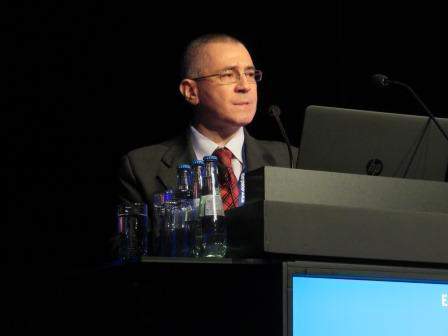
Cristian Guja, MD, of the Carol Davila University of Medicine and Pharmacy in Bucharest, Romania, presented the findings of DURATION-8, a 28-week randomized, double-blinded study at 109 sites in six countries, Sept. 16 at the annual meeting of the European Association for the Study of Diabetes. The study was published simultaneously in the Lancet Diabetes & Endocrinology.
In 2014 and 2015, 695 adults with type 2 diabetes and insufficient glycemic control, defined as HbA1c 8%-12%, were randomly assigned to one of three groups: exenatide plus dapagliflozin (n = 231), exenatide alone (n = 231; n = 1 untreated), or dapagliflozin alone (n = 233). All were given placebo pills or injections for 1 week before randomization.
Patients’ average age was 54-55 years, most were white (82%-85%), with 37%-42% reporting Hispanic heritage. Men and women were nearly equally represented. The average body mass index was 32-33 kg/m2. Patients took basal insulin on a rescue basis as needed and were continued on prescribed blood pressure and cholesterol medications.
At 28 weeks, baseline HbA1c in the combination group fell by 2.0% (9.3% to 7.3%), compared with 1.6% (9.3% to 7.6%) in the exenatide-only group and by 1.4% (9.3% to 7.8%) in the dapagliflozin-only group.
Combination therapy was “significantly superior” to either drug alone for all secondary endpoints, including reduced fasting plasma and postprandial glucose, more patients with an HbA1c less than 7.0%, weight-related measures, and lowered systolic blood pressure (Lancet Diabetes Endocrinol. 2016 Sep 16. doi. org/10.1016/S2213-8587(16)30267-4).
Specifically, the percentage of patients with an HbA1c less than 7.0% was 45% for the combination group, 27% for the exenatide group, and 19% for the dapagliflozin group at 28 weeks. A third of patients taking the combination lost more than 5% of their weight, compared with 14% of those on exenatide alone and 20% of those on dapagliflozin.
The side-effect rate (57%) was highest in the combination group and lower (54% and 52%, respectively) for the exenatide and dapagliflozin groups.
Diarrhea, injection-site nodules, nausea, and urinary tract infection occurred in 5% or more of patients in at least one of the three groups. Across all study groups, 2%-5% of patients discontinued treatment because of side effects; discontinuation was greatest (5%) in the exenatide group. Three patients in the combination group died, as did one patient in each of the solo-treatment groups.
This study “provides high-quality evidence that the combination of exenatide and dapagliflozin is more effective than either drug alone in patients with inadequate response to metformin monotherapy,” Dr. Guja and his colleagues noted.
In a commentary, Michael A. Nauck, MD, and Juris J. Meier, MD, both of St. Josef Hospital in Bochum, Germany, called the percentage of patients (45%) who reached an HbA1c level of less than 7.0% in the combination treatment group “disappointing.” They suggested that GLP-1 receptor agonists (i.e., exenatide) may be more effective when combined with SGLT2 inhibitors (i.e., dapagliflozin) than when used with insulin treatment.
The study was funded by AstraZeneca, maker of both exenatide and dapagliflozin.
The combination of exenatide and dapagliflozin produced better results than did either drug alone in patients with type 2 diabetes whose glucose levels aren’t controlled effectively by metformin.
At 28 weeks, HbA1c levels were lower in the combination group, where a third of patients lost more than 5% of their body weight, and 45% reached HbA1c levels under 7.0%, outpacing those on the solo treatments.
Cristian Guja, MD, of the Carol Davila University of Medicine and Pharmacy in Bucharest, Romania, presented the findings of DURATION-8, a 28-week randomized, double-blinded study at 109 sites in six countries, Sept. 16 at the annual meeting of the European Association for the Study of Diabetes. The study was published simultaneously in the Lancet Diabetes & Endocrinology.
In 2014 and 2015, 695 adults with type 2 diabetes and insufficient glycemic control, defined as HbA1c 8%-12%, were randomly assigned to one of three groups: exenatide plus dapagliflozin (n = 231), exenatide alone (n = 231; n = 1 untreated), or dapagliflozin alone (n = 233). All were given placebo pills or injections for 1 week before randomization.
Patients’ average age was 54-55 years, most were white (82%-85%), with 37%-42% reporting Hispanic heritage. Men and women were nearly equally represented. The average body mass index was 32-33 kg/m2. Patients took basal insulin on a rescue basis as needed and were continued on prescribed blood pressure and cholesterol medications.
At 28 weeks, baseline HbA1c in the combination group fell by 2.0% (9.3% to 7.3%), compared with 1.6% (9.3% to 7.6%) in the exenatide-only group and by 1.4% (9.3% to 7.8%) in the dapagliflozin-only group.
Combination therapy was “significantly superior” to either drug alone for all secondary endpoints, including reduced fasting plasma and postprandial glucose, more patients with an HbA1c less than 7.0%, weight-related measures, and lowered systolic blood pressure (Lancet Diabetes Endocrinol. 2016 Sep 16. doi. org/10.1016/S2213-8587(16)30267-4).
Specifically, the percentage of patients with an HbA1c less than 7.0% was 45% for the combination group, 27% for the exenatide group, and 19% for the dapagliflozin group at 28 weeks. A third of patients taking the combination lost more than 5% of their weight, compared with 14% of those on exenatide alone and 20% of those on dapagliflozin.
The side-effect rate (57%) was highest in the combination group and lower (54% and 52%, respectively) for the exenatide and dapagliflozin groups.
Diarrhea, injection-site nodules, nausea, and urinary tract infection occurred in 5% or more of patients in at least one of the three groups. Across all study groups, 2%-5% of patients discontinued treatment because of side effects; discontinuation was greatest (5%) in the exenatide group. Three patients in the combination group died, as did one patient in each of the solo-treatment groups.
This study “provides high-quality evidence that the combination of exenatide and dapagliflozin is more effective than either drug alone in patients with inadequate response to metformin monotherapy,” Dr. Guja and his colleagues noted.
In a commentary, Michael A. Nauck, MD, and Juris J. Meier, MD, both of St. Josef Hospital in Bochum, Germany, called the percentage of patients (45%) who reached an HbA1c level of less than 7.0% in the combination treatment group “disappointing.” They suggested that GLP-1 receptor agonists (i.e., exenatide) may be more effective when combined with SGLT2 inhibitors (i.e., dapagliflozin) than when used with insulin treatment.
The study was funded by AstraZeneca, maker of both exenatide and dapagliflozin.
The combination of exenatide and dapagliflozin produced better results than did either drug alone in patients with type 2 diabetes whose glucose levels aren’t controlled effectively by metformin.
At 28 weeks, HbA1c levels were lower in the combination group, where a third of patients lost more than 5% of their body weight, and 45% reached HbA1c levels under 7.0%, outpacing those on the solo treatments.
Cristian Guja, MD, of the Carol Davila University of Medicine and Pharmacy in Bucharest, Romania, presented the findings of DURATION-8, a 28-week randomized, double-blinded study at 109 sites in six countries, Sept. 16 at the annual meeting of the European Association for the Study of Diabetes. The study was published simultaneously in the Lancet Diabetes & Endocrinology.
In 2014 and 2015, 695 adults with type 2 diabetes and insufficient glycemic control, defined as HbA1c 8%-12%, were randomly assigned to one of three groups: exenatide plus dapagliflozin (n = 231), exenatide alone (n = 231; n = 1 untreated), or dapagliflozin alone (n = 233). All were given placebo pills or injections for 1 week before randomization.
Patients’ average age was 54-55 years, most were white (82%-85%), with 37%-42% reporting Hispanic heritage. Men and women were nearly equally represented. The average body mass index was 32-33 kg/m2. Patients took basal insulin on a rescue basis as needed and were continued on prescribed blood pressure and cholesterol medications.
At 28 weeks, baseline HbA1c in the combination group fell by 2.0% (9.3% to 7.3%), compared with 1.6% (9.3% to 7.6%) in the exenatide-only group and by 1.4% (9.3% to 7.8%) in the dapagliflozin-only group.
Combination therapy was “significantly superior” to either drug alone for all secondary endpoints, including reduced fasting plasma and postprandial glucose, more patients with an HbA1c less than 7.0%, weight-related measures, and lowered systolic blood pressure (Lancet Diabetes Endocrinol. 2016 Sep 16. doi. org/10.1016/S2213-8587(16)30267-4).
Specifically, the percentage of patients with an HbA1c less than 7.0% was 45% for the combination group, 27% for the exenatide group, and 19% for the dapagliflozin group at 28 weeks. A third of patients taking the combination lost more than 5% of their weight, compared with 14% of those on exenatide alone and 20% of those on dapagliflozin.
The side-effect rate (57%) was highest in the combination group and lower (54% and 52%, respectively) for the exenatide and dapagliflozin groups.
Diarrhea, injection-site nodules, nausea, and urinary tract infection occurred in 5% or more of patients in at least one of the three groups. Across all study groups, 2%-5% of patients discontinued treatment because of side effects; discontinuation was greatest (5%) in the exenatide group. Three patients in the combination group died, as did one patient in each of the solo-treatment groups.
This study “provides high-quality evidence that the combination of exenatide and dapagliflozin is more effective than either drug alone in patients with inadequate response to metformin monotherapy,” Dr. Guja and his colleagues noted.
In a commentary, Michael A. Nauck, MD, and Juris J. Meier, MD, both of St. Josef Hospital in Bochum, Germany, called the percentage of patients (45%) who reached an HbA1c level of less than 7.0% in the combination treatment group “disappointing.” They suggested that GLP-1 receptor agonists (i.e., exenatide) may be more effective when combined with SGLT2 inhibitors (i.e., dapagliflozin) than when used with insulin treatment.
The study was funded by AstraZeneca, maker of both exenatide and dapagliflozin.
FROM EASD 2016
Key clinical point: An exenatide-dapagliflozin combination may be better than either drug alone for patients with type 2 diabetes who have not responded to metformin.
Major finding: At 28 weeks, baseline HbA1c was reduced by 2.0% to 7.3% in the combination group, compared with 1.6% to 7.6% in the exenatide-only group and by 1.4% to 7.8% in the dapagliflozin-only group.
Data source: Double-blind, randomized, active-controlled phase III trial of 685 patients (611 completed) over 28 weeks.
Disclosures: The study was funded by AstraZeneca. Study authors reported grants and similar funding from many pharmaceutical companies including AstraZeneca.
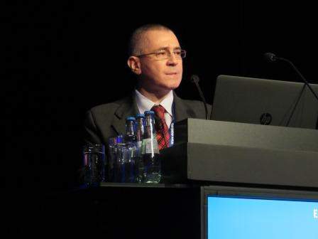
Experts: Insulin apps can pose major risks
SAN DIEGO – How much damage could a diabetes app do? Plenty. That was the word from a Food and Drug Administration official and a technology guru who spoke to a crowd of diabetes educators about the dangers lurking in apps that promise to track things like insulin dosing.
An app could jeopardize the safety of a patient and provide inaccurate data, advised Molly McElwee, RN, CDE, head of Patient Engagement at TypeZero Technologies. “Vet the app that your patient is using or wants to use. Really vet your apps for these patients.”

Ms. McElwee told an audience at the annual meeting of the American Association of Diabetes Educators that more than 84 “bolus calculator” apps are available for iPhone and Android. But only one, the Accu-Check Connect app, is cleared by the FDA for use with a prescription, she confirmed in an interview. Some app makers claim that their technology has been cleared in their countries of origin, but that raises questions about whether the technology has gone through proper vetting, she said.
“The idea you’d let someone with no medical experience just design [an app], put it out there for you, and then tell you how to dose seems a bit odd,” but that’s the way things work in the “murky, uncharted territory” of apps, she said.
Why is there a need for diabetes apps in the first place? As Ms. McElwee noted, many diabetes patients make crucial decisions about their insulin injections each day. “There aren’t a lot of other diseases where you’re dosing something that could kill you if you dose it incorrectly.” That is exactly why “people look for things like an app that can help them,” she noted.
The FDA does indeed regulate medical apps but far from all of them. It doesn’t regulate apps such as calorie counters and exercise trackers because they’re considered to be low risk, said Courtney Lias, PhD, director of the FDA’s Division of Chemistry and Toxicology Devices. “They don’t give advice or tell patients what action to take,” she said. Nor does the FDA regulate apps that retrospectively analyze data from insulin pumps, she said.
The FDA regulates apps such as insulin-dosing calculators and those used to calculate the levels of insulin injections via a pump, she said. But Apple’s App Store and Google’s Google Play, both of which sell apps, aren’t required to take down apps that don’t comply with FDA regulations, according to Ms. McElwee. Indeed, the FDA says it doesn’t regulate app stores, nor does it regulate manufacturers of devices such as smartphones, although the stores may set their own policies. Instead, “the onus is on the company, the developer, to take the correct regulatory pathway,” Ms. McElwee said.
What’s next? Dr. Lias noted that the FDA will monitor apps that command and control insulin pumps, especially as automated insulin delivery systems loom on the horizon. But there are a variety of technical challenges. How will operating systems be updated? What will happen to critical functions when a phone call interrupts the operation of a diabetes app? What about warnings when the phone is muted? Dr. Lias wondered.
One thing is clear, she said: “The explosion of apps is directly related to how much they’re needed.”
Ms. McElwee and Dr. Lias reported having no relevant financial disclosures.
SAN DIEGO – How much damage could a diabetes app do? Plenty. That was the word from a Food and Drug Administration official and a technology guru who spoke to a crowd of diabetes educators about the dangers lurking in apps that promise to track things like insulin dosing.
An app could jeopardize the safety of a patient and provide inaccurate data, advised Molly McElwee, RN, CDE, head of Patient Engagement at TypeZero Technologies. “Vet the app that your patient is using or wants to use. Really vet your apps for these patients.”
Ms. McElwee told an audience at the annual meeting of the American Association of Diabetes Educators that more than 84 “bolus calculator” apps are available for iPhone and Android. But only one, the Accu-Check Connect app, is cleared by the FDA for use with a prescription, she confirmed in an interview. Some app makers claim that their technology has been cleared in their countries of origin, but that raises questions about whether the technology has gone through proper vetting, she said.
“The idea you’d let someone with no medical experience just design [an app], put it out there for you, and then tell you how to dose seems a bit odd,” but that’s the way things work in the “murky, uncharted territory” of apps, she said.
Why is there a need for diabetes apps in the first place? As Ms. McElwee noted, many diabetes patients make crucial decisions about their insulin injections each day. “There aren’t a lot of other diseases where you’re dosing something that could kill you if you dose it incorrectly.” That is exactly why “people look for things like an app that can help them,” she noted.
The FDA does indeed regulate medical apps but far from all of them. It doesn’t regulate apps such as calorie counters and exercise trackers because they’re considered to be low risk, said Courtney Lias, PhD, director of the FDA’s Division of Chemistry and Toxicology Devices. “They don’t give advice or tell patients what action to take,” she said. Nor does the FDA regulate apps that retrospectively analyze data from insulin pumps, she said.
The FDA regulates apps such as insulin-dosing calculators and those used to calculate the levels of insulin injections via a pump, she said. But Apple’s App Store and Google’s Google Play, both of which sell apps, aren’t required to take down apps that don’t comply with FDA regulations, according to Ms. McElwee. Indeed, the FDA says it doesn’t regulate app stores, nor does it regulate manufacturers of devices such as smartphones, although the stores may set their own policies. Instead, “the onus is on the company, the developer, to take the correct regulatory pathway,” Ms. McElwee said.
What’s next? Dr. Lias noted that the FDA will monitor apps that command and control insulin pumps, especially as automated insulin delivery systems loom on the horizon. But there are a variety of technical challenges. How will operating systems be updated? What will happen to critical functions when a phone call interrupts the operation of a diabetes app? What about warnings when the phone is muted? Dr. Lias wondered.
One thing is clear, she said: “The explosion of apps is directly related to how much they’re needed.”
Ms. McElwee and Dr. Lias reported having no relevant financial disclosures.
SAN DIEGO – How much damage could a diabetes app do? Plenty. That was the word from a Food and Drug Administration official and a technology guru who spoke to a crowd of diabetes educators about the dangers lurking in apps that promise to track things like insulin dosing.
An app could jeopardize the safety of a patient and provide inaccurate data, advised Molly McElwee, RN, CDE, head of Patient Engagement at TypeZero Technologies. “Vet the app that your patient is using or wants to use. Really vet your apps for these patients.”
Ms. McElwee told an audience at the annual meeting of the American Association of Diabetes Educators that more than 84 “bolus calculator” apps are available for iPhone and Android. But only one, the Accu-Check Connect app, is cleared by the FDA for use with a prescription, she confirmed in an interview. Some app makers claim that their technology has been cleared in their countries of origin, but that raises questions about whether the technology has gone through proper vetting, she said.
“The idea you’d let someone with no medical experience just design [an app], put it out there for you, and then tell you how to dose seems a bit odd,” but that’s the way things work in the “murky, uncharted territory” of apps, she said.
Why is there a need for diabetes apps in the first place? As Ms. McElwee noted, many diabetes patients make crucial decisions about their insulin injections each day. “There aren’t a lot of other diseases where you’re dosing something that could kill you if you dose it incorrectly.” That is exactly why “people look for things like an app that can help them,” she noted.
The FDA does indeed regulate medical apps but far from all of them. It doesn’t regulate apps such as calorie counters and exercise trackers because they’re considered to be low risk, said Courtney Lias, PhD, director of the FDA’s Division of Chemistry and Toxicology Devices. “They don’t give advice or tell patients what action to take,” she said. Nor does the FDA regulate apps that retrospectively analyze data from insulin pumps, she said.
The FDA regulates apps such as insulin-dosing calculators and those used to calculate the levels of insulin injections via a pump, she said. But Apple’s App Store and Google’s Google Play, both of which sell apps, aren’t required to take down apps that don’t comply with FDA regulations, according to Ms. McElwee. Indeed, the FDA says it doesn’t regulate app stores, nor does it regulate manufacturers of devices such as smartphones, although the stores may set their own policies. Instead, “the onus is on the company, the developer, to take the correct regulatory pathway,” Ms. McElwee said.
What’s next? Dr. Lias noted that the FDA will monitor apps that command and control insulin pumps, especially as automated insulin delivery systems loom on the horizon. But there are a variety of technical challenges. How will operating systems be updated? What will happen to critical functions when a phone call interrupts the operation of a diabetes app? What about warnings when the phone is muted? Dr. Lias wondered.
One thing is clear, she said: “The explosion of apps is directly related to how much they’re needed.”
Ms. McElwee and Dr. Lias reported having no relevant financial disclosures.
AT AADE 16

Sleep doctor: Less than 7 hours can worsen diabetes
SAN DIEGO – A sleep specialist told an audience of diabetes educators that quality sleep is “profoundly important” to the health of their patients, and regularly sleeping fewer than 7 hours a night can wreak havoc on glucose levels and insulin resistance.
There’s even evidence of a link between lack of sleep, diabetes, and heart disease, according to Terese C. Hammond, MD, medical director of the Keck Hospital of the University of Southern California Sleep Disorders Center in Los Angeles. “We don’t know which way the cause goes, but we know there’s a connection,” she said.

Dr. Hammond, who spoke at the annual meeting of the American Association of Diabetes Educators, noted that the vast majority of adults need 7-9 hours of sleep a night.
If you repeatedly don’t reach 7 hours, “there is ample evidence that some metabolic and biologic things happen to your body. Glucose goes up and insulin is secreted. Leptin decreases – the protein that tells us when we’re sated, when our hunger is satisfied. It’s a pretty potent combination, and this is associated with increases in weight and carbohydrate intake,” Dr. Hammond said.
Indeed, findings from one small study suggest a link between chronic insufficient sleep to consumption of more calories, mostly carbohydrates. The study authors speculated this may be because people eat extra food to account for being awake more but take in more than they need (Proc Natl Acad Sci USA. 2013 Apr;110[14]:5695-700).
It is possible for people to pay off a “sleep debt” by catching up on sleep, Dr. Hammond said, but only if the sleep is lost temporarily, as during a heavy medical rotation or after a personal tragedy. When sleep loss is chronic, she said, “it’s becoming apparent that the end-organ consequences are not fully recoverable.”
In regard to diabetes specifically, she said, research has linked insufficient sleep to higher rates of central obesity, higher rates of diabetes in blacks and whites, impaired glucose tolerance, decreased insulin, metabolic syndrome, and high lipids. Too much sleep has also been linked to poor health outcomes.
How can medical professionals help patients improve sleep? “The most important thing to remember about sleep is that it’s a very primitive process,” according to Dr. Hammond. “You can’t think yourself into better sleep. You have to act yourself into better sleep through light, temperature, sound, and repetitive behaviors.”
Controversial research has linked sleeping pills to higher mortality rates, she says.“I try to avoid them long term for insomnia, and not only because there’s probably an increased risk. They stop working eventually.”
Another approach, cognitive behavioral therapy (CBT), is “the most potent way to change behavior,” she said.
“CBT improves sleep efficiency in a vast majority of chronic medical diseases,” she said. “It does just as well as drugs and better than most sleeping pills.”
According to Dr. Hammond, CBT focuses on several strategies:
• Education about sleep hygiene, such as limiting the bedroom to nighttime sleep and intimacy, going to bed at the same time every night, and focusing on a dark, cool, quiet environment.
• Increasing the “sleep drive” through temporary sleep deprivation.
• Relaxation training through techniques like guided imagery, biofeedback, progressive muscle relaxation, and self-hypnosis.
Dr. Hammond reported having no relevant financial disclosures.
SAN DIEGO – A sleep specialist told an audience of diabetes educators that quality sleep is “profoundly important” to the health of their patients, and regularly sleeping fewer than 7 hours a night can wreak havoc on glucose levels and insulin resistance.
There’s even evidence of a link between lack of sleep, diabetes, and heart disease, according to Terese C. Hammond, MD, medical director of the Keck Hospital of the University of Southern California Sleep Disorders Center in Los Angeles. “We don’t know which way the cause goes, but we know there’s a connection,” she said.
Dr. Hammond, who spoke at the annual meeting of the American Association of Diabetes Educators, noted that the vast majority of adults need 7-9 hours of sleep a night.
If you repeatedly don’t reach 7 hours, “there is ample evidence that some metabolic and biologic things happen to your body. Glucose goes up and insulin is secreted. Leptin decreases – the protein that tells us when we’re sated, when our hunger is satisfied. It’s a pretty potent combination, and this is associated with increases in weight and carbohydrate intake,” Dr. Hammond said.
Indeed, findings from one small study suggest a link between chronic insufficient sleep to consumption of more calories, mostly carbohydrates. The study authors speculated this may be because people eat extra food to account for being awake more but take in more than they need (Proc Natl Acad Sci USA. 2013 Apr;110[14]:5695-700).
It is possible for people to pay off a “sleep debt” by catching up on sleep, Dr. Hammond said, but only if the sleep is lost temporarily, as during a heavy medical rotation or after a personal tragedy. When sleep loss is chronic, she said, “it’s becoming apparent that the end-organ consequences are not fully recoverable.”
In regard to diabetes specifically, she said, research has linked insufficient sleep to higher rates of central obesity, higher rates of diabetes in blacks and whites, impaired glucose tolerance, decreased insulin, metabolic syndrome, and high lipids. Too much sleep has also been linked to poor health outcomes.
How can medical professionals help patients improve sleep? “The most important thing to remember about sleep is that it’s a very primitive process,” according to Dr. Hammond. “You can’t think yourself into better sleep. You have to act yourself into better sleep through light, temperature, sound, and repetitive behaviors.”
Controversial research has linked sleeping pills to higher mortality rates, she says.“I try to avoid them long term for insomnia, and not only because there’s probably an increased risk. They stop working eventually.”
Another approach, cognitive behavioral therapy (CBT), is “the most potent way to change behavior,” she said.
“CBT improves sleep efficiency in a vast majority of chronic medical diseases,” she said. “It does just as well as drugs and better than most sleeping pills.”
According to Dr. Hammond, CBT focuses on several strategies:
• Education about sleep hygiene, such as limiting the bedroom to nighttime sleep and intimacy, going to bed at the same time every night, and focusing on a dark, cool, quiet environment.
• Increasing the “sleep drive” through temporary sleep deprivation.
• Relaxation training through techniques like guided imagery, biofeedback, progressive muscle relaxation, and self-hypnosis.
Dr. Hammond reported having no relevant financial disclosures.
SAN DIEGO – A sleep specialist told an audience of diabetes educators that quality sleep is “profoundly important” to the health of their patients, and regularly sleeping fewer than 7 hours a night can wreak havoc on glucose levels and insulin resistance.
There’s even evidence of a link between lack of sleep, diabetes, and heart disease, according to Terese C. Hammond, MD, medical director of the Keck Hospital of the University of Southern California Sleep Disorders Center in Los Angeles. “We don’t know which way the cause goes, but we know there’s a connection,” she said.
Dr. Hammond, who spoke at the annual meeting of the American Association of Diabetes Educators, noted that the vast majority of adults need 7-9 hours of sleep a night.
If you repeatedly don’t reach 7 hours, “there is ample evidence that some metabolic and biologic things happen to your body. Glucose goes up and insulin is secreted. Leptin decreases – the protein that tells us when we’re sated, when our hunger is satisfied. It’s a pretty potent combination, and this is associated with increases in weight and carbohydrate intake,” Dr. Hammond said.
Indeed, findings from one small study suggest a link between chronic insufficient sleep to consumption of more calories, mostly carbohydrates. The study authors speculated this may be because people eat extra food to account for being awake more but take in more than they need (Proc Natl Acad Sci USA. 2013 Apr;110[14]:5695-700).
It is possible for people to pay off a “sleep debt” by catching up on sleep, Dr. Hammond said, but only if the sleep is lost temporarily, as during a heavy medical rotation or after a personal tragedy. When sleep loss is chronic, she said, “it’s becoming apparent that the end-organ consequences are not fully recoverable.”
In regard to diabetes specifically, she said, research has linked insufficient sleep to higher rates of central obesity, higher rates of diabetes in blacks and whites, impaired glucose tolerance, decreased insulin, metabolic syndrome, and high lipids. Too much sleep has also been linked to poor health outcomes.
How can medical professionals help patients improve sleep? “The most important thing to remember about sleep is that it’s a very primitive process,” according to Dr. Hammond. “You can’t think yourself into better sleep. You have to act yourself into better sleep through light, temperature, sound, and repetitive behaviors.”
Controversial research has linked sleeping pills to higher mortality rates, she says.“I try to avoid them long term for insomnia, and not only because there’s probably an increased risk. They stop working eventually.”
Another approach, cognitive behavioral therapy (CBT), is “the most potent way to change behavior,” she said.
“CBT improves sleep efficiency in a vast majority of chronic medical diseases,” she said. “It does just as well as drugs and better than most sleeping pills.”
According to Dr. Hammond, CBT focuses on several strategies:
• Education about sleep hygiene, such as limiting the bedroom to nighttime sleep and intimacy, going to bed at the same time every night, and focusing on a dark, cool, quiet environment.
• Increasing the “sleep drive” through temporary sleep deprivation.
• Relaxation training through techniques like guided imagery, biofeedback, progressive muscle relaxation, and self-hypnosis.
Dr. Hammond reported having no relevant financial disclosures.
AT AADE 16

Endocrinologist links nighttime hypoglycemia to many ills
SAN DIEGO – A top endocrinologist cautioned diabetes educators that research is linking nighttime hypoglycemia to a variety of ills, and technology isn’t providing much hope – yet.
Patients with nocturnal low blood sugar “say this is the hardest thing they have to deal with. It upsets their whole day and they feel terrible,” said Anthony L. McCall, MD, PhD, James M. Moss Professor of Diabetes at the University of Virginia, Charlottesville, and vice president of clinical science with the Endocrine Society.
Dr. McCall told an audience at the annual meeting of the American Association of Diabetes Educators that half of hypoglycemia is nocturnal and unrecognized despite its dangers. According to him, hypoglycemia represents a blood glucose level of at or under 70 mg/dL (3.9 mmol/L). This is higher than the threshold for hypoglycemia in nondiabetics and those with well controlled diabetes.
Even as few as two values in a week in the range of the 60s (mg/dL) can go unrecognized and lead to full-blown hypoglycemia-associated autonomic failure, he said. There are other possible risks: “impaired sleep quality, daytime drowsiness, mood changes, risk for nocturnal falls,” he said.
Cognitive dysfunction is possible, especially in children, he added. “Neurological dysfunction may be temporary, but those who can answer simple questions may not be OK.”
There’s a potential for a vicious cycle here, he said, because people with diabetes can also develop impaired hypoglycemia awareness, making it less likely they’ll notice the low blood sugar levels that contribute to autonomic failure.
Dr. McCall reported that nighttime hypoglycemia may also:
• Trigger neurologic symptoms like those of strokes or temporary ischemic attacks. “Someone’s got check to their blood sugar,” he says.
• Lengthen the QT interval and boost the risk of irregular heartbeats.
• Contribute to “dead in bed” syndrome in which young people with type 1 diabetes are discovered dead despite not having any complications or showing signs of convulsion.
What can be done to help these patients? One approach is to combat impaired hypoglycemia awareness. Bedtime snacks, caffeine, and uncooked cornstarch are among the many nutrition supplements (and medications) that have shown inconsistent results at best on this front, Dr. McCall said. If they work, he said, they often lead to hyperglycemia.
Another strategy is to look for factors that raise the risk of nighttime hypoglycemia, such as basal insulin overtreatment, long periods between meals, delayed effects of exercise, and higher insulin sensitivity overnight.
Insulin pumps may be helpful, he said, and he generally favors their use. However, he cautioned that it’s hard to show that they reduce hypoglycemia, and some patients don’t use them properly.
Data have been mixed until recently regarding real-time continuous glucose monitoring, he said, and the devices must be worn 75%-85% of the time to show benefit. As for sensor-augmented insulin pumps, he said they’ve shown mixed results.
Dr. McCall said the artificial pancreas, once it makes it to market, could mark the beginning of a new era. “This was around the corner 40 years ago. But it’s closer now,” he said. “I have great hope that we’re going to do better.”
Dr. McCall reported being a consultant to Sanofi regarding new insulin studies and serving on the advisory board of DexCom/Google regarding the use of continuous glucose monitoring.
SAN DIEGO – A top endocrinologist cautioned diabetes educators that research is linking nighttime hypoglycemia to a variety of ills, and technology isn’t providing much hope – yet.
Patients with nocturnal low blood sugar “say this is the hardest thing they have to deal with. It upsets their whole day and they feel terrible,” said Anthony L. McCall, MD, PhD, James M. Moss Professor of Diabetes at the University of Virginia, Charlottesville, and vice president of clinical science with the Endocrine Society.
Dr. McCall told an audience at the annual meeting of the American Association of Diabetes Educators that half of hypoglycemia is nocturnal and unrecognized despite its dangers. According to him, hypoglycemia represents a blood glucose level of at or under 70 mg/dL (3.9 mmol/L). This is higher than the threshold for hypoglycemia in nondiabetics and those with well controlled diabetes.
Even as few as two values in a week in the range of the 60s (mg/dL) can go unrecognized and lead to full-blown hypoglycemia-associated autonomic failure, he said. There are other possible risks: “impaired sleep quality, daytime drowsiness, mood changes, risk for nocturnal falls,” he said.
Cognitive dysfunction is possible, especially in children, he added. “Neurological dysfunction may be temporary, but those who can answer simple questions may not be OK.”
There’s a potential for a vicious cycle here, he said, because people with diabetes can also develop impaired hypoglycemia awareness, making it less likely they’ll notice the low blood sugar levels that contribute to autonomic failure.
Dr. McCall reported that nighttime hypoglycemia may also:
• Trigger neurologic symptoms like those of strokes or temporary ischemic attacks. “Someone’s got check to their blood sugar,” he says.
• Lengthen the QT interval and boost the risk of irregular heartbeats.
• Contribute to “dead in bed” syndrome in which young people with type 1 diabetes are discovered dead despite not having any complications or showing signs of convulsion.
What can be done to help these patients? One approach is to combat impaired hypoglycemia awareness. Bedtime snacks, caffeine, and uncooked cornstarch are among the many nutrition supplements (and medications) that have shown inconsistent results at best on this front, Dr. McCall said. If they work, he said, they often lead to hyperglycemia.
Another strategy is to look for factors that raise the risk of nighttime hypoglycemia, such as basal insulin overtreatment, long periods between meals, delayed effects of exercise, and higher insulin sensitivity overnight.
Insulin pumps may be helpful, he said, and he generally favors their use. However, he cautioned that it’s hard to show that they reduce hypoglycemia, and some patients don’t use them properly.
Data have been mixed until recently regarding real-time continuous glucose monitoring, he said, and the devices must be worn 75%-85% of the time to show benefit. As for sensor-augmented insulin pumps, he said they’ve shown mixed results.
Dr. McCall said the artificial pancreas, once it makes it to market, could mark the beginning of a new era. “This was around the corner 40 years ago. But it’s closer now,” he said. “I have great hope that we’re going to do better.”
Dr. McCall reported being a consultant to Sanofi regarding new insulin studies and serving on the advisory board of DexCom/Google regarding the use of continuous glucose monitoring.
SAN DIEGO – A top endocrinologist cautioned diabetes educators that research is linking nighttime hypoglycemia to a variety of ills, and technology isn’t providing much hope – yet.
Patients with nocturnal low blood sugar “say this is the hardest thing they have to deal with. It upsets their whole day and they feel terrible,” said Anthony L. McCall, MD, PhD, James M. Moss Professor of Diabetes at the University of Virginia, Charlottesville, and vice president of clinical science with the Endocrine Society.
Dr. McCall told an audience at the annual meeting of the American Association of Diabetes Educators that half of hypoglycemia is nocturnal and unrecognized despite its dangers. According to him, hypoglycemia represents a blood glucose level of at or under 70 mg/dL (3.9 mmol/L). This is higher than the threshold for hypoglycemia in nondiabetics and those with well controlled diabetes.
Even as few as two values in a week in the range of the 60s (mg/dL) can go unrecognized and lead to full-blown hypoglycemia-associated autonomic failure, he said. There are other possible risks: “impaired sleep quality, daytime drowsiness, mood changes, risk for nocturnal falls,” he said.
Cognitive dysfunction is possible, especially in children, he added. “Neurological dysfunction may be temporary, but those who can answer simple questions may not be OK.”
There’s a potential for a vicious cycle here, he said, because people with diabetes can also develop impaired hypoglycemia awareness, making it less likely they’ll notice the low blood sugar levels that contribute to autonomic failure.
Dr. McCall reported that nighttime hypoglycemia may also:
• Trigger neurologic symptoms like those of strokes or temporary ischemic attacks. “Someone’s got check to their blood sugar,” he says.
• Lengthen the QT interval and boost the risk of irregular heartbeats.
• Contribute to “dead in bed” syndrome in which young people with type 1 diabetes are discovered dead despite not having any complications or showing signs of convulsion.
What can be done to help these patients? One approach is to combat impaired hypoglycemia awareness. Bedtime snacks, caffeine, and uncooked cornstarch are among the many nutrition supplements (and medications) that have shown inconsistent results at best on this front, Dr. McCall said. If they work, he said, they often lead to hyperglycemia.
Another strategy is to look for factors that raise the risk of nighttime hypoglycemia, such as basal insulin overtreatment, long periods between meals, delayed effects of exercise, and higher insulin sensitivity overnight.
Insulin pumps may be helpful, he said, and he generally favors their use. However, he cautioned that it’s hard to show that they reduce hypoglycemia, and some patients don’t use them properly.
Data have been mixed until recently regarding real-time continuous glucose monitoring, he said, and the devices must be worn 75%-85% of the time to show benefit. As for sensor-augmented insulin pumps, he said they’ve shown mixed results.
Dr. McCall said the artificial pancreas, once it makes it to market, could mark the beginning of a new era. “This was around the corner 40 years ago. But it’s closer now,” he said. “I have great hope that we’re going to do better.”
Dr. McCall reported being a consultant to Sanofi regarding new insulin studies and serving on the advisory board of DexCom/Google regarding the use of continuous glucose monitoring.
AT AADE 16