User login
A persistently swollen lip
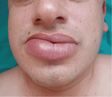
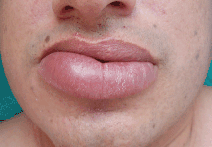
A 44-year-old man is referred for evaluation of asymptomatic swelling of the lower lip that has persisted for 10 months. He has been treated unsuccessfully with oral antihistamines for suspected chronic angioedema. He has no other symptoms and appears to be well otherwise. He has no history of applied irritants or local trauma, and his medical history is unremarkable.
Results of the laboratory evaluation, including serum angiotensin-converting enzyme level, are normal. Patch tests to detect contact sensitivity to food additives are negative. Biopsy of the affected lip reveals dense infiltrate of the submucosal connective tissue with focal nonnecrotizing granulomas. Imaging and endoscopic studies show no evidence of sarcoidosis or Crohn disease.
Q: Given what we know so far, which of the following is the most likely diagnosis of the persistent lip swelling?
- Melkersson-Rosenthal syndrome
- Amyloidosis
- Quincke edema
- Cheilitis granulomatosa
- Cutaneous tuberculosis
A: From what we know so far, the correct answer is cheilitis granulomatosa. While this rare condition may be a feature of Melkersson-Rosenthal syndrome and amyloidosis, at this point in the evaluation these have not been confirmed. Quincke edema (ie, angioedema) is unlikely, given the ineffectiveness of previous treatment with oral antihistamines. Cutaneous tuberculosis usually presents as “lupus vulgaris,” which is characterized by solitary, small, sharply marginated, red-brown papules of gelatinous consistency (“apple-jelly nodules”), mainly on the head and neck.
Cheilitis granulomatosa is a rare inflammatory disorder1 that primarily affects young adults. Its key feature is recurrent or persistent painless swelling of one or both lips. It may occur without other signs of disease, but it is also a manifestation of Melkersson-Rosenthal syndrome and it may be a presenting symptom of Crohn disease or, rarely, sarcoidosis.2 The term “orofacial granulomatosis” was introduced to encompass the broad spectrum of nonnecrotizing granulomatous inflammation in the orofacial region, including cheilitis granulomatosa, the complete Melkersson-Rosenthal syndrome, sarcoidosis, Crohn disease, and infectious disorders such as tuberculosis.1
The cause of cheilitis granulomatosa is unknown. Specific T-cell clonality has been identified in several patients with orofacial granulomatosis, suggesting a delayed hypersensitivity response. Moreover, the HLA haplotypes HLA-A2 and HLA-A11 have been found in 25% of patients with orofacial granulomatosis, suggesting a viral etiology. A genetic predisposition may exist in Melkersson- Rosenthal syndrome: siblings have been affected, and otherwise unaffected relatives may have a fissured tongue (lingua plicata).
Melkersson-Rosenthal syndrome, a rare condition, is characterized by a classic triad of recurrent swelling of the lips or face (or both), fissured tongue, and relapsing peripheral facial nerve paralysis. It is an unusual cause of facial swelling that can be confused with angioedema.3 This syndrome can be ruled out in this patient because he has only one of the three classic signs. Contact antigens are sometimes implicated.
DIFFERENTIAL DIAGNOSIS OF CHEILITIS GRANULOMATOSA
The differential diagnosis of cheilitis granulomatosa is extensive and includes amyloidosis, cheilitis glandularis, sarcoidosis, Crohn disease, actinic cheilitis, neoplasms, and infections, such as tuberculosis, syphilis, and leprosy.1
As many as 11% of patients with Crohn disease may develop mucocutaneous lesions. Oral lesions of Crohn disease include apthae, cobblestoning of the buccal mucosa, swelling of one or both lips (soft or rubbery), vertical clefts of the lips, or hypertrophic gingivitis. Only 5% of patients with Crohn disease ever develop cheilitis granulomatosa, though most cases occur in children.
Ultimately, the diagnosis of cheilitis granulomatosa is made by correlating the patient’s history and clinical features, usually supported by histopathologic findings of nonnecrotic granulomas extending into the deep dermis, composed of histiocytes and giant cells and associated with a lymphomonocytic infiltrate.
TREATMENT
Treatment of cheilitis granulomatosa is difficult because the cause is unknown and the rate of recurrence is high. Response to treatment is often late and unpredictable. Corticosteroids, clofazimine (Lamprene), and surgical intervention such as cheiloplasty have been described as treatment options. Other treatment options include thalidomide (Thalomid), sulfasalazine (Sulfazine), erythromycin, azathioprine (Imuran), and cyclosporine (Sandimmune). Infliximab (Remicade) has been recently reported as a new alternative treatment, in particular for Melkersson-Rosenthal syndrome.4
In our patient, twice-monthly injections of 1 mL of triamcinolone acetonide 10 mg/mL into the affected lip brought acceptable improvement at 3 months. The patient is on maintenance treatment with twice-monthly triamcinolone injections and has had no relapses after 2 years.
- van der Waal RI, Schulten EA, van de Scheur MR, Wauters IM, Starink TM, van der Waal I. Cheilitis granulomatosa. J Eur Acad Dermatol Venereol 2001; 15:519–523.
- van der Waal RI, Schulten EA, van der Meij EH, van de Scheur MR, Starink TM, van der Waal I. Cheilitis granulomatosa: overview of 13 patients with long-term follow-up—results of management. Int J Dermatol 2002; 41:225–229.
- Kakimoto C, Sparks C, White AA. Melkersson-Rosenthal syndrome: a form of pseudoangioedema. Ann Allergy Asthma Immunol 2007; 99:185–189.
- Ratzinger G, Sepp N, Vogetseder W, Tilg H. Cheilitis granulomatosa and Melkersson-Rosenthal syndrome: evaluation of gastrointestinal involvement and therapeutic regimens in a series of 14 patients. J Eur Acad Dermatol Venereol 2007; 21:1065–1070.
A 44-year-old man is referred for evaluation of asymptomatic swelling of the lower lip that has persisted for 10 months. He has been treated unsuccessfully with oral antihistamines for suspected chronic angioedema. He has no other symptoms and appears to be well otherwise. He has no history of applied irritants or local trauma, and his medical history is unremarkable.
Results of the laboratory evaluation, including serum angiotensin-converting enzyme level, are normal. Patch tests to detect contact sensitivity to food additives are negative. Biopsy of the affected lip reveals dense infiltrate of the submucosal connective tissue with focal nonnecrotizing granulomas. Imaging and endoscopic studies show no evidence of sarcoidosis or Crohn disease.
Q: Given what we know so far, which of the following is the most likely diagnosis of the persistent lip swelling?
- Melkersson-Rosenthal syndrome
- Amyloidosis
- Quincke edema
- Cheilitis granulomatosa
- Cutaneous tuberculosis
A: From what we know so far, the correct answer is cheilitis granulomatosa. While this rare condition may be a feature of Melkersson-Rosenthal syndrome and amyloidosis, at this point in the evaluation these have not been confirmed. Quincke edema (ie, angioedema) is unlikely, given the ineffectiveness of previous treatment with oral antihistamines. Cutaneous tuberculosis usually presents as “lupus vulgaris,” which is characterized by solitary, small, sharply marginated, red-brown papules of gelatinous consistency (“apple-jelly nodules”), mainly on the head and neck.
Cheilitis granulomatosa is a rare inflammatory disorder1 that primarily affects young adults. Its key feature is recurrent or persistent painless swelling of one or both lips. It may occur without other signs of disease, but it is also a manifestation of Melkersson-Rosenthal syndrome and it may be a presenting symptom of Crohn disease or, rarely, sarcoidosis.2 The term “orofacial granulomatosis” was introduced to encompass the broad spectrum of nonnecrotizing granulomatous inflammation in the orofacial region, including cheilitis granulomatosa, the complete Melkersson-Rosenthal syndrome, sarcoidosis, Crohn disease, and infectious disorders such as tuberculosis.1
The cause of cheilitis granulomatosa is unknown. Specific T-cell clonality has been identified in several patients with orofacial granulomatosis, suggesting a delayed hypersensitivity response. Moreover, the HLA haplotypes HLA-A2 and HLA-A11 have been found in 25% of patients with orofacial granulomatosis, suggesting a viral etiology. A genetic predisposition may exist in Melkersson- Rosenthal syndrome: siblings have been affected, and otherwise unaffected relatives may have a fissured tongue (lingua plicata).
Melkersson-Rosenthal syndrome, a rare condition, is characterized by a classic triad of recurrent swelling of the lips or face (or both), fissured tongue, and relapsing peripheral facial nerve paralysis. It is an unusual cause of facial swelling that can be confused with angioedema.3 This syndrome can be ruled out in this patient because he has only one of the three classic signs. Contact antigens are sometimes implicated.
DIFFERENTIAL DIAGNOSIS OF CHEILITIS GRANULOMATOSA
The differential diagnosis of cheilitis granulomatosa is extensive and includes amyloidosis, cheilitis glandularis, sarcoidosis, Crohn disease, actinic cheilitis, neoplasms, and infections, such as tuberculosis, syphilis, and leprosy.1
As many as 11% of patients with Crohn disease may develop mucocutaneous lesions. Oral lesions of Crohn disease include apthae, cobblestoning of the buccal mucosa, swelling of one or both lips (soft or rubbery), vertical clefts of the lips, or hypertrophic gingivitis. Only 5% of patients with Crohn disease ever develop cheilitis granulomatosa, though most cases occur in children.
Ultimately, the diagnosis of cheilitis granulomatosa is made by correlating the patient’s history and clinical features, usually supported by histopathologic findings of nonnecrotic granulomas extending into the deep dermis, composed of histiocytes and giant cells and associated with a lymphomonocytic infiltrate.
TREATMENT
Treatment of cheilitis granulomatosa is difficult because the cause is unknown and the rate of recurrence is high. Response to treatment is often late and unpredictable. Corticosteroids, clofazimine (Lamprene), and surgical intervention such as cheiloplasty have been described as treatment options. Other treatment options include thalidomide (Thalomid), sulfasalazine (Sulfazine), erythromycin, azathioprine (Imuran), and cyclosporine (Sandimmune). Infliximab (Remicade) has been recently reported as a new alternative treatment, in particular for Melkersson-Rosenthal syndrome.4
In our patient, twice-monthly injections of 1 mL of triamcinolone acetonide 10 mg/mL into the affected lip brought acceptable improvement at 3 months. The patient is on maintenance treatment with twice-monthly triamcinolone injections and has had no relapses after 2 years.
- van der Waal RI, Schulten EA, van de Scheur MR, Wauters IM, Starink TM, van der Waal I. Cheilitis granulomatosa. J Eur Acad Dermatol Venereol 2001; 15:519–523.
- van der Waal RI, Schulten EA, van der Meij EH, van de Scheur MR, Starink TM, van der Waal I. Cheilitis granulomatosa: overview of 13 patients with long-term follow-up—results of management. Int J Dermatol 2002; 41:225–229.
- Kakimoto C, Sparks C, White AA. Melkersson-Rosenthal syndrome: a form of pseudoangioedema. Ann Allergy Asthma Immunol 2007; 99:185–189.
- Ratzinger G, Sepp N, Vogetseder W, Tilg H. Cheilitis granulomatosa and Melkersson-Rosenthal syndrome: evaluation of gastrointestinal involvement and therapeutic regimens in a series of 14 patients. J Eur Acad Dermatol Venereol 2007; 21:1065–1070.
Black hairy tongue
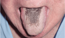
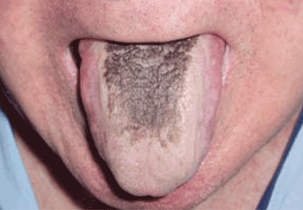
A 71-year-old man presents for evaluation of an asymptomatic black discoloration of the tongue that he noticed several days earlier. The tongue does not itch or hurt, and the patient is otherwise well, although he is concerned about potential malignancy.
He has a history of hypertension, hyperuricemia, and type 2 diabetes treated with oral glucose-lowering drugs, and he has had no recent changes in his medications. He drinks coffee and uses tobacco. His oral hygiene is poor, with intense halitosis.
Q: What is the most likely diagnosis?
- Oral leukoplakia
- Epidermoid carcinoma of the tongue
- Malignant melanoma of the tongue
- Mucosal candidiasis
- Black hairy tongue
A: Black hairy tongue is correct. A simple treatment consisting of brushing the tongue daily with a soft toothbrush enhanced by previous application of 30% urea is recommended to the patient, and the discoloration resolves completely within 4 weeks. He is educated on correct oral hygiene and discontinues smoking, with no clinical relapses after 2 years of follow-up.
THE CAUSES AND THE COURSE
Black hairy tongue, also known as lingua villosa nigra, is a painless, benign disorder caused by defective desquamation and reactive hypertrophy of the filiform papillae of the tongue. The hairy appearance is due to elongation of keratinized filiform papillae, which may have different colors, varying from white to yellowish brown to black depending on extrinsic factors (eg, tobacco, coffee, tea, food) and intrinsic factors (ie, chromogenic organisms in normal flora).1
The exact pathogenesis is unclear. Precipitating factors include poor oral hygiene, use of the antipsychotic drug olanzapine1 (Zyprexa) or a broad-spectrum antibiotic such as erythromycin,2 and therapeutic radiation of the head and the neck. Tobacco use and drinking coffee and tea are also contributory factors. Neurologic conditions such as trigeminal neuropathy may be associated. 3 Manabe et al4 applied a panel of antikeratin probes, showing that defective desquamation of the cells in the central column of filiform papillae resulted in the formation of highly elongated, cornified spines or “hairs”—the hallmark of lingua villosa nigra.
PRESENTATION AND DIAGNOSIS
Black hairy tongue is usually asymptomatic. However, symptoms such as altered (metallic) taste, nausea, or halitosis may be noted. Most patients with hairy tongue drink coffee or tea, often in addition to tobacco use.
The diagnosis is based on filiform papillae that are elongated more than 3 mm on the dorsal surface of the tongue. Cultures may be taken to rule out a superimposed oral candidiasis or other suspected oral infection.
MANAGEMENT
Although frightening to the patient, black hairy tongue is completely harmless. In most cases, treatment does not require drugs. If fungal overgrowth is present, a topical antifungal can be used when the condition is symptomatic.
Empirical approaches such as brushing or scraping the tongue, improving oral hygiene, and eliminating potential offending factors (eg, tobacco, candies, strong mouthwashes, antibiotics) is usually sufficient to resolve the lesions.5
In our experience, educating the patient about proper oral hygiene (including discontinuing smoking) and encouraging routine tongue brushing are the best preventive and therapeutic measures.
- Tamam L, Annagur BB. Black hairy tongue associated with olanzapine treatment: a case report. Mt Sinai J Med 2006; 73:891–894.
- Pigatto PD, Spadari F, Meroni L, Guzzi G. Black hairy tongue associated with long-term oral erythromycin use. J Eur Acad Dermatol Venereol 2008; 22:1269–1270.
- Chesire WP. Unilateral black hairy tongue in trigeminal neuralgia. Headache 2004; 44:908–910.
- Manabe M, Lim HW, Winzer M, Loomis CA. Architectural organization of filiform papillae in normal and black hairy tongue epithelium: dissection of differentiation pathways in a complex human epithelium according to their patterns of keratin expression. Arch Dermatol 1999; 135:177–181.
- Sarti GM, Haddy RI, Schaffer D, Kihm J. Black hairy tongue. Am Fam Physician 1990; 41:1751–1755.
A 71-year-old man presents for evaluation of an asymptomatic black discoloration of the tongue that he noticed several days earlier. The tongue does not itch or hurt, and the patient is otherwise well, although he is concerned about potential malignancy.
He has a history of hypertension, hyperuricemia, and type 2 diabetes treated with oral glucose-lowering drugs, and he has had no recent changes in his medications. He drinks coffee and uses tobacco. His oral hygiene is poor, with intense halitosis.
Q: What is the most likely diagnosis?
- Oral leukoplakia
- Epidermoid carcinoma of the tongue
- Malignant melanoma of the tongue
- Mucosal candidiasis
- Black hairy tongue
A: Black hairy tongue is correct. A simple treatment consisting of brushing the tongue daily with a soft toothbrush enhanced by previous application of 30% urea is recommended to the patient, and the discoloration resolves completely within 4 weeks. He is educated on correct oral hygiene and discontinues smoking, with no clinical relapses after 2 years of follow-up.
THE CAUSES AND THE COURSE
Black hairy tongue, also known as lingua villosa nigra, is a painless, benign disorder caused by defective desquamation and reactive hypertrophy of the filiform papillae of the tongue. The hairy appearance is due to elongation of keratinized filiform papillae, which may have different colors, varying from white to yellowish brown to black depending on extrinsic factors (eg, tobacco, coffee, tea, food) and intrinsic factors (ie, chromogenic organisms in normal flora).1
The exact pathogenesis is unclear. Precipitating factors include poor oral hygiene, use of the antipsychotic drug olanzapine1 (Zyprexa) or a broad-spectrum antibiotic such as erythromycin,2 and therapeutic radiation of the head and the neck. Tobacco use and drinking coffee and tea are also contributory factors. Neurologic conditions such as trigeminal neuropathy may be associated. 3 Manabe et al4 applied a panel of antikeratin probes, showing that defective desquamation of the cells in the central column of filiform papillae resulted in the formation of highly elongated, cornified spines or “hairs”—the hallmark of lingua villosa nigra.
PRESENTATION AND DIAGNOSIS
Black hairy tongue is usually asymptomatic. However, symptoms such as altered (metallic) taste, nausea, or halitosis may be noted. Most patients with hairy tongue drink coffee or tea, often in addition to tobacco use.
The diagnosis is based on filiform papillae that are elongated more than 3 mm on the dorsal surface of the tongue. Cultures may be taken to rule out a superimposed oral candidiasis or other suspected oral infection.
MANAGEMENT
Although frightening to the patient, black hairy tongue is completely harmless. In most cases, treatment does not require drugs. If fungal overgrowth is present, a topical antifungal can be used when the condition is symptomatic.
Empirical approaches such as brushing or scraping the tongue, improving oral hygiene, and eliminating potential offending factors (eg, tobacco, candies, strong mouthwashes, antibiotics) is usually sufficient to resolve the lesions.5
In our experience, educating the patient about proper oral hygiene (including discontinuing smoking) and encouraging routine tongue brushing are the best preventive and therapeutic measures.
- Tamam L, Annagur BB. Black hairy tongue associated with olanzapine treatment: a case report. Mt Sinai J Med 2006; 73:891–894.
- Pigatto PD, Spadari F, Meroni L, Guzzi G. Black hairy tongue associated with long-term oral erythromycin use. J Eur Acad Dermatol Venereol 2008; 22:1269–1270.
- Chesire WP. Unilateral black hairy tongue in trigeminal neuralgia. Headache 2004; 44:908–910.
- Manabe M, Lim HW, Winzer M, Loomis CA. Architectural organization of filiform papillae in normal and black hairy tongue epithelium: dissection of differentiation pathways in a complex human epithelium according to their patterns of keratin expression. Arch Dermatol 1999; 135:177–181.
- Sarti GM, Haddy RI, Schaffer D, Kihm J. Black hairy tongue. Am Fam Physician 1990; 41:1751–1755.


