User login
Postmenopausal AUB: Rule out endometrial cancer first
- Uterine bleeding or spotting after the initiation of hormone replacement therapy (HRT) is not unusual.
- Endometrial evaluation is called for in women not taking HRT who develop uterine bleeding after more than 1 year of amenorrhea. It also is indicated in postmenopausal women on HRT for more than 6 months with persistent uterine bleeding, and previously amenorrheic women on HRT who begin bleeding without apparent cause.
- Screening asymptomatic women for endometrial cancer through transvaginal ultrasound or endometrial biopsy is not recommended.
- If endometrial thickness is greater than 4 mm, sonohysterography should be performed.
Approximately 1 of every 8 post-menopausal women who present with abnormal uterine bleeding (AUB) will be diagnosed with endometrial cancer, making this one of the most troubling symptoms clinicians encounter in gynecologic practice. Because a wide variety of pathophysiologic problems can cause AUB in post-menopausal women, these patients require prompt evaluation. At a minimum, this should include a clinical history and physical examination, as well as endometrial sampling or evaluation by ultrasound—especially for women not taking hormone replacement therapy (HRT). This process enables clinicians to detect endometrial cancer at an early stage, before it has spread beyond the uterus. Early detection is associated with an expected survival rate of 90%.1
Over the past decade, the evaluation and treatment of postmenopausal bleeding has evolved significantly, thanks to the availability of office-based transvaginal ultrasound (TVUS) and saline-infusion sonohysterography, as well as the advent of endometrial ablation (TABLE 1). Here, I discuss diagnostic testing and review conditions that can lead to postmenopausal bleeding. Once the etiology is identified, the choice of therapeutic intervention usually is self-evident.
TABLE 1
Tests for evaluating postmenopausal AUB
|
Pathophysiology
Our understanding of the pathophysiology of AUB is in its infancy. Here’s what we do know: During a woman’s reproductive years, her endometrium constantly remodels itself under the influence of estrogen, which stimulates cellular growth, and progesterone, which antagonizes the estrogen’s growth effects. When a woman is anovulatory, however, the endometrium can be stimulated by continual estrogen exposure, resulting in endometrial proliferation. This leads to endometrial instability and, in turn, uterine bleeding. Several cellular signaling molecules—such as cytokines, growth factors, and matrix metalloproteinases—are involved. Once menopause occurs, estrogen and progesterone are no longer produced by the ovaries; nor are they produced in any appreciable amounts by the liver and fat. The endometrium regresses to some degree, and no further bleeding should occur. When bleeding does resume, therefore, abnormal pathologies must be investigated.
Endometrial hyperplasia and cancer
Endometrial cancer is the most common gynecologic cancer in the United States, affecting 21 of every 100,000 women, according to the National Cancer Institute’s (NCI) Surveillance, Epidemiology, and End Results (SEER) Program.2 Fortunately, because AUB is an early harbinger of the disease, endometrial cancer generally has a favorable prognosis: When all endometrial cancer cases are considered, the 5-year survival rate is 84%, though the prognosis for individual patients depends on a variety of factors, including the initial stage of the endometrial cancer as well as the cellular differentiation.
Over the past 2 decades, the rate of endometrial cancer has decreased by more than 26%.2 This may be because many women are entering menopause who previously used combination oral contraceptives (OCs) containing progestin, which is associated with a reduced risk of endometrial hyperplasia and cancer. The increased use of progestin in HRT regimens in recent years also likely plays a role.
Risk factors for endometrial cancer are conditions typically associated with chronic elevations of endogenous estrogen levels or increased estrogen action at the level of the endometrium. These include obesity, history of chronic anovulation, diabetes mellitus, estrogen-secreting tumors, exogenous estrogen unopposed by progesterone or progestin, tamoxifen use, and a family history of Lynch type II syndrome (hereditary nonpolyposis colorectal, ovarian, or endometrial cancer). Since endometrial cancer may occur in the absence of risk factors, however, they should not be the sole means of identifying patients.
Since the cost-effectiveness of endometrial biopsy and transvaginal ultrasound are comparable, the choice of modality rests with the physician.
Diagnostic tests. Endometrial evaluation is called for when any menopausal woman not taking HRT develops uterine bleeding after more than 1 year of amenorrhea. It also is indicated in any postmenopausal woman on HRT for 6 months or more with persistent uterine bleeding, and any previously amenorrheic woman on HRT who begins bleeding without apparent cause.
The measurement of endometrial thickness by TVUS is now almost standard in the evaluation of postmenopausal bleeding. However, screening asymptomatic women for endometrial cancer with TVUS is not recommended. Although the test is very specific (the number of false positives is exceedingly small when the endometrial thickness is set at less than 5 mm), it isn’t sensitive. Many women without endometrial cancer will have an endometrial thickness of 5 mm or more.
Endometrial biopsy is another option for patients with postmenopausal AUB, though the American College of Obstetricians and Gynecologists (ACOG) does not recommend this method for screening asymptomatic menopausal women not on HRT.3 The evidence: Korhonen et al evaluated screening by endometrial biopsy in 2,964 peri- and post-menopausal women, ages 40 to 66 (mean age, 52), who were not on HRT. They identified 19 samples with simple endometrial hyperplasia without cytologic atypia (0.6%) and only 2 cases of well-differentiated adenocarcinoma of the endometrium (0.07%). The majority of the endometrial samples (68.7%) were atrophic, and approximately 25% were proliferative.4
Since the cost-effectiveness of endometrial biopsy and TVUS of the endometrium are comparable, the choice of modality rests with the physician. If TVUS is performed, an endometrial thickness of 4 mm or less is associated with endometrial cancer in about 0.6% of cases.5 If the endometrial thickness is greater than 4 mm, a sonohysterogram should be performed to determine whether the endometrium is symmetrically thickened. If it is, an endometrial biopsy also should be obtained. If an asymmetrical filling defect is found, hysteroscopy and directed biopsy are recommended (FIGURE 1). In women with continued bleeding after a negative initial evaluation, further testing with hysteroscopically directed biopsy is essential, since approximately 4% of these patients will have underlying pathology detected on subsequent evaluation.6
FIGURE 1 Initial management of postmenopausal bleeding
HRT = hormone replacement therapy
*Endometrial thickness acts as a surrogate marker for the combined biologic estrogenic and progestogenic effect on the endometrium.
Anatomic problems
In up to 30% of cases, postmenopausal bleeding is associated with an anatomic abnormality (TABLE 2).7 TVUS and saline-infusion sonohysterography are helpful in identifying these abnormalities. Uterine fibroids and adenomyomas generally are apparent on ultrasound. Uterine polyps may appear as a thickened endometrial stripe, but these and submucous myomas can be clearly identified as filling defects when a sonohysterography is performed.
Tamoxifen is associated with a two- to threefold increased risk of endometrial cancer in postmenopausal women.
A Pap smear should be obtained if the patient has not had 1 within the past year, although cervical disease is an uncommon cause of postmenopausal bleeding. Still, atypical endometrial cells occasionally are noted on a Pap smear. This should prompt an endometrial biopsy or other histologic examination.
TABLE 2
Anatomic causes of postmenopausal bleeding
| UTERUS | CERVIX | OVARY | VAGINA/VULVA |
|---|---|---|---|
| Submucosal fibroid | Cervical polyps | Estrogen-secreting tumor | Atrophic vaginitis |
| Endometrial polyp | Cervical erosion/cervicitis | Vaginal neoplasm | |
| Endometrial hyperplasia | Cervical cancer | Vulvar carcinoma | |
| Adenomyosis | |||
| Endometritis | |||
| Adenocarcinoma | |||
| Sarcoma |
Systemic conditions
Abnormalities of the hematologic system also must be considered as a possible cause of post menopausal bleeding (TABLE 3). On rare occasions, AUB will be the first sign of leukemia or a blood dyscrasia. By the menopausal years, a woman should know whether she has von Willebrand’s disease, factor IX deficiency, an inherited clotting disorder, or other disorders that may cause menorrhagia in premenopausal women but are unlikely to be the reason for postmenopausal bleeding. Overuse of anticoagulant medications such as aspirin, heparin, and warfarin—which are taken with greater frequency by patients in this age group—may contribute to post-menopausal bleeding, although this accounts for a minority of cases. Still, uterine bleeding that occurs in conjunction with these agents may unmask an underlying uterine problem. Thus, in this scenario, it is worthwhile to evaluate the endometrium with TVUS or saline-infusion sonohysterography.
TABLE 3
Nonanatomic causes of postmenopausal bleeding
| HRT USERS | NON-HRT USERS |
|---|---|
| Improper use of HRT | Atrophic endometrium |
| Poor absorption of HRT due to ulcerative colitis or Crohn’s disease | Endometrial polyp or neoplasm related to tamoxifen therapy |
| Drug interactions with hepatic enzyme inducers (SSRIs, phenytoin, barbiturates, griseofulvin) | |
| Coagulation defects:* | |
| Thrombocytopenia | |
| von Willebrand’s disease | |
| Leukemia | |
| Anticoagulant excess | |
| Inadequate progestational response at the endometrium | |
| HRT = hormone replacement therapy; SSRI = selective serotonin reuptake inhibitor | |
| *Uterine bleeding may occur in any women with these conditions, though it is more likely with HRT use. | |
Tamoxifen use
Tamoxifen therapy is associated with a two-to threefold increased risk of endometrial cancer in postmenopausal women.8 TVUS of patients on this therapy typically shows an increased endometrial thickness. Risk appears to increase with higher cumulative doses of tamoxifen and longer duration of treatment. For these reasons, uterine bleeding in women aking tamoxifen should be evaluated with saline-infusion sonohysterography. If the endometrium is symmetrically thickened, an endometrial biopsy should be performed. If an endometrial filling defect, such as a polyp, is identified, hysteroscopically directed biopsy is in order.
Postmenopausal bleeding and HRT
The occurrence of uterine bleeding or spotting after the initiation of HRT is not unusual. More than half of HRT users will have some spotting or bleeding at the beginning of therapy. Usually such bleeding is lighter than a menstrual period and lessens with time; after 6 months, it stops completely in most women.9 The dose and type of progestin used in the HRT regimen, as well as the length of time since menopause, also affect the occurrence of bleeding. Women more than 3 years beyond menopause when starting HRT have fewer episodes of bleeding and are more likely to be amenorrheic after 3 months of treatment than women who start HRT within 3 years of menopause.10
Anatomic problems. Bleeding that persists after 6 months of continuous combined HRT often is related to an anatomic problem in the uterus, such as a uterine polyp, or a submucous fibroid or adenomyoma impinging on the endometrial cavity. Endometrial hyperplasia also could be to blame, although this is unlikely when progestins are used continuously—unless the condition was preexisting. The addition of progestin to the HRT regimen reduces the risk of endometrial cancer in postmenopausal women to less than that experienced by women not taking HRT, though only if it is used continuously, rather than cyclically or sporadically. In fact, the use of cyclic progestin for less than 10 days a month has been associated with an increased risk of endometrial cancer after 5 years, compared with regimens in which progestin is taken more frequently.11
Perimenopausalwomen placed on HRT during an episode of ovarian resistance may later experience erratic bleeding.
Hormonal factors. Other causes of post-menopausal bleeding in women taking HRT often relate to hormonal factors, such as those that occur when an HRT patch does not adhere properly or is not replaced as directed, or when the patient fails to take her medications correctly.12 This is becoming less common, however, now that some HRT preparations contain the estrogen and progestin components in 1 pill. In addition, the packaging of some preparations is now similar to that of OCs, with punch cards listing the days of the week, making it easier to tell if the appropriate pill has been taken.
On rare occasions, other medications may interfere with the absorption of HRT or increase its hepatic metabolism. This will result in lower levels of available estrogen and, consequently, atrophic bleeding. It is therefore helpful to obtain an estradiol level for women taking HRT preparations. In addition, endometrial atrophy should be apparent on the TVUS of the endometrium (endometrial stripe less than or equal to 4 mm) or from the endometrial biopsy. Occasionally, the endometrium’s response to the progestin is insufficient to obtain a secretory endometrium. These women will have a proliferative histology on endometrial biopsy, and should achieveces-sation of bleeding with an increase in the progestin dose.
Once menopause has occurred, uterine bleeding in the absence of exogenous HRT should be considered abnormal.
Sometimes perimenopausal women are placed on HRT during an episode of ovarian resistance, based on the assumption that they are menopausal. These patients may later experience erratic bleeding once their ovarian function returns, since the dose of estrogen in HRT is insufficient to suppress ovarian function. Low-dose OCs may offer these women better control of uterine bleeding and menopausal symptoms.13 Once menopause—defined by the absence of menstruation for 1 year—has occurred, uterine bleeding in the absence of exogenous HRT should be considered abnormal.
Conclusion
Because endometrial cancer is present in 1 of 8 postmenopausal women who present with AUB, ruling out this malignancy should be the first step in evaluation. Fortunately, since AUB is one the first harbingers, endomtrial cancer usually can be detected early in its course. Among the modalities useful in evaluating postmenopausal bleeding are TVUS, endometrial biopsy, saline-infusion sonohys-terography, and hysteroscopy with directed biopsy. The circumstances and characteristics of the individual patient help determine the best route of exploration and treatment.
Dr. Eisenberg reports that she serves on the Speakers Bureau at Pfizer.
1. Paley PJ. Screening for the major malignancies affecting women: current guidelines. Am J Obstet Gynecol. 2001;184(5):1021-1030.
2. National Cancer Institute. Endometrial cancer. Available at: http://www3.can-cer.gov/admin/fmb/2001sigitems/endometrial.html Accessed August 26, 2002.
3. American College of Obstetricians and Gynecologists. Routine cancer screening. Committee Opinion #247. Washington, DC: ACOG; December 2000.
4. Korhonen MO, Symons JP, Hyde BM, Rowan JP, Wilborn WH. Histologic classification and pathologic findings for endometrial biopsy specimens obtained from 2,964 perimenopausal and postmenopausal women undergoing screening for continuous hormones as replacement therapy (CHART 2 Study). Am J Obstet Gynecol. 1997;176:377-380.
5. Gull B, Carlsson SA, Karlsson B, et al. Transvaginal ultrasonography of the endometrium in women with postmenopausal bleeding: Is it always necessary to perform an endometrial biopsy? Am J Obstet Gynecol. 2000;182:509-515.
6. Feldman S, Shapter A, Welch WR, Berkowitz RS. Two-year followup of 263 patients with post/perimenopausal vaginal bleeding and negative initial biopsy. Gynecol Oncol. 1994;55:56-59.
7. Akkad AA, Habiba MA, Ismail N, Abrams K, Al-Azzawi F. Abnormal bleeding on hormone replacement: the importance of intrauterine structural abnormalities. Obstet Gynecol. 1995;86:330-334.
8. Sismondi P, Biglia N, Volpi E, Giai M, de Grandis T. Tamoxifen and endometrial cancer. Ann NY Acad Sci. 1994;734:310-332.
9. Nand SL, Webster MA, Baber R, O’Connor V. Bleeding pattern and endometrial changes during continuous combined hormone replacement therapy. Obstet Gynecol. 1998;91:678-684.
10. Archer DF, Pickar JH. Hormone replacement therapy: effect of progestin dose and time since menopause on endometrial bleeding. Obstet Gynecol. 2000;96:899-905.
11. Weiderpass E, Adami HO, Baron JA, et al. Risk of endometrial cancer following estrogen replacement with and without progestin. J Natl Cancer Inst. 1999;91:1131-1137.
12. Spencer CP, Cooper AJ, Whitehead MI. Fortnightly review: management of abnormal bleeding in women receiving hormone replacement therapy. BMJ. 1997;315:37-42.
13. Kaunitz AM. Oral contraceptive use in perimenopause. Am J Obstet Gynecol. 2001;185:S32-S37.
- Uterine bleeding or spotting after the initiation of hormone replacement therapy (HRT) is not unusual.
- Endometrial evaluation is called for in women not taking HRT who develop uterine bleeding after more than 1 year of amenorrhea. It also is indicated in postmenopausal women on HRT for more than 6 months with persistent uterine bleeding, and previously amenorrheic women on HRT who begin bleeding without apparent cause.
- Screening asymptomatic women for endometrial cancer through transvaginal ultrasound or endometrial biopsy is not recommended.
- If endometrial thickness is greater than 4 mm, sonohysterography should be performed.
Approximately 1 of every 8 post-menopausal women who present with abnormal uterine bleeding (AUB) will be diagnosed with endometrial cancer, making this one of the most troubling symptoms clinicians encounter in gynecologic practice. Because a wide variety of pathophysiologic problems can cause AUB in post-menopausal women, these patients require prompt evaluation. At a minimum, this should include a clinical history and physical examination, as well as endometrial sampling or evaluation by ultrasound—especially for women not taking hormone replacement therapy (HRT). This process enables clinicians to detect endometrial cancer at an early stage, before it has spread beyond the uterus. Early detection is associated with an expected survival rate of 90%.1
Over the past decade, the evaluation and treatment of postmenopausal bleeding has evolved significantly, thanks to the availability of office-based transvaginal ultrasound (TVUS) and saline-infusion sonohysterography, as well as the advent of endometrial ablation (TABLE 1). Here, I discuss diagnostic testing and review conditions that can lead to postmenopausal bleeding. Once the etiology is identified, the choice of therapeutic intervention usually is self-evident.
TABLE 1
Tests for evaluating postmenopausal AUB
|
Pathophysiology
Our understanding of the pathophysiology of AUB is in its infancy. Here’s what we do know: During a woman’s reproductive years, her endometrium constantly remodels itself under the influence of estrogen, which stimulates cellular growth, and progesterone, which antagonizes the estrogen’s growth effects. When a woman is anovulatory, however, the endometrium can be stimulated by continual estrogen exposure, resulting in endometrial proliferation. This leads to endometrial instability and, in turn, uterine bleeding. Several cellular signaling molecules—such as cytokines, growth factors, and matrix metalloproteinases—are involved. Once menopause occurs, estrogen and progesterone are no longer produced by the ovaries; nor are they produced in any appreciable amounts by the liver and fat. The endometrium regresses to some degree, and no further bleeding should occur. When bleeding does resume, therefore, abnormal pathologies must be investigated.
Endometrial hyperplasia and cancer
Endometrial cancer is the most common gynecologic cancer in the United States, affecting 21 of every 100,000 women, according to the National Cancer Institute’s (NCI) Surveillance, Epidemiology, and End Results (SEER) Program.2 Fortunately, because AUB is an early harbinger of the disease, endometrial cancer generally has a favorable prognosis: When all endometrial cancer cases are considered, the 5-year survival rate is 84%, though the prognosis for individual patients depends on a variety of factors, including the initial stage of the endometrial cancer as well as the cellular differentiation.
Over the past 2 decades, the rate of endometrial cancer has decreased by more than 26%.2 This may be because many women are entering menopause who previously used combination oral contraceptives (OCs) containing progestin, which is associated with a reduced risk of endometrial hyperplasia and cancer. The increased use of progestin in HRT regimens in recent years also likely plays a role.
Risk factors for endometrial cancer are conditions typically associated with chronic elevations of endogenous estrogen levels or increased estrogen action at the level of the endometrium. These include obesity, history of chronic anovulation, diabetes mellitus, estrogen-secreting tumors, exogenous estrogen unopposed by progesterone or progestin, tamoxifen use, and a family history of Lynch type II syndrome (hereditary nonpolyposis colorectal, ovarian, or endometrial cancer). Since endometrial cancer may occur in the absence of risk factors, however, they should not be the sole means of identifying patients.
Since the cost-effectiveness of endometrial biopsy and transvaginal ultrasound are comparable, the choice of modality rests with the physician.
Diagnostic tests. Endometrial evaluation is called for when any menopausal woman not taking HRT develops uterine bleeding after more than 1 year of amenorrhea. It also is indicated in any postmenopausal woman on HRT for 6 months or more with persistent uterine bleeding, and any previously amenorrheic woman on HRT who begins bleeding without apparent cause.
The measurement of endometrial thickness by TVUS is now almost standard in the evaluation of postmenopausal bleeding. However, screening asymptomatic women for endometrial cancer with TVUS is not recommended. Although the test is very specific (the number of false positives is exceedingly small when the endometrial thickness is set at less than 5 mm), it isn’t sensitive. Many women without endometrial cancer will have an endometrial thickness of 5 mm or more.
Endometrial biopsy is another option for patients with postmenopausal AUB, though the American College of Obstetricians and Gynecologists (ACOG) does not recommend this method for screening asymptomatic menopausal women not on HRT.3 The evidence: Korhonen et al evaluated screening by endometrial biopsy in 2,964 peri- and post-menopausal women, ages 40 to 66 (mean age, 52), who were not on HRT. They identified 19 samples with simple endometrial hyperplasia without cytologic atypia (0.6%) and only 2 cases of well-differentiated adenocarcinoma of the endometrium (0.07%). The majority of the endometrial samples (68.7%) were atrophic, and approximately 25% were proliferative.4
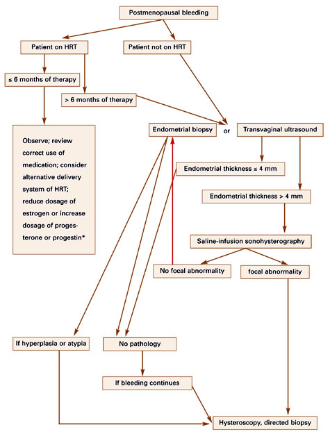
Since the cost-effectiveness of endometrial biopsy and TVUS of the endometrium are comparable, the choice of modality rests with the physician. If TVUS is performed, an endometrial thickness of 4 mm or less is associated with endometrial cancer in about 0.6% of cases.5 If the endometrial thickness is greater than 4 mm, a sonohysterogram should be performed to determine whether the endometrium is symmetrically thickened. If it is, an endometrial biopsy also should be obtained. If an asymmetrical filling defect is found, hysteroscopy and directed biopsy are recommended (FIGURE 1). In women with continued bleeding after a negative initial evaluation, further testing with hysteroscopically directed biopsy is essential, since approximately 4% of these patients will have underlying pathology detected on subsequent evaluation.6
FIGURE 1 Initial management of postmenopausal bleeding
HRT = hormone replacement therapy
*Endometrial thickness acts as a surrogate marker for the combined biologic estrogenic and progestogenic effect on the endometrium.
Anatomic problems
In up to 30% of cases, postmenopausal bleeding is associated with an anatomic abnormality (TABLE 2).7 TVUS and saline-infusion sonohysterography are helpful in identifying these abnormalities. Uterine fibroids and adenomyomas generally are apparent on ultrasound. Uterine polyps may appear as a thickened endometrial stripe, but these and submucous myomas can be clearly identified as filling defects when a sonohysterography is performed.
Tamoxifen is associated with a two- to threefold increased risk of endometrial cancer in postmenopausal women.
A Pap smear should be obtained if the patient has not had 1 within the past year, although cervical disease is an uncommon cause of postmenopausal bleeding. Still, atypical endometrial cells occasionally are noted on a Pap smear. This should prompt an endometrial biopsy or other histologic examination.
TABLE 2
Anatomic causes of postmenopausal bleeding
| UTERUS | CERVIX | OVARY | VAGINA/VULVA |
|---|---|---|---|
| Submucosal fibroid | Cervical polyps | Estrogen-secreting tumor | Atrophic vaginitis |
| Endometrial polyp | Cervical erosion/cervicitis | Vaginal neoplasm | |
| Endometrial hyperplasia | Cervical cancer | Vulvar carcinoma | |
| Adenomyosis | |||
| Endometritis | |||
| Adenocarcinoma | |||
| Sarcoma |
Systemic conditions
Abnormalities of the hematologic system also must be considered as a possible cause of post menopausal bleeding (TABLE 3). On rare occasions, AUB will be the first sign of leukemia or a blood dyscrasia. By the menopausal years, a woman should know whether she has von Willebrand’s disease, factor IX deficiency, an inherited clotting disorder, or other disorders that may cause menorrhagia in premenopausal women but are unlikely to be the reason for postmenopausal bleeding. Overuse of anticoagulant medications such as aspirin, heparin, and warfarin—which are taken with greater frequency by patients in this age group—may contribute to post-menopausal bleeding, although this accounts for a minority of cases. Still, uterine bleeding that occurs in conjunction with these agents may unmask an underlying uterine problem. Thus, in this scenario, it is worthwhile to evaluate the endometrium with TVUS or saline-infusion sonohysterography.
TABLE 3
Nonanatomic causes of postmenopausal bleeding
| HRT USERS | NON-HRT USERS |
|---|---|
| Improper use of HRT | Atrophic endometrium |
| Poor absorption of HRT due to ulcerative colitis or Crohn’s disease | Endometrial polyp or neoplasm related to tamoxifen therapy |
| Drug interactions with hepatic enzyme inducers (SSRIs, phenytoin, barbiturates, griseofulvin) | |
| Coagulation defects:* | |
| Thrombocytopenia | |
| von Willebrand’s disease | |
| Leukemia | |
| Anticoagulant excess | |
| Inadequate progestational response at the endometrium | |
| HRT = hormone replacement therapy; SSRI = selective serotonin reuptake inhibitor | |
| *Uterine bleeding may occur in any women with these conditions, though it is more likely with HRT use. | |
Tamoxifen use
Tamoxifen therapy is associated with a two-to threefold increased risk of endometrial cancer in postmenopausal women.8 TVUS of patients on this therapy typically shows an increased endometrial thickness. Risk appears to increase with higher cumulative doses of tamoxifen and longer duration of treatment. For these reasons, uterine bleeding in women aking tamoxifen should be evaluated with saline-infusion sonohysterography. If the endometrium is symmetrically thickened, an endometrial biopsy should be performed. If an endometrial filling defect, such as a polyp, is identified, hysteroscopically directed biopsy is in order.
Postmenopausal bleeding and HRT
The occurrence of uterine bleeding or spotting after the initiation of HRT is not unusual. More than half of HRT users will have some spotting or bleeding at the beginning of therapy. Usually such bleeding is lighter than a menstrual period and lessens with time; after 6 months, it stops completely in most women.9 The dose and type of progestin used in the HRT regimen, as well as the length of time since menopause, also affect the occurrence of bleeding. Women more than 3 years beyond menopause when starting HRT have fewer episodes of bleeding and are more likely to be amenorrheic after 3 months of treatment than women who start HRT within 3 years of menopause.10
Anatomic problems. Bleeding that persists after 6 months of continuous combined HRT often is related to an anatomic problem in the uterus, such as a uterine polyp, or a submucous fibroid or adenomyoma impinging on the endometrial cavity. Endometrial hyperplasia also could be to blame, although this is unlikely when progestins are used continuously—unless the condition was preexisting. The addition of progestin to the HRT regimen reduces the risk of endometrial cancer in postmenopausal women to less than that experienced by women not taking HRT, though only if it is used continuously, rather than cyclically or sporadically. In fact, the use of cyclic progestin for less than 10 days a month has been associated with an increased risk of endometrial cancer after 5 years, compared with regimens in which progestin is taken more frequently.11
Perimenopausalwomen placed on HRT during an episode of ovarian resistance may later experience erratic bleeding.
Hormonal factors. Other causes of post-menopausal bleeding in women taking HRT often relate to hormonal factors, such as those that occur when an HRT patch does not adhere properly or is not replaced as directed, or when the patient fails to take her medications correctly.12 This is becoming less common, however, now that some HRT preparations contain the estrogen and progestin components in 1 pill. In addition, the packaging of some preparations is now similar to that of OCs, with punch cards listing the days of the week, making it easier to tell if the appropriate pill has been taken.
On rare occasions, other medications may interfere with the absorption of HRT or increase its hepatic metabolism. This will result in lower levels of available estrogen and, consequently, atrophic bleeding. It is therefore helpful to obtain an estradiol level for women taking HRT preparations. In addition, endometrial atrophy should be apparent on the TVUS of the endometrium (endometrial stripe less than or equal to 4 mm) or from the endometrial biopsy. Occasionally, the endometrium’s response to the progestin is insufficient to obtain a secretory endometrium. These women will have a proliferative histology on endometrial biopsy, and should achieveces-sation of bleeding with an increase in the progestin dose.
Once menopause has occurred, uterine bleeding in the absence of exogenous HRT should be considered abnormal.
Sometimes perimenopausal women are placed on HRT during an episode of ovarian resistance, based on the assumption that they are menopausal. These patients may later experience erratic bleeding once their ovarian function returns, since the dose of estrogen in HRT is insufficient to suppress ovarian function. Low-dose OCs may offer these women better control of uterine bleeding and menopausal symptoms.13 Once menopause—defined by the absence of menstruation for 1 year—has occurred, uterine bleeding in the absence of exogenous HRT should be considered abnormal.
Conclusion
Because endometrial cancer is present in 1 of 8 postmenopausal women who present with AUB, ruling out this malignancy should be the first step in evaluation. Fortunately, since AUB is one the first harbingers, endomtrial cancer usually can be detected early in its course. Among the modalities useful in evaluating postmenopausal bleeding are TVUS, endometrial biopsy, saline-infusion sonohys-terography, and hysteroscopy with directed biopsy. The circumstances and characteristics of the individual patient help determine the best route of exploration and treatment.
Dr. Eisenberg reports that she serves on the Speakers Bureau at Pfizer.
- Uterine bleeding or spotting after the initiation of hormone replacement therapy (HRT) is not unusual.
- Endometrial evaluation is called for in women not taking HRT who develop uterine bleeding after more than 1 year of amenorrhea. It also is indicated in postmenopausal women on HRT for more than 6 months with persistent uterine bleeding, and previously amenorrheic women on HRT who begin bleeding without apparent cause.
- Screening asymptomatic women for endometrial cancer through transvaginal ultrasound or endometrial biopsy is not recommended.
- If endometrial thickness is greater than 4 mm, sonohysterography should be performed.
Approximately 1 of every 8 post-menopausal women who present with abnormal uterine bleeding (AUB) will be diagnosed with endometrial cancer, making this one of the most troubling symptoms clinicians encounter in gynecologic practice. Because a wide variety of pathophysiologic problems can cause AUB in post-menopausal women, these patients require prompt evaluation. At a minimum, this should include a clinical history and physical examination, as well as endometrial sampling or evaluation by ultrasound—especially for women not taking hormone replacement therapy (HRT). This process enables clinicians to detect endometrial cancer at an early stage, before it has spread beyond the uterus. Early detection is associated with an expected survival rate of 90%.1
Over the past decade, the evaluation and treatment of postmenopausal bleeding has evolved significantly, thanks to the availability of office-based transvaginal ultrasound (TVUS) and saline-infusion sonohysterography, as well as the advent of endometrial ablation (TABLE 1). Here, I discuss diagnostic testing and review conditions that can lead to postmenopausal bleeding. Once the etiology is identified, the choice of therapeutic intervention usually is self-evident.
TABLE 1
Tests for evaluating postmenopausal AUB
|
Pathophysiology
Our understanding of the pathophysiology of AUB is in its infancy. Here’s what we do know: During a woman’s reproductive years, her endometrium constantly remodels itself under the influence of estrogen, which stimulates cellular growth, and progesterone, which antagonizes the estrogen’s growth effects. When a woman is anovulatory, however, the endometrium can be stimulated by continual estrogen exposure, resulting in endometrial proliferation. This leads to endometrial instability and, in turn, uterine bleeding. Several cellular signaling molecules—such as cytokines, growth factors, and matrix metalloproteinases—are involved. Once menopause occurs, estrogen and progesterone are no longer produced by the ovaries; nor are they produced in any appreciable amounts by the liver and fat. The endometrium regresses to some degree, and no further bleeding should occur. When bleeding does resume, therefore, abnormal pathologies must be investigated.
Endometrial hyperplasia and cancer
Endometrial cancer is the most common gynecologic cancer in the United States, affecting 21 of every 100,000 women, according to the National Cancer Institute’s (NCI) Surveillance, Epidemiology, and End Results (SEER) Program.2 Fortunately, because AUB is an early harbinger of the disease, endometrial cancer generally has a favorable prognosis: When all endometrial cancer cases are considered, the 5-year survival rate is 84%, though the prognosis for individual patients depends on a variety of factors, including the initial stage of the endometrial cancer as well as the cellular differentiation.
Over the past 2 decades, the rate of endometrial cancer has decreased by more than 26%.2 This may be because many women are entering menopause who previously used combination oral contraceptives (OCs) containing progestin, which is associated with a reduced risk of endometrial hyperplasia and cancer. The increased use of progestin in HRT regimens in recent years also likely plays a role.
Risk factors for endometrial cancer are conditions typically associated with chronic elevations of endogenous estrogen levels or increased estrogen action at the level of the endometrium. These include obesity, history of chronic anovulation, diabetes mellitus, estrogen-secreting tumors, exogenous estrogen unopposed by progesterone or progestin, tamoxifen use, and a family history of Lynch type II syndrome (hereditary nonpolyposis colorectal, ovarian, or endometrial cancer). Since endometrial cancer may occur in the absence of risk factors, however, they should not be the sole means of identifying patients.
Since the cost-effectiveness of endometrial biopsy and transvaginal ultrasound are comparable, the choice of modality rests with the physician.
Diagnostic tests. Endometrial evaluation is called for when any menopausal woman not taking HRT develops uterine bleeding after more than 1 year of amenorrhea. It also is indicated in any postmenopausal woman on HRT for 6 months or more with persistent uterine bleeding, and any previously amenorrheic woman on HRT who begins bleeding without apparent cause.
The measurement of endometrial thickness by TVUS is now almost standard in the evaluation of postmenopausal bleeding. However, screening asymptomatic women for endometrial cancer with TVUS is not recommended. Although the test is very specific (the number of false positives is exceedingly small when the endometrial thickness is set at less than 5 mm), it isn’t sensitive. Many women without endometrial cancer will have an endometrial thickness of 5 mm or more.
Endometrial biopsy is another option for patients with postmenopausal AUB, though the American College of Obstetricians and Gynecologists (ACOG) does not recommend this method for screening asymptomatic menopausal women not on HRT.3 The evidence: Korhonen et al evaluated screening by endometrial biopsy in 2,964 peri- and post-menopausal women, ages 40 to 66 (mean age, 52), who were not on HRT. They identified 19 samples with simple endometrial hyperplasia without cytologic atypia (0.6%) and only 2 cases of well-differentiated adenocarcinoma of the endometrium (0.07%). The majority of the endometrial samples (68.7%) were atrophic, and approximately 25% were proliferative.4
Since the cost-effectiveness of endometrial biopsy and TVUS of the endometrium are comparable, the choice of modality rests with the physician. If TVUS is performed, an endometrial thickness of 4 mm or less is associated with endometrial cancer in about 0.6% of cases.5 If the endometrial thickness is greater than 4 mm, a sonohysterogram should be performed to determine whether the endometrium is symmetrically thickened. If it is, an endometrial biopsy also should be obtained. If an asymmetrical filling defect is found, hysteroscopy and directed biopsy are recommended (FIGURE 1). In women with continued bleeding after a negative initial evaluation, further testing with hysteroscopically directed biopsy is essential, since approximately 4% of these patients will have underlying pathology detected on subsequent evaluation.6
FIGURE 1 Initial management of postmenopausal bleeding
HRT = hormone replacement therapy
*Endometrial thickness acts as a surrogate marker for the combined biologic estrogenic and progestogenic effect on the endometrium.
Anatomic problems
In up to 30% of cases, postmenopausal bleeding is associated with an anatomic abnormality (TABLE 2).7 TVUS and saline-infusion sonohysterography are helpful in identifying these abnormalities. Uterine fibroids and adenomyomas generally are apparent on ultrasound. Uterine polyps may appear as a thickened endometrial stripe, but these and submucous myomas can be clearly identified as filling defects when a sonohysterography is performed.
Tamoxifen is associated with a two- to threefold increased risk of endometrial cancer in postmenopausal women.
A Pap smear should be obtained if the patient has not had 1 within the past year, although cervical disease is an uncommon cause of postmenopausal bleeding. Still, atypical endometrial cells occasionally are noted on a Pap smear. This should prompt an endometrial biopsy or other histologic examination.
TABLE 2
Anatomic causes of postmenopausal bleeding
| UTERUS | CERVIX | OVARY | VAGINA/VULVA |
|---|---|---|---|
| Submucosal fibroid | Cervical polyps | Estrogen-secreting tumor | Atrophic vaginitis |
| Endometrial polyp | Cervical erosion/cervicitis | Vaginal neoplasm | |
| Endometrial hyperplasia | Cervical cancer | Vulvar carcinoma | |
| Adenomyosis | |||
| Endometritis | |||
| Adenocarcinoma | |||
| Sarcoma |
Systemic conditions
Abnormalities of the hematologic system also must be considered as a possible cause of post menopausal bleeding (TABLE 3). On rare occasions, AUB will be the first sign of leukemia or a blood dyscrasia. By the menopausal years, a woman should know whether she has von Willebrand’s disease, factor IX deficiency, an inherited clotting disorder, or other disorders that may cause menorrhagia in premenopausal women but are unlikely to be the reason for postmenopausal bleeding. Overuse of anticoagulant medications such as aspirin, heparin, and warfarin—which are taken with greater frequency by patients in this age group—may contribute to post-menopausal bleeding, although this accounts for a minority of cases. Still, uterine bleeding that occurs in conjunction with these agents may unmask an underlying uterine problem. Thus, in this scenario, it is worthwhile to evaluate the endometrium with TVUS or saline-infusion sonohysterography.
TABLE 3
Nonanatomic causes of postmenopausal bleeding
| HRT USERS | NON-HRT USERS |
|---|---|
| Improper use of HRT | Atrophic endometrium |
| Poor absorption of HRT due to ulcerative colitis or Crohn’s disease | Endometrial polyp or neoplasm related to tamoxifen therapy |
| Drug interactions with hepatic enzyme inducers (SSRIs, phenytoin, barbiturates, griseofulvin) | |
| Coagulation defects:* | |
| Thrombocytopenia | |
| von Willebrand’s disease | |
| Leukemia | |
| Anticoagulant excess | |
| Inadequate progestational response at the endometrium | |
| HRT = hormone replacement therapy; SSRI = selective serotonin reuptake inhibitor | |
| *Uterine bleeding may occur in any women with these conditions, though it is more likely with HRT use. | |
Tamoxifen use
Tamoxifen therapy is associated with a two-to threefold increased risk of endometrial cancer in postmenopausal women.8 TVUS of patients on this therapy typically shows an increased endometrial thickness. Risk appears to increase with higher cumulative doses of tamoxifen and longer duration of treatment. For these reasons, uterine bleeding in women aking tamoxifen should be evaluated with saline-infusion sonohysterography. If the endometrium is symmetrically thickened, an endometrial biopsy should be performed. If an endometrial filling defect, such as a polyp, is identified, hysteroscopically directed biopsy is in order.
Postmenopausal bleeding and HRT
The occurrence of uterine bleeding or spotting after the initiation of HRT is not unusual. More than half of HRT users will have some spotting or bleeding at the beginning of therapy. Usually such bleeding is lighter than a menstrual period and lessens with time; after 6 months, it stops completely in most women.9 The dose and type of progestin used in the HRT regimen, as well as the length of time since menopause, also affect the occurrence of bleeding. Women more than 3 years beyond menopause when starting HRT have fewer episodes of bleeding and are more likely to be amenorrheic after 3 months of treatment than women who start HRT within 3 years of menopause.10
Anatomic problems. Bleeding that persists after 6 months of continuous combined HRT often is related to an anatomic problem in the uterus, such as a uterine polyp, or a submucous fibroid or adenomyoma impinging on the endometrial cavity. Endometrial hyperplasia also could be to blame, although this is unlikely when progestins are used continuously—unless the condition was preexisting. The addition of progestin to the HRT regimen reduces the risk of endometrial cancer in postmenopausal women to less than that experienced by women not taking HRT, though only if it is used continuously, rather than cyclically or sporadically. In fact, the use of cyclic progestin for less than 10 days a month has been associated with an increased risk of endometrial cancer after 5 years, compared with regimens in which progestin is taken more frequently.11
Perimenopausalwomen placed on HRT during an episode of ovarian resistance may later experience erratic bleeding.
Hormonal factors. Other causes of post-menopausal bleeding in women taking HRT often relate to hormonal factors, such as those that occur when an HRT patch does not adhere properly or is not replaced as directed, or when the patient fails to take her medications correctly.12 This is becoming less common, however, now that some HRT preparations contain the estrogen and progestin components in 1 pill. In addition, the packaging of some preparations is now similar to that of OCs, with punch cards listing the days of the week, making it easier to tell if the appropriate pill has been taken.
On rare occasions, other medications may interfere with the absorption of HRT or increase its hepatic metabolism. This will result in lower levels of available estrogen and, consequently, atrophic bleeding. It is therefore helpful to obtain an estradiol level for women taking HRT preparations. In addition, endometrial atrophy should be apparent on the TVUS of the endometrium (endometrial stripe less than or equal to 4 mm) or from the endometrial biopsy. Occasionally, the endometrium’s response to the progestin is insufficient to obtain a secretory endometrium. These women will have a proliferative histology on endometrial biopsy, and should achieveces-sation of bleeding with an increase in the progestin dose.
Once menopause has occurred, uterine bleeding in the absence of exogenous HRT should be considered abnormal.
Sometimes perimenopausal women are placed on HRT during an episode of ovarian resistance, based on the assumption that they are menopausal. These patients may later experience erratic bleeding once their ovarian function returns, since the dose of estrogen in HRT is insufficient to suppress ovarian function. Low-dose OCs may offer these women better control of uterine bleeding and menopausal symptoms.13 Once menopause—defined by the absence of menstruation for 1 year—has occurred, uterine bleeding in the absence of exogenous HRT should be considered abnormal.
Conclusion
Because endometrial cancer is present in 1 of 8 postmenopausal women who present with AUB, ruling out this malignancy should be the first step in evaluation. Fortunately, since AUB is one the first harbingers, endomtrial cancer usually can be detected early in its course. Among the modalities useful in evaluating postmenopausal bleeding are TVUS, endometrial biopsy, saline-infusion sonohys-terography, and hysteroscopy with directed biopsy. The circumstances and characteristics of the individual patient help determine the best route of exploration and treatment.
Dr. Eisenberg reports that she serves on the Speakers Bureau at Pfizer.
1. Paley PJ. Screening for the major malignancies affecting women: current guidelines. Am J Obstet Gynecol. 2001;184(5):1021-1030.
2. National Cancer Institute. Endometrial cancer. Available at: http://www3.can-cer.gov/admin/fmb/2001sigitems/endometrial.html Accessed August 26, 2002.
3. American College of Obstetricians and Gynecologists. Routine cancer screening. Committee Opinion #247. Washington, DC: ACOG; December 2000.
4. Korhonen MO, Symons JP, Hyde BM, Rowan JP, Wilborn WH. Histologic classification and pathologic findings for endometrial biopsy specimens obtained from 2,964 perimenopausal and postmenopausal women undergoing screening for continuous hormones as replacement therapy (CHART 2 Study). Am J Obstet Gynecol. 1997;176:377-380.
5. Gull B, Carlsson SA, Karlsson B, et al. Transvaginal ultrasonography of the endometrium in women with postmenopausal bleeding: Is it always necessary to perform an endometrial biopsy? Am J Obstet Gynecol. 2000;182:509-515.
6. Feldman S, Shapter A, Welch WR, Berkowitz RS. Two-year followup of 263 patients with post/perimenopausal vaginal bleeding and negative initial biopsy. Gynecol Oncol. 1994;55:56-59.
7. Akkad AA, Habiba MA, Ismail N, Abrams K, Al-Azzawi F. Abnormal bleeding on hormone replacement: the importance of intrauterine structural abnormalities. Obstet Gynecol. 1995;86:330-334.
8. Sismondi P, Biglia N, Volpi E, Giai M, de Grandis T. Tamoxifen and endometrial cancer. Ann NY Acad Sci. 1994;734:310-332.
9. Nand SL, Webster MA, Baber R, O’Connor V. Bleeding pattern and endometrial changes during continuous combined hormone replacement therapy. Obstet Gynecol. 1998;91:678-684.
10. Archer DF, Pickar JH. Hormone replacement therapy: effect of progestin dose and time since menopause on endometrial bleeding. Obstet Gynecol. 2000;96:899-905.
11. Weiderpass E, Adami HO, Baron JA, et al. Risk of endometrial cancer following estrogen replacement with and without progestin. J Natl Cancer Inst. 1999;91:1131-1137.
12. Spencer CP, Cooper AJ, Whitehead MI. Fortnightly review: management of abnormal bleeding in women receiving hormone replacement therapy. BMJ. 1997;315:37-42.
13. Kaunitz AM. Oral contraceptive use in perimenopause. Am J Obstet Gynecol. 2001;185:S32-S37.