User login
Transient Benign Neonatal Skin Findings
Review the PDF of the fact sheet on transient benign neonatal skin findings with board-relevant, easy-to-review material. This fact sheet lists benign findings that can be seen in neonates and infants.
Practice Questions
1. The parents of a 2-month-old infant present with their child. They are worried because the infant has “acne” that is not going away. Friends told them to try gentle cleansers and they have avoided using lotions or cream on her face. However, the bumps will not go away. On examination she has papules and pustules. Comedones cannot be identified. What are your next steps?
a. adapalene cream 0.1% every night at bedtime
b. benzoyl peroxide cream 4%
c. benzoyl peroxide wash 2.5%
d. erythromycin gel 2%
e. ketoconazole cream 2% twice daily
2. While in the newborn nursery prior to discharge, the attending pediatrician notices a rash on a 2-day-old neonate who is otherwise completely healthy. The pediatrician consults a dermatologist for his/her opinion. The dermatologist sees erythematous macules with central pustules located predominately on the trunk and proximal extremities. A pustule is unroofed with a blade, the contents smeared on a glass slide, and a Giemsa stain is performed. What is the predominant cell type you would expect to see on histological examination?
a. eosinophils
b. Langerhans cells
c. lymphocytes
d. neutrophils
e. no cells are visualized
3. Shortly after delivery, the pediatricians notice that the baby has numerous hyperpigmented macules on the back. No other primary lesions are seen. The neonate is otherwise normal in appearance and nontoxic appearing. A dermatologist is consulted for a recommendation for further workup or potential biopsy. The dermatologist examines the newborn. He is a well-appearing black boy with skin that is otherwise intact. A few pustules on the back are present that have a collarette of scale. The dermatologist reviews the mother’s prenatal history and the review shows that she was screened for syphilis and had a negative screening test with no other history of infectious diseases. What is the most appropriate next step to confirm your suspicions?
a. do a swab of a pustule and send it for viral culture
b. have his blood drawn and check for signs of neonatal herpes simplex virus infection
c. perform a biopsy of a pustule
d. perform a Giemsa stain on a smear of the pustule
e. start treatment with permethrin
4. Which intraoral cysts occur on the alveolar ridge of a neonate?
a. Bohn nodule
b. branchial cleft cyst
c. Epstein pearls
d. median raphe cyst
e. palatal cysts of the newborn
5. Miliaria rubra is associated with inflammation of the sweat glands in what portion of the skin?
a. basement membrane zone
b. dermis
c. dermoepidermal junction
d. intraepidermal
e. subcutis
Answers to practice questions provided on next page
Practice Question Answers
1. The parents of a 2-month-old infant present with their child. They are worried because the infant has “acne” that is not going away. Friends told them to try gentle cleansers and they have avoided using lotions or cream on her face. However, the bumps will not go away. On examination she has papules and pustules. Comedones cannot be identified. What are your next steps?
a. adapalene cream 0.1% every night at bedtime
b. benzoyl peroxide cream 4%
c. benzoyl peroxide wash 2.5%
d. erythromycin gel 2%
e. ketoconazole cream 2% twice daily
2. While in the newborn nursery prior to discharge, the attending pediatrician notices a rash on a 2-day-old neonate who is otherwise completely healthy. The pediatrician consults a dermatologist for his/her opinion. The dermatologist sees erythematous macules with central pustules located predominately on the trunk and proximal extremities. A pustule is unroofed with a blade, the contents smeared on a glass slide, and a Giemsa stain is performed. What is the predominant cell type you would expect to see on histological examination?
a. eosinophils
b. Langerhans cells
c. lymphocytes
d. neutrophils
e. no cells are visualized
3. Shortly after delivery, the pediatricians notice that the baby has numerous hyperpigmented macules on the back. No other primary lesions are seen. The neonate is otherwise normal in appearance and nontoxic appearing. A dermatologist is consulted for a recommendation for further workup or potential biopsy. The dermatologist examines the newborn. He is a well-appearing black boy with skin that is otherwise intact. A few pustules on the back are present that have a collarette of scale. The dermatologist reviews the mother’s prenatal history and the review shows that she was screened for syphilis and had a negative screening test with no other history of infectious diseases. What is the most appropriate next step to confirm your suspicions?
a. do a swab of a pustule and send it for viral culture
b. have his blood drawn and check for signs of neonatal herpes simplex virus infection
c. perform a biopsy of a pustule
d. perform a Giemsa stain on a smear of the pustule
e. start treatment with permethrin
4. Which intraoral cysts occur on the alveolar ridge of a neonate?
a. Bohn nodule
b. branchial cleft cyst
c. Epstein pearls
d. median raphe cyst
e. palatal cysts of the newborn
5. Miliaria rubra is associated with inflammation of the sweat glands in what portion of the skin?
a. basement membrane zone
b. dermis
c. dermoepidermal junction
d. intraepidermal
e. subcutis
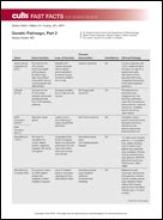
Genetic Pathways, Part 2
After, test your knowledge by answering the 5 practice questions.
Practice Questions
1. A 6-month-old male infant presented to your dermatology clinic with an ash-leaf macule on the right back. What is the most common gene defect seen in this condition?
a. tuberin
b. merlin
c. neurofibromin
d. smoothened
e. hamartin
2. Bilateral acoustic neuromas are associated with what gene mutation?
a. NF1 (neurofibromin 1)
b. NF2 (neurofibromin 2)
c. PTCH1 (patched 1)
d. TSC1 (tuberous sclerosis 1)
e. TSC2 (tuberous sclerosis 2)
3. Which of the following would least likely be seen in neurofibromatosis types 1 or 2?
a. angiofibromas
b. café au lait macules
c. gliomas
d. Lisch nodules
e. neurofibromas
4. What protein is the patched 1 gene a receptor for?
a. fused
b. glioma-associated oncogene
c. smoothened
d. sonic hedgehog
e. suppressor of fused
5. A 20-year-old woman presented to your dermatology clinic with a history of numerous basal cell carcinomas. On physical examination, it is noted that she has numerous palmar pits. What finding could you find from radiograph of the head?
a. calcification of the dura
b. calcifications of the temporal lobe
c. cysts of the mandible
d. thickening of the corpus callosum
e. tumor of the cerebellum
Genetic Pathways, Part 1
After, test your knowledge by answering the 5 practice questions.
Practice Questions
1. Which keratinization disorder is characterized by drastically lower levels of lamellar bodies?
a. Harlequin ichthyosis
b. ichthyosis vulgaris
c. lamellar ichthyosis
d. nonbullous congenital ichthyosiform erythroderma
e. X-linked ichthyosis
2. Mutation of this enzyme leads to uncontrolled proteolytic activity causing degradation of lamellar body lipid processing enzymes.
a. FALDH (fatty aldehyde dehydrogenase)
b. LEKTI (lympho-epithelial Kazal-type-related inhibitor)
c. PEX7 (perioxsomal biogenesis factor 7)
d. PHYH (phytanoyl-CoA hydroxylase)
e. NSDHL (NAD[P] dependent steroid dehydrogenase-like)
3. A 30-year-old man presented for evaluation of abnormal nails. Physical examination revealed a red streak with distal V-shaped nicking. Numerous keratotic papules on the hands and chest and oral papules also were noted. The gene responsible for these findings encodes a Ca2+-ATPase responsible for a Ca2+ influx into what cellular structure?
a. cytoplasm
b. endoplasmic reticulum
c. golgi
d. nucleus
e. ribosome
4. A female neonate aged 2 weeks presented with linear and whorled vesicles on the thighs and trunk. The delivery was uncomplicated. The patient was afebrile, but her mother said she has been “doing well” at home. On pathology, what do you expect to see?
a. apoptosis of epidermal cells
b. cell-poor blister
c. molding and margination of chromatin as well as multinucleated giant cells
d. numerous pseudohyphae
e. spongiosis of epidermal cells
5. A young child presents with linear atrophic plaques with fat herniation and raspberrylike oral papillomas. What signal transduction pathway is altered in this syndrome?
a. ABCA12 (ATP-binding cassette, sub-family A, member 12)
b. adenylate cyclase
c. ß-catenin
d. LEKTI (lympho-epithelial Kazal-type-related inhibitor)
e. nuclear factor κ light chain enhancer of activated B cells
The answers appear on the next page.
Practice Question Answers
1. Which keratinization disorder is characterized by drastically lower levels of lamellar bodies?
a. Harlequin ichthyosis
b. ichthyosis vulgaris
c. lamellar ichthyosis
d. nonbullous congenital ichthyosiform erythroderma
e. X-linked ichthyosis
2. Mutation of this enzyme leads to uncontrolled proteolytic activity causing degradation of lamellar body lipid processing enzymes.
a. FALDH (fatty aldehyde dehydrogenase)
b. LEKTI (lympho-epithelial Kazal-type-related inhibitor)
c. PEX7 (perioxsomal biogenesis factor 7)
d. PHYH (phytanoyl-CoA hydroxylase)
e. NSDHL (NAD[P] dependent steroid dehydrogenase-like)
3. A 30-year-old man presented for evaluation of abnormal nails. Physical examination revealed a red streak with distal V-shaped nicking. Numerous keratotic papules on the hands and chest and oral papules also were noted. The gene responsible for these findings encodes a Ca2+-ATPase responsible for a Ca2+ influx into what cellular structure?
a. cytoplasm
b. endoplasmic reticulum
c. golgi
d. nucleus
e. ribosome
4. A female neonate aged 2 weeks presented with linear and whorled vesicles on the thighs and trunk. The delivery was uncomplicated. The patient was afebrile, but her mother said she has been “doing well” at home. On pathology, what do you expect to see?
a. apoptosis of epidermal cells
b. cell-poor blister
c. molding and margination of chromatin as well as multinucleated giant cells
d. numerous pseudohyphae
e. spongiosis of epidermal cells
5. A young child presents with linear atrophic plaques with fat herniation and raspberrylike oral papillomas. What signal transduction pathway is altered in this syndrome?
a. ABCA12 (ATP-binding cassette, sub-family A, member 12)
b. adenylate cyclase
c. ß-catenin
d. LEKTI (lympho-epithelial Kazal-type-related inhibitor)
e. nuclear factor κ light chain enhancer of activated B cells


