Allowed Publications
Slot System
Featured Buckets
Featured Buckets Admin

42-year-old woman with abnormal uterine bleeding

Article Type
Changed
Tue, 08/28/2018 - 11:09
Display Headline
42-year-old woman with abnormal uterine bleeding
What's the diagnosis based on pelvic ultrasonography?

A) Endometrial polyp INCORRECT
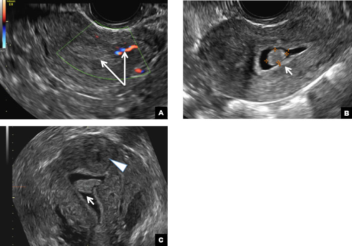
Endometrial polyps on ultrasonography appear as focal echogenic (hyperechoic) masses or as nonspecific endometrial thickening.1 Color Doppler often demonstrates a vascular stalk, which is a nonspecific finding that also can be seen in submucosal fibroids and endometrial cancer.2 On sonohysterography (SHG), endometrial polyps typically appear as well-defined echogenic/hyperechoic polypoid lesions (tissue appearance similar to that of normal endometrium) protruding into the endometrial canal but still preserving the endometrial−myometrial interface.2,3

Endometrial polyp. (A) Transvaginal pelvic ultrasound of the uterus with color Doppler demonstrates a focal echogenic lesion with a vascular stalk (long arrows). (B) SHG shows a well-defined polypoid lesion, isoechoic to the endometrium, and protruding into the endometrial canal but still preserving the endometrial−myometrial interface (short arrow). (C) 3D SHG imaging shows the echogenic endometrial polyp (short arrow) and an incidental intramural fibroid in the fundus (arrowhead).


B) Submucosal fibroid CORRECT
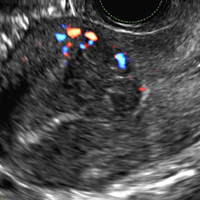
Submucosal fibroids on ultrasonography appear as heterogeneous hypoechoic lesions distorting the endometrial cavity.1 In contrast to endometrial polyps, which involve the endometrium only, submucosal (intracavitary) fibroids originate in the myometrium, as clarified in Figures 3A & B. SHG demonstrates a broad-based mixed hypoechoic/isoechoic lesion protruding into the endometrial canal but preserving the echogenic endometrium, distinguishing myometrial from endometrial lesions. Submucosal fibroids often distort the endometrial myometrial interface and demonstrate acoustic shadowing.2,3

Submucosal fibroid. (A) Transvaginal pelvic ultrasound with color Doppler demonstrates a heterogeneous hypoechoic/isoechoic lesion arising from the myometrium (long arrow) and distorting the endometrial cavity. (B) SHG demonstrates a broad-based hypoechoic lesion protruding into the endometrial canal with areas of acoustic shadowing (arrowhead) but preserving the echogenic endometrium (short arrow).


C) Endometrial carcinoma INCORRECT
Endometrial carcinoma on SHG appears as an irregular inhomogeneous lobulated vascular mass distorting the endometrial−myometrial interface.3 Additionally, irregular frond-like projections can be seen extending from the mass into the endometrial cavity, which are distended with echogenic fluid.2

Endometrial carcinoma. (A) Transvaginal pelvic ultrasound demonstrates a retroverted uterus with the endometrial cavity distended by tissue and fluid. An irregular inhomogeneous lobulated mass with frond-like projections (long arrow) distorts the endometrial−myometrial interface (short arrow). (B) Color Doppler image shows marked vascularity within the mass and echogenic fluid in the endometrial canal (arrowhead).


D) Endometrial hyperplasia INCORRECT
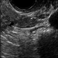
On ultrasonography, endometrial hyperplasia has a nonspecific appearance, often presenting as diffuse smooth endometrial thickening.1 SHG typically demonstrates diffuse thickening of the echogenic endometrial stripe without a focal lesion and, when a focal lesion is present, can mimic a broad-based endometrial polyp.2,3

Endometrial hyperplasia. (A) Transvaginal pelvic ultrasound demonstrates diffuse endometrial thickening (long arrow). (B) SHG shows diffuse thickening of the echogenic endometrial stripe without a focal lesion (arrow heads).

References
  1. Nalaboff KM, Pellerito JS, Ben-Levi E. Imaging the endometrium: disease and normal variants. Radiographics. 2001;21(6):1409-1424.  
  2. Davis PC, O'Neill MJ, Yoder IC, Lee SI, Mueller PR. Sonohysterographic findings of endometrial and subendometrial conditions. Radiographics. 2002;22(4):803- 816.  
  3. Yang T, Pandya A, Marcal L, et al. Sonohysterography: principles, technique and role in diagnosis of endometrial pathology. World J Radiol. 2013;5(3):81-87.
Author and Disclosure Information

Dr. Kanmaniraja is Assistant Professor and Chief, Division of Abdominal Imaging, Department of Radiology, University of Florida College of Medicine–Jacksonville.

Dr. Kaunitz is University of Florida Research Foundation Professor and Associate Chairman, Department of Obstetrics and Gynecology, University of Florida College of Medicine–Jacksonville. He is Medical Director and Director of Menopause and Gynecologic Ultrasound Services at UF Women’s Health Specialists–Emerson. He also serves on the OBG Management Board of Editors.

The authors report no financial relationships relevant to this quiz.

Issue
OBG Management - 29(3)
Publications
Topics
Sections
Author and Disclosure Information

Dr. Kanmaniraja is Assistant Professor and Chief, Division of Abdominal Imaging, Department of Radiology, University of Florida College of Medicine–Jacksonville.

Dr. Kaunitz is University of Florida Research Foundation Professor and Associate Chairman, Department of Obstetrics and Gynecology, University of Florida College of Medicine–Jacksonville. He is Medical Director and Director of Menopause and Gynecologic Ultrasound Services at UF Women’s Health Specialists–Emerson. He also serves on the OBG Management Board of Editors.

The authors report no financial relationships relevant to this quiz.

What's the diagnosis based on pelvic ultrasonography?

A) Endometrial polyp INCORRECT
Endometrial polyps on ultrasonography appear as focal echogenic (hyperechoic) masses or as nonspecific endometrial thickening.1 Color Doppler often demonstrates a vascular stalk, which is a nonspecific finding that also can be seen in submucosal fibroids and endometrial cancer.2 On sonohysterography (SHG), endometrial polyps typically appear as well-defined echogenic/hyperechoic polypoid lesions (tissue appearance similar to that of normal endometrium) protruding into the endometrial canal but still preserving the endometrial−myometrial interface.2,3

Endometrial polyp. (A) Transvaginal pelvic ultrasound of the uterus with color Doppler demonstrates a focal echogenic lesion with a vascular stalk (long arrows). (B) SHG shows a well-defined polypoid lesion, isoechoic to the endometrium, and protruding into the endometrial canal but still preserving the endometrial−myometrial interface (short arrow). (C) 3D SHG imaging shows the echogenic endometrial polyp (short arrow) and an incidental intramural fibroid in the fundus (arrowhead).


B) Submucosal fibroid CORRECT
Submucosal fibroids on ultrasonography appear as heterogeneous hypoechoic lesions distorting the endometrial cavity.1 In contrast to endometrial polyps, which involve the endometrium only, submucosal (intracavitary) fibroids originate in the myometrium, as clarified in Figures 3A & B. SHG demonstrates a broad-based mixed hypoechoic/isoechoic lesion protruding into the endometrial canal but preserving the echogenic endometrium, distinguishing myometrial from endometrial lesions. Submucosal fibroids often distort the endometrial myometrial interface and demonstrate acoustic shadowing.2,3

Submucosal fibroid. (A) Transvaginal pelvic ultrasound with color Doppler demonstrates a heterogeneous hypoechoic/isoechoic lesion arising from the myometrium (long arrow) and distorting the endometrial cavity. (B) SHG demonstrates a broad-based hypoechoic lesion protruding into the endometrial canal with areas of acoustic shadowing (arrowhead) but preserving the echogenic endometrium (short arrow).


C) Endometrial carcinoma INCORRECT
Endometrial carcinoma on SHG appears as an irregular inhomogeneous lobulated vascular mass distorting the endometrial−myometrial interface.3 Additionally, irregular frond-like projections can be seen extending from the mass into the endometrial cavity, which are distended with echogenic fluid.2

Endometrial carcinoma. (A) Transvaginal pelvic ultrasound demonstrates a retroverted uterus with the endometrial cavity distended by tissue and fluid. An irregular inhomogeneous lobulated mass with frond-like projections (long arrow) distorts the endometrial−myometrial interface (short arrow). (B) Color Doppler image shows marked vascularity within the mass and echogenic fluid in the endometrial canal (arrowhead).


D) Endometrial hyperplasia INCORRECT
On ultrasonography, endometrial hyperplasia has a nonspecific appearance, often presenting as diffuse smooth endometrial thickening.1 SHG typically demonstrates diffuse thickening of the echogenic endometrial stripe without a focal lesion and, when a focal lesion is present, can mimic a broad-based endometrial polyp.2,3

Endometrial hyperplasia. (A) Transvaginal pelvic ultrasound demonstrates diffuse endometrial thickening (long arrow). (B) SHG shows diffuse thickening of the echogenic endometrial stripe without a focal lesion (arrow heads).

References
  1. Nalaboff KM, Pellerito JS, Ben-Levi E. Imaging the endometrium: disease and normal variants. Radiographics. 2001;21(6):1409-1424.  
  2. Davis PC, O'Neill MJ, Yoder IC, Lee SI, Mueller PR. Sonohysterographic findings of endometrial and subendometrial conditions. Radiographics. 2002;22(4):803- 816.  
  3. Yang T, Pandya A, Marcal L, et al. Sonohysterography: principles, technique and role in diagnosis of endometrial pathology. World J Radiol. 2013;5(3):81-87.
Issue
OBG Management - 29(3)
Issue
OBG Management - 29(3)
Publications
Publications
Topics
Article Type
Display Headline
42-year-old woman with abnormal uterine bleeding
Display Headline
42-year-old woman with abnormal uterine bleeding
Sections
Questionnaire Body

A 42-year-old woman presents to her gynecologist's office with abnormal uterine bleeding. Pelvic ultrasonography of the uterus is performed with color Doppler (A) and subsequent sonohysterogram (SHG)/saline infusion sonohysterography (SIS) (B). Figures shown above.

Disallow All Ads
Content Gating
No Gating (article Unlocked/Free)
Alternative CME

32-year-old woman with pelvic pain and irregular menstrual periods

Article Type
Changed
Tue, 08/28/2018 - 11:08
Display Headline
32-year-old woman with pelvic pain and irregular menstrual periods
What’s the diagnosis?

(A) Paratubal cyst CORRECT

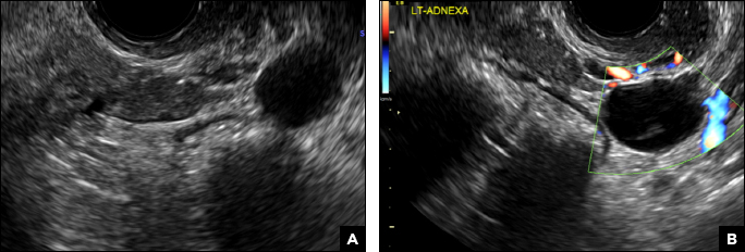
Paratubal, or paraovarian, cysts typically are round or oval avascular hypoechoic cysts (long arrow) separate from the adjacent ovary (short arrow). Since they are congenital remnants of the Wolffian duct, they arise from the mesosalpinx, specifically the broad ligament or fallopian tube.1,2 They usually are seen in close proximity to but separate from the ovary without distorting the ovary’s architecture.1,2

Paratubal cyst. (A) Transvaginal pelvic ultrasound of the left adnexa demonstrates an oval hypoechoic cyst (long arrow) separate from the adjacent ovary (short arrow). (B) The paratubal cyst is avascular on color Doppler.


B) Hydrosalpinx INCORRECT

A hydrosalpinx appears as an elongated C- or S-shaped, thin-walled tubular serpiginous cystic lesion separate from the ovary. It often has incomplete septations that are infolding of the tube on itself (long arrow).3 Other findings include diametrically opposed indentations (short arrows) of the wall (Waist sign) and short linear mucosal or submucosal folds (arrowhead) that when viewed in cross section appear similar to the spokes of a cogwheel (Cogwheel sign).1–3 Prior tubal infection or gynecologic surgery represent risk factors for hydrosalpinx.
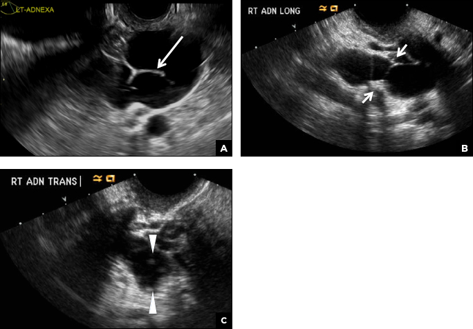
Hydrosalpinx. (A) Transvaginal pelvic ultrasound of the left adnexa demonstrates an elongated C- or S-shaped, thin-walled tubular serpiginous cystic lesion with incomplete septations (long arrow). (B) Longitudinal image of the right adnexa shows the dilated fallopian tube with diametrically opposed indentations of the wall consistent with the Waist sign (short arrows). (C) Transverse image of the dilated fallopian tube viewed in cross section has the appearance of several short mural nodules similar to the spokes of a cogwheel (arrowheads).


C) Peritoneal inclusion cyst INCORRECT

A peritoneal inclusion cyst appears as an anechoic cystic mass that conforms passively to the shape of the peritoneal cavity/pelvic sidewall (long arrow) and may contain entrapped ovaries (short arrow)  within or along the periphery of the fluid collection.1,2 Septations within it are likely from peritoneal adhesions (arrowhead) and may show vascularity.2 Prior (often multiple) gynecologic surgeries represent a risk factor for peritoneal inclusion cysts.

Peritoneal inclusion cyst. (A) Longitudinal transvaginal pelvic ultrasound of the left adnexa demonstrating an anechoic cystic lesion that conforms passively to the shape of the peritoneal cavity/pelvic sidewall (long arrow) with a thick septation (arrowhead). (B) Transverse image demonstrates the left ovary entrapped within the fluid collection (short arrow).


D) Dilated pelvic veins INCORRECT

Dilated pelvic veins appear on sonography as a cluster of elongated and tubular cystic lesions in the adnexa along the broad ligament and demonstrate low level echoes due to sluggish flow (long arrow) and visible red blood cell rouleaux formation. This can be confirmed on color Doppler images (short arrow) and help differentiate it from hydrosalpinx.
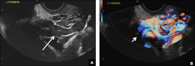
Dilated pelvic veins. (A) Transvaginal pelvic ultrasound of the left adnexa reveals a cluster of elongated and tubular cystic lesions that demonstrate low level echoes due to sluggish flow (long arrow). (B) Color Doppler ultrasound confirms vascularity within these dilated pelvic veins (short arrow).

References
  1. Laing FC, Allison SF. US of the ovary and adnexa: to worry or not to worry? Radiographics. 2012:32(6):1621−1639.
  2. Moyle PL, Kataoka MY, Nakai A, Takahata A, Reinhold C, Sala E. Nonovarian cystic lesions of the pelvis. Radiographics. 2010;30(4):921−938.
  3. Rezvani M, Shaaban AM. Fallopian tube disease in the nonpregnant patient. Radiographics. 2011;31(2):527−548.
Author and Disclosure Information

Dr. Kanmaniraja is Assistant Professor and Chief, Division of Abdominal Imaging, Department of Radiology, University of Florida College of Medicine–Jacksonville.

Dr. Kaunitz is University of Florida Research Foundation Professor and Associate Chairman, Department of Obstetrics and Gynecology, University of Florida College of Medicine–Jacksonville. He is Medical Director and Director of Menopause and Gynecologic Ultrasound Services at UF Women’s Health Specialists–Emerson. He also serves on the OBG Management Board of Editors.

The authors report no financial relationships relevant to this quiz.

Issue
OBG Management - 29(2)
Publications
Topics
Sections
Author and Disclosure Information

Dr. Kanmaniraja is Assistant Professor and Chief, Division of Abdominal Imaging, Department of Radiology, University of Florida College of Medicine–Jacksonville.

Dr. Kaunitz is University of Florida Research Foundation Professor and Associate Chairman, Department of Obstetrics and Gynecology, University of Florida College of Medicine–Jacksonville. He is Medical Director and Director of Menopause and Gynecologic Ultrasound Services at UF Women’s Health Specialists–Emerson. He also serves on the OBG Management Board of Editors.

The authors report no financial relationships relevant to this quiz.

What’s the diagnosis?

(A) Paratubal cyst CORRECT

Paratubal, or paraovarian, cysts typically are round or oval avascular hypoechoic cysts (long arrow) separate from the adjacent ovary (short arrow). Since they are congenital remnants of the Wolffian duct, they arise from the mesosalpinx, specifically the broad ligament or fallopian tube.1,2 They usually are seen in close proximity to but separate from the ovary without distorting the ovary’s architecture.1,2

Paratubal cyst. (A) Transvaginal pelvic ultrasound of the left adnexa demonstrates an oval hypoechoic cyst (long arrow) separate from the adjacent ovary (short arrow). (B) The paratubal cyst is avascular on color Doppler.


B) Hydrosalpinx INCORRECT

A hydrosalpinx appears as an elongated C- or S-shaped, thin-walled tubular serpiginous cystic lesion separate from the ovary. It often has incomplete septations that are infolding of the tube on itself (long arrow).3 Other findings include diametrically opposed indentations (short arrows) of the wall (Waist sign) and short linear mucosal or submucosal folds (arrowhead) that when viewed in cross section appear similar to the spokes of a cogwheel (Cogwheel sign).1–3 Prior tubal infection or gynecologic surgery represent risk factors for hydrosalpinx.

Hydrosalpinx. (A) Transvaginal pelvic ultrasound of the left adnexa demonstrates an elongated C- or S-shaped, thin-walled tubular serpiginous cystic lesion with incomplete septations (long arrow). (B) Longitudinal image of the right adnexa shows the dilated fallopian tube with diametrically opposed indentations of the wall consistent with the Waist sign (short arrows). (C) Transverse image of the dilated fallopian tube viewed in cross section has the appearance of several short mural nodules similar to the spokes of a cogwheel (arrowheads).


C) Peritoneal inclusion cyst INCORRECT

A peritoneal inclusion cyst appears as an anechoic cystic mass that conforms passively to the shape of the peritoneal cavity/pelvic sidewall (long arrow) and may contain entrapped ovaries (short arrow)  within or along the periphery of the fluid collection.1,2 Septations within it are likely from peritoneal adhesions (arrowhead) and may show vascularity.2 Prior (often multiple) gynecologic surgeries represent a risk factor for peritoneal inclusion cysts.

Peritoneal inclusion cyst. (A) Longitudinal transvaginal pelvic ultrasound of the left adnexa demonstrating an anechoic cystic lesion that conforms passively to the shape of the peritoneal cavity/pelvic sidewall (long arrow) with a thick septation (arrowhead). (B) Transverse image demonstrates the left ovary entrapped within the fluid collection (short arrow).


D) Dilated pelvic veins INCORRECT

Dilated pelvic veins appear on sonography as a cluster of elongated and tubular cystic lesions in the adnexa along the broad ligament and demonstrate low level echoes due to sluggish flow (long arrow) and visible red blood cell rouleaux formation. This can be confirmed on color Doppler images (short arrow) and help differentiate it from hydrosalpinx.

Dilated pelvic veins. (A) Transvaginal pelvic ultrasound of the left adnexa reveals a cluster of elongated and tubular cystic lesions that demonstrate low level echoes due to sluggish flow (long arrow). (B) Color Doppler ultrasound confirms vascularity within these dilated pelvic veins (short arrow).

(A) Paratubal cyst CORRECT

Paratubal, or paraovarian, cysts typically are round or oval avascular hypoechoic cysts (long arrow) separate from the adjacent ovary (short arrow). Since they are congenital remnants of the Wolffian duct, they arise from the mesosalpinx, specifically the broad ligament or fallopian tube.1,2 They usually are seen in close proximity to but separate from the ovary without distorting the ovary’s architecture.1,2

Paratubal cyst. (A) Transvaginal pelvic ultrasound of the left adnexa demonstrates an oval hypoechoic cyst (long arrow) separate from the adjacent ovary (short arrow). (B) The paratubal cyst is avascular on color Doppler.


B) Hydrosalpinx INCORRECT

A hydrosalpinx appears as an elongated C- or S-shaped, thin-walled tubular serpiginous cystic lesion separate from the ovary. It often has incomplete septations that are infolding of the tube on itself (long arrow).3 Other findings include diametrically opposed indentations (short arrows) of the wall (Waist sign) and short linear mucosal or submucosal folds (arrowhead) that when viewed in cross section appear similar to the spokes of a cogwheel (Cogwheel sign).1–3 Prior tubal infection or gynecologic surgery represent risk factors for hydrosalpinx.

Hydrosalpinx. (A) Transvaginal pelvic ultrasound of the left adnexa demonstrates an elongated C- or S-shaped, thin-walled tubular serpiginous cystic lesion with incomplete septations (long arrow). (B) Longitudinal image of the right adnexa shows the dilated fallopian tube with diametrically opposed indentations of the wall consistent with the Waist sign (short arrows). (C) Transverse image of the dilated fallopian tube viewed in cross section has the appearance of several short mural nodules similar to the spokes of a cogwheel (arrowheads).


C) Peritoneal inclusion cyst INCORRECT

A peritoneal inclusion cyst appears as an anechoic cystic mass that conforms passively to the shape of the peritoneal cavity/pelvic sidewall (long arrow) and may contain entrapped ovaries (short arrow)  within or along the periphery of the fluid collection.1,2 Septations within it are likely from peritoneal adhesions (arrowhead) and may show vascularity.2 Prior (often multiple) gynecologic surgeries represent a risk factor for peritoneal inclusion cysts.

Peritoneal inclusion cyst. (A) Longitudinal transvaginal pelvic ultrasound of the left adnexa demonstrating an anechoic cystic lesion that conforms passively to the shape of the peritoneal cavity/pelvic sidewall (long arrow) with a thick septation (arrowhead). (B) Transverse image demonstrates the left ovary entrapped within the fluid collection (short arrow).


D) Dilated pelvic veins INCORRECT

Dilated pelvic veins appear on sonography as a cluster of elongated and tubular cystic lesions in the adnexa along the broad ligament and demonstrate low level echoes due to sluggish flow (long arrow) and visible red blood cell rouleaux formation. This can be confirmed on color Doppler images (short arrow) and help differentiate it from hydrosalpinx.

Dilated pelvic veins. (A) Transvaginal pelvic ultrasound of the left adnexa reveals a cluster of elongated and tubular cystic lesions that demonstrate low level echoes due to sluggish flow (long arrow). (B) Color Doppler ultrasound confirms vascularity within these dilated pelvic veins (short arrow).

References
  1. Laing FC, Allison SF. US of the ovary and adnexa: to worry or not to worry? Radiographics. 2012:32(6):1621−1639.
  2. Moyle PL, Kataoka MY, Nakai A, Takahata A, Reinhold C, Sala E. Nonovarian cystic lesions of the pelvis. Radiographics. 2010;30(4):921−938.
  3. Rezvani M, Shaaban AM. Fallopian tube disease in the nonpregnant patient. Radiographics. 2011;31(2):527−548.
Issue
OBG Management - 29(2)
Issue
OBG Management - 29(2)
Publications
Publications
Topics
Article Type
Display Headline
32-year-old woman with pelvic pain and irregular menstrual periods
Display Headline
32-year-old woman with pelvic pain and irregular menstrual periods
Sections
Questionnaire Body

A 32-year-old women presents to her gynecologist’s office reporting pelvic pain and irregular menstrual periods. Results of a urine pregnancy test are negative. Pelvic ultrasonography is performed, with gray scale ( A ) and color Doppler ( B ) images of the left adnexa obtained. Figures shown above.

Disallow All Ads
Content Gating
No Gating (article Unlocked/Free)
Alternative CME

2-week left-sided pelvic pain

Article Type
Changed
Tue, 08/28/2018 - 11:08
Display Headline
2-week left-sided pelvic pain
What’s the diagnosis?

(A) Simple ovarian cyst INCORRECT
Here is an example of a well circumscribed round or oval anechoic, avascular simple ovarian cyst with posterior acoustic enhancement and thin smooth walls.1 No septations or solid components are identified.

Simple ovarian cyst. Transvaginal pelvic ultrasound of the left ovary demonstrates well circumscribed oval anechoic, avascular cyst with posterior acoustic enhancement and thin smooth walls. No septations or solid components identified.

(B) Hemorrhagic cyst CORRECT
This type of cyst is well circumscribed and hypoechoic, with posterior acoustic enhancement and demonstrates a lacy reticular pattern of internal echoes due to fibrin strands (long arrow). The internal echoes also may be solid appearing with concave margins (short arrow) due to retractile hemorrhagic clot.1 The absence of internal vascular flow on color Doppler helps differentiate it from the solid components seen in ovarian neoplasm.
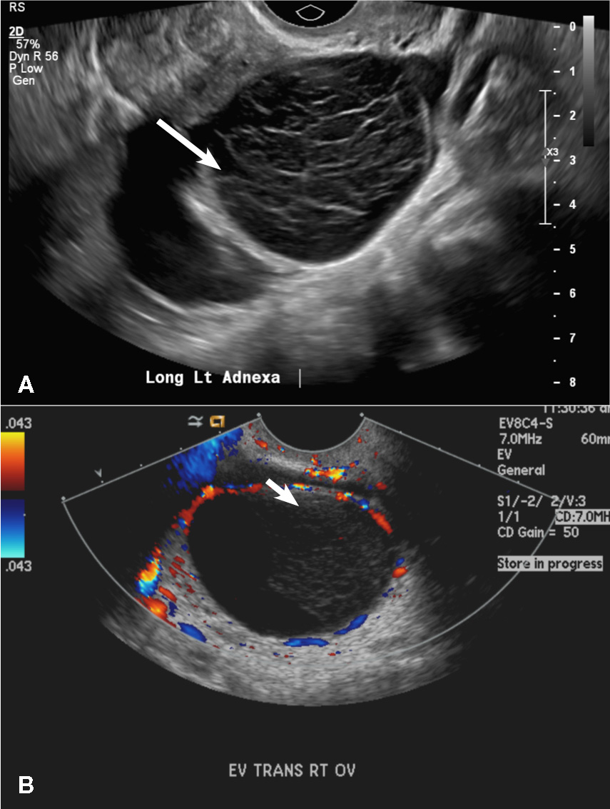
Hemorrhagic cysts. (A) Transvaginal pelvic ultrasound of the left ovary demonstrates a well-circumscribed hypoechoic cyst with posterior acoustic enhancement and demonstrates a lacy reticular pattern of internal echoes (long arrow). (B) Transvaginal pelvic ultrasound of the right ovary demonstrates a well-circumscribed hypoechoic cyst with solid appearing retractile hemorrhagic clot with concave margins (short arrow) and no vascular flow on color Doppler.

(C) Endometrioma INCORRECT
This mass is a well-circumscribed hypoechoic cyst with homogeneous ground glass or low level echoes and increased through transmission.1 It is also avascular without solid components.

Endometrioma. Transvaginal pelvic ultrasound of the right ovary demonstrates a well-circumscribed hypoechoic cyst with homogeneous ground glass or low level echoes and increased through transmission. It is also avascular without solid components.

(D) Dermoid INCORRECT
This common benign ovarian tumor has varying appearances. The most common appearance is a cystic lesion with a focal echogenic nodule (long arrow) protruding into the cyst (Rokitansky nodule).2 The second most common appearance is a focal or diffuse hyperechoic mass with areas of acoustic shadowing (arrowhead) from the sebaceous material and hair (tip of the iceberg sign). A third common appearance is a cystic lesion with multiple thin echogenic bands (lines and dots), which are hair floating within the cyst (short arrow). There is no internal vascular flow identified.

Dermoid cysts. (A) Transvaginal pelvic ultrasound of the right adnexa demonstrates a cystic lesion with a focal echogenic nodule protruding into the cyst compatible with a Rokitansky nodule (long arrow). Also seen are multiple thin echogenic lines and dots (short arrow). (B) Transvaginal pelvic ultrasound of the left adnexa shows a diffuse hyperechoic mass with areas of acoustic shadowing (arrowhead) from the sebaceous material and hair (tip of the iceberg sign).

(E) Cystic ovarian neoplasm INCORRECT
These are large complex masses with both cystic and solid components demonstrating internal vascular flow. They usually demonstrate a thick irregular wall, multiple septations, and nodular papillary projections.3
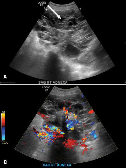
Borderline ovarian neoplasm. (A) Transvaginal pelvic ultrasound of the right adnexa demonstrates a large complex cystic and solid mass with thick irregular wall, multiple septations (arrow) and nodular papillary projections. (B) The mass demonstrates internal vascular flow on color Doppler images.

References
  1. Levine D, Brown DL, Andreotti RF, et al. Management of asymptomatic ovarian and other adnexal cysts imaged at US Society of Radiologists in Ultrasound consensus conference statement. Radiology. 2010;256:(3):943−954.
  2. Outwater EK, Siegelman ES, Hunt JL. Ovarian teratomas: tumor types and imaging characteristics. Radiographics. 2001;21(2):475−490.
  3. Wasnik AP, Menias CO, Platt JF, Lalchandani UR, Bedi DG, Elsayes KM. Multimodality imaging of ovarian cystic lesions: review with an imaging based algorithmic approach. World J Radiol. 2013;5(3):113−125.
Author and Disclosure Information

Dr. Kanmaniraja is Assistant Professor and Chief, Division of Abdominal Imaging, Department of Radiology, University of Florida College of Medicine–Jacksonville.

Dr. Kaunitz is University of Florida Research Foundation Professor and Associate Chairman, Department of Obstetrics and Gynecology, University of Florida College of Medicine–Jacksonville. He is Medical Director and Director of Menopause and Gynecologic Ultrasound Services at UF Women’s Health Specialists–Emerson. He also serves on the OBG Management Board of Editors.

The authors report no financial relationships relevant to this quiz.

Issue
OBG Management - 29(1)
Publications
Sections
Author and Disclosure Information

Dr. Kanmaniraja is Assistant Professor and Chief, Division of Abdominal Imaging, Department of Radiology, University of Florida College of Medicine–Jacksonville.

Dr. Kaunitz is University of Florida Research Foundation Professor and Associate Chairman, Department of Obstetrics and Gynecology, University of Florida College of Medicine–Jacksonville. He is Medical Director and Director of Menopause and Gynecologic Ultrasound Services at UF Women’s Health Specialists–Emerson. He also serves on the OBG Management Board of Editors.

The authors report no financial relationships relevant to this quiz.

What’s the diagnosis?

(A) Simple ovarian cyst INCORRECT
Here is an example of a well circumscribed round or oval anechoic, avascular simple ovarian cyst with posterior acoustic enhancement and thin smooth walls.1 No septations or solid components are identified.

Simple ovarian cyst. Transvaginal pelvic ultrasound of the left ovary demonstrates well circumscribed oval anechoic, avascular cyst with posterior acoustic enhancement and thin smooth walls. No septations or solid components identified.

(B) Hemorrhagic cyst CORRECT
This type of cyst is well circumscribed and hypoechoic, with posterior acoustic enhancement and demonstrates a lacy reticular pattern of internal echoes due to fibrin strands (long arrow). The internal echoes also may be solid appearing with concave margins (short arrow) due to retractile hemorrhagic clot.1 The absence of internal vascular flow on color Doppler helps differentiate it from the solid components seen in ovarian neoplasm.

Hemorrhagic cysts. (A) Transvaginal pelvic ultrasound of the left ovary demonstrates a well-circumscribed hypoechoic cyst with posterior acoustic enhancement and demonstrates a lacy reticular pattern of internal echoes (long arrow). (B) Transvaginal pelvic ultrasound of the right ovary demonstrates a well-circumscribed hypoechoic cyst with solid appearing retractile hemorrhagic clot with concave margins (short arrow) and no vascular flow on color Doppler.

(C) Endometrioma INCORRECT
This mass is a well-circumscribed hypoechoic cyst with homogeneous ground glass or low level echoes and increased through transmission.1 It is also avascular without solid components.

Endometrioma. Transvaginal pelvic ultrasound of the right ovary demonstrates a well-circumscribed hypoechoic cyst with homogeneous ground glass or low level echoes and increased through transmission. It is also avascular without solid components.

(D) Dermoid INCORRECT
This common benign ovarian tumor has varying appearances. The most common appearance is a cystic lesion with a focal echogenic nodule (long arrow) protruding into the cyst (Rokitansky nodule).2 The second most common appearance is a focal or diffuse hyperechoic mass with areas of acoustic shadowing (arrowhead) from the sebaceous material and hair (tip of the iceberg sign). A third common appearance is a cystic lesion with multiple thin echogenic bands (lines and dots), which are hair floating within the cyst (short arrow). There is no internal vascular flow identified.

Dermoid cysts. (A) Transvaginal pelvic ultrasound of the right adnexa demonstrates a cystic lesion with a focal echogenic nodule protruding into the cyst compatible with a Rokitansky nodule (long arrow). Also seen are multiple thin echogenic lines and dots (short arrow). (B) Transvaginal pelvic ultrasound of the left adnexa shows a diffuse hyperechoic mass with areas of acoustic shadowing (arrowhead) from the sebaceous material and hair (tip of the iceberg sign).

(E) Cystic ovarian neoplasm INCORRECT
These are large complex masses with both cystic and solid components demonstrating internal vascular flow. They usually demonstrate a thick irregular wall, multiple septations, and nodular papillary projections.3

Borderline ovarian neoplasm. (A) Transvaginal pelvic ultrasound of the right adnexa demonstrates a large complex cystic and solid mass with thick irregular wall, multiple septations (arrow) and nodular papillary projections. (B) The mass demonstrates internal vascular flow on color Doppler images.

(A) Simple ovarian cyst INCORRECT
Here is an example of a well circumscribed round or oval anechoic, avascular simple ovarian cyst with posterior acoustic enhancement and thin smooth walls.1 No septations or solid components are identified.

Simple ovarian cyst. Transvaginal pelvic ultrasound of the left ovary demonstrates well circumscribed oval anechoic, avascular cyst with posterior acoustic enhancement and thin smooth walls. No septations or solid components identified.

(B) Hemorrhagic cyst CORRECT
This type of cyst is well circumscribed and hypoechoic, with posterior acoustic enhancement and demonstrates a lacy reticular pattern of internal echoes due to fibrin strands (long arrow). The internal echoes also may be solid appearing with concave margins (short arrow) due to retractile hemorrhagic clot.1 The absence of internal vascular flow on color Doppler helps differentiate it from the solid components seen in ovarian neoplasm.

Hemorrhagic cysts. (A) Transvaginal pelvic ultrasound of the left ovary demonstrates a well-circumscribed hypoechoic cyst with posterior acoustic enhancement and demonstrates a lacy reticular pattern of internal echoes (long arrow). (B) Transvaginal pelvic ultrasound of the right ovary demonstrates a well-circumscribed hypoechoic cyst with solid appearing retractile hemorrhagic clot with concave margins (short arrow) and no vascular flow on color Doppler.

(C) Endometrioma INCORRECT
This mass is a well-circumscribed hypoechoic cyst with homogeneous ground glass or low level echoes and increased through transmission.1 It is also avascular without solid components.

Endometrioma. Transvaginal pelvic ultrasound of the right ovary demonstrates a well-circumscribed hypoechoic cyst with homogeneous ground glass or low level echoes and increased through transmission. It is also avascular without solid components.

(D) Dermoid INCORRECT
This common benign ovarian tumor has varying appearances. The most common appearance is a cystic lesion with a focal echogenic nodule (long arrow) protruding into the cyst (Rokitansky nodule).2 The second most common appearance is a focal or diffuse hyperechoic mass with areas of acoustic shadowing (arrowhead) from the sebaceous material and hair (tip of the iceberg sign). A third common appearance is a cystic lesion with multiple thin echogenic bands (lines and dots), which are hair floating within the cyst (short arrow). There is no internal vascular flow identified.

Dermoid cysts. (A) Transvaginal pelvic ultrasound of the right adnexa demonstrates a cystic lesion with a focal echogenic nodule protruding into the cyst compatible with a Rokitansky nodule (long arrow). Also seen are multiple thin echogenic lines and dots (short arrow). (B) Transvaginal pelvic ultrasound of the left adnexa shows a diffuse hyperechoic mass with areas of acoustic shadowing (arrowhead) from the sebaceous material and hair (tip of the iceberg sign).

(E) Cystic ovarian neoplasm INCORRECT
These are large complex masses with both cystic and solid components demonstrating internal vascular flow. They usually demonstrate a thick irregular wall, multiple septations, and nodular papillary projections.3

Borderline ovarian neoplasm. (A) Transvaginal pelvic ultrasound of the right adnexa demonstrates a large complex cystic and solid mass with thick irregular wall, multiple septations (arrow) and nodular papillary projections. (B) The mass demonstrates internal vascular flow on color Doppler images.

References
  1. Levine D, Brown DL, Andreotti RF, et al. Management of asymptomatic ovarian and other adnexal cysts imaged at US Society of Radiologists in Ultrasound consensus conference statement. Radiology. 2010;256:(3):943−954.
  2. Outwater EK, Siegelman ES, Hunt JL. Ovarian teratomas: tumor types and imaging characteristics. Radiographics. 2001;21(2):475−490.
  3. Wasnik AP, Menias CO, Platt JF, Lalchandani UR, Bedi DG, Elsayes KM. Multimodality imaging of ovarian cystic lesions: review with an imaging based algorithmic approach. World J Radiol. 2013;5(3):113−125.
Issue
OBG Management - 29(1)
Issue
OBG Management - 29(1)
Publications
Publications
Article Type
Display Headline
2-week left-sided pelvic pain
Display Headline
2-week left-sided pelvic pain
Sections
Questionnaire Body

A 37-year-old woman presents to the emergency department reporting left-sided pelvic pain for 2 weeks duration. She has a negative urine pregnancy test. Pelvic ultrasonography of the left adnexa is performed with gray scale (A) and color Doppler images (B). Figures shown above.
 

Disallow All Ads
Alternative CME