User login
Population Health Prevails at Two Institutions
Population health—a movement to improve the health of an entire population—is a growing trend driven by the U.S. government. Many health systems are already on board, as healthcare shifts from a fee-for-service system to a value-based system.
One group of Premier Health hospitals and health systems has been collaborating since 2011 to build capabilities to become clinically integrated care networks that are accountable for the health of defined populations within their communities, according to Joseph Damore, vice president of population health management for the Charlotte, N.C-based company.
Damore says Premier has developed a comprehensive framework for the activities and capabilities necessary for successful population health management. Building blocks include:
- Patient-centered foundation (greater patient engagement and involvement in clinical decisions);
- Health home (a primary care medical home);
- High-value network (a set of providers who deliver quality care at an efficient price and whose performance is measured in the areas of cost, quality, and satisfaction);
- Payer partnership (care delivery network providers working with payers to create aligned financial incentives consistent with providing high-value care);
- Population health data management (collecting, analyzing, and reporting data covering all of the care the network’s patient population receives); and
- Network leadership (systematic governance and administration) focused on improving health, managing and coordinating care, and managing per capita cost.
“We’re also working with health systems on initiatives to establish patient-centered foundations and medical homes and create clinically integrated networks, providing our members with a direct roadmap to follow to successfully transition to this new value-based model,” Damore says.
At Jackson Memorial Hospital, one of the nation’s largest safety net hospitals, managing population health is ingrained in staff from day one. Nonetheless, Joshua D. Lenchus, DO, RPh, FACP, SFHM, president of Jackson Health System Medical Staff, says there are opportunities for improvement.
“A more team-based, collaborative approach is being piloted on some floors of our hospital, with specific physician groups,” he says. “Armed with the knowledge of these interventions, we can work on bolstering the pearls and rectifying the pitfalls as we move forward.
“One of our biggest obstacles to success is our patients’ general socioeconomic status.”
A current initiative at Jackson includes piloting a physician-led, multidisciplinary approach to address some of the health determinants. Furthermore, the health system is building additional satellite community clinics and urgent care centers, as it attempts to address disease earlier in the process. Additionally, there is a renewed emphasis on reinforcing the primary care infrastructure to facilitate patient appointment needs, Dr. Lenchus says. TH
Population health—a movement to improve the health of an entire population—is a growing trend driven by the U.S. government. Many health systems are already on board, as healthcare shifts from a fee-for-service system to a value-based system.
One group of Premier Health hospitals and health systems has been collaborating since 2011 to build capabilities to become clinically integrated care networks that are accountable for the health of defined populations within their communities, according to Joseph Damore, vice president of population health management for the Charlotte, N.C-based company.
Damore says Premier has developed a comprehensive framework for the activities and capabilities necessary for successful population health management. Building blocks include:
- Patient-centered foundation (greater patient engagement and involvement in clinical decisions);
- Health home (a primary care medical home);
- High-value network (a set of providers who deliver quality care at an efficient price and whose performance is measured in the areas of cost, quality, and satisfaction);
- Payer partnership (care delivery network providers working with payers to create aligned financial incentives consistent with providing high-value care);
- Population health data management (collecting, analyzing, and reporting data covering all of the care the network’s patient population receives); and
- Network leadership (systematic governance and administration) focused on improving health, managing and coordinating care, and managing per capita cost.
“We’re also working with health systems on initiatives to establish patient-centered foundations and medical homes and create clinically integrated networks, providing our members with a direct roadmap to follow to successfully transition to this new value-based model,” Damore says.
At Jackson Memorial Hospital, one of the nation’s largest safety net hospitals, managing population health is ingrained in staff from day one. Nonetheless, Joshua D. Lenchus, DO, RPh, FACP, SFHM, president of Jackson Health System Medical Staff, says there are opportunities for improvement.
“A more team-based, collaborative approach is being piloted on some floors of our hospital, with specific physician groups,” he says. “Armed with the knowledge of these interventions, we can work on bolstering the pearls and rectifying the pitfalls as we move forward.
“One of our biggest obstacles to success is our patients’ general socioeconomic status.”
A current initiative at Jackson includes piloting a physician-led, multidisciplinary approach to address some of the health determinants. Furthermore, the health system is building additional satellite community clinics and urgent care centers, as it attempts to address disease earlier in the process. Additionally, there is a renewed emphasis on reinforcing the primary care infrastructure to facilitate patient appointment needs, Dr. Lenchus says. TH
Population health—a movement to improve the health of an entire population—is a growing trend driven by the U.S. government. Many health systems are already on board, as healthcare shifts from a fee-for-service system to a value-based system.
One group of Premier Health hospitals and health systems has been collaborating since 2011 to build capabilities to become clinically integrated care networks that are accountable for the health of defined populations within their communities, according to Joseph Damore, vice president of population health management for the Charlotte, N.C-based company.
Damore says Premier has developed a comprehensive framework for the activities and capabilities necessary for successful population health management. Building blocks include:
- Patient-centered foundation (greater patient engagement and involvement in clinical decisions);
- Health home (a primary care medical home);
- High-value network (a set of providers who deliver quality care at an efficient price and whose performance is measured in the areas of cost, quality, and satisfaction);
- Payer partnership (care delivery network providers working with payers to create aligned financial incentives consistent with providing high-value care);
- Population health data management (collecting, analyzing, and reporting data covering all of the care the network’s patient population receives); and
- Network leadership (systematic governance and administration) focused on improving health, managing and coordinating care, and managing per capita cost.
“We’re also working with health systems on initiatives to establish patient-centered foundations and medical homes and create clinically integrated networks, providing our members with a direct roadmap to follow to successfully transition to this new value-based model,” Damore says.
At Jackson Memorial Hospital, one of the nation’s largest safety net hospitals, managing population health is ingrained in staff from day one. Nonetheless, Joshua D. Lenchus, DO, RPh, FACP, SFHM, president of Jackson Health System Medical Staff, says there are opportunities for improvement.
“A more team-based, collaborative approach is being piloted on some floors of our hospital, with specific physician groups,” he says. “Armed with the knowledge of these interventions, we can work on bolstering the pearls and rectifying the pitfalls as we move forward.
“One of our biggest obstacles to success is our patients’ general socioeconomic status.”
A current initiative at Jackson includes piloting a physician-led, multidisciplinary approach to address some of the health determinants. Furthermore, the health system is building additional satellite community clinics and urgent care centers, as it attempts to address disease earlier in the process. Additionally, there is a renewed emphasis on reinforcing the primary care infrastructure to facilitate patient appointment needs, Dr. Lenchus says. TH
Five Strategies for Early Career, Academic Hospitalists
One of the challenges a new academic hospitalist faces is trying to become a valued member of the teaching faculty. You are intent on becoming that next great attending you have always aspired to be; however, no one has clearly guided you on making this transition. In our experience, a handful of common teaching pitfalls frequently unravel the best efforts of young hospitalists. Below, you will find five strategies to take your teaching to the next level.
1. Don’t Try to Be Attending AND Resident.
Being a hospitalist attending is distinctly different than being a resident. It certainly is not being a “super-resident,” PGY-4, or PGY-17 for the team. When challenged with a new role and greater responsibility, it is natural to default to a more comfortable position (i.e., the hands-on mindset of the resident), but by doing so, you encroach on the work of the actual resident on your team. Adult learning theory teaches that it is responsibility that is the cornerstone of adult motivation. Trainees must have a chance to perform the work expected of their position without interference.

Let the resident be the resident. To reach this goal, set clear expectations and discuss the expectations up front. Make sure that you draw a sharp line between where the responsibility of the resident ends and yours begins. Have your resident help set her own expectations for the team, as well. This empowers the resident and also gives you insight into her view of the team dynamics. Your expectations should vary with the ability of the learner and the time of year. For example, the degree of autonomy that you may give a second-year resident in July is much less than what you might give a ready-to-graduate, third-year resident in June.
If granting autonomy makes you uncomfortable, observe your trainees from a distance for your own reassurance. Read the electronic medical record in depth; follow up on orders after attending rounds. If you don’t like a treatment decision, step in without inducing undue shame. After all, this is why residents are still in training, and it gives you the chance to demonstrate how to turn good care into great care.
2. Make Sure You Get to Know Your Trainees
Getting to know your trainees seems so simple that it often gets overlooked. Yet this may be the way your teaching and role modeling make their greatest impact. For adults to thrive in a learning environment, inclusion is key. There is no better way to feel included than to feel known. Inclusion allows learners to feel comfortable with being vulnerable by answering questions, asking questions, interacting, and participating in a meaningful manner on rounds. Consider your own behavior: How comfortable are you asking a question among a large audience of strangers versus asking the same question in a small group of friends? Inclusion will affect behavior.
Engage in “biographic rounds” near the start of your time together. As the attending, set the example by telling your trainees about yourself. Let them know where you are from, where you trained, what led you to choose hospital medicine, and some details about what you do in your spare time. Personal information will help break down the artificial walls that separate attendings from trainees.
Allow every member of your team to tell his or her story. It may not seem like much to you, but the effect on learners has been well established. If you invest in them, they will invest in the team.
3. Make Time to Observe Your Learners
You are responsible for evaluation and feedback of all your learners. Many factors contribute to poor feedback, but one of the most important is that new attendings often do not make a conscious effort to observe their learners. These attendings struggle to give meaningful feedback.

Take time and take notes:
- Take the time to watch your resident respond when the student is presenting her patient.
- Take time to allow the resident or intern to conduct bedside rounds on his patient.
- Take time to stop by on call to watch a student, intern, or resident take a history and perform a physical exam.
Even if you are unable to observe the whole encounter, there is little that gives you as much insight into your trainees as seeing them perform even part of a history and physical exam on a new patient. With time, a series of small observations will add up to a large number of specific comments.
Take notes on your trainees’ actions as you might do for your patients. This way, you have a record of what they did well and what needs work. Specific feedback will show that you paid attention and took the time to care about them as you would your patients.
4. Don’t Keep Your Thoughts to Yourself

Your job is akin to that of a syndicated columnist who is paid to give an informed narrative on the facts of the day. You must explain how the facts actually matter to patient care.
Think out loud. Explain your thoughts as much as possible. Do not assume that even your most senior trainees understand why you recommend a certain test or treatment. It is like algebra, where the teacher would never accept your answer unless you showed your work. For the sake of your learners, you must always “show your thinking.” They will learn as much from your clinical reasoning as they can from any canned talk on a subject.
5. Explicitly Plan Time for Teaching
“Thinking out loud” is a great way to teach, but a prepared talk can go into more depth on a topic. Yet how to find the time? The demands of a busy clinical service can overwhelm the best of intentions. Preparation is key. Good teaching does not happen by chance.
Set aside time for formal teaching outside of rounds. Be explicit as to when this will happen. Tell your team a day before, so they can prepare themselves or clear their time.
Have a handful of “canned” talks that you can give on topics related to common situations encountered in the hospital. They need not last more than 10 minutes. Always leave time for questions, and do your best to make them interactive. Even on a busy service, 10-15 minutes is reasonable for a brief, focused teaching session.
In Sum
Being an academic hospitalist with teaching responsibilities is highly rewarding. But becoming that next “great attending” requires an ongoing commitment to acquiring and developing your teaching skills.
Consider attending the SHM annual meeting or the Academic Hospitalist Academy to gain further knowledge on how to enhance your teaching career. In the meantime, try and practice some of the above tips—your learners may thank you. TH
Dr. Burger is associate program director of internal medicine residency in the Department of Medicine at Mount Sinai Beth Israel and assistant professor of medicine at Icahn School of Medicine at Mount Sinai, both in New York City. Dr. Miller is chief of the division of general internal medicine, associate chair of education, and associate professor in the Department of Internal Medicine at Saint Louis University.
One of the challenges a new academic hospitalist faces is trying to become a valued member of the teaching faculty. You are intent on becoming that next great attending you have always aspired to be; however, no one has clearly guided you on making this transition. In our experience, a handful of common teaching pitfalls frequently unravel the best efforts of young hospitalists. Below, you will find five strategies to take your teaching to the next level.
1. Don’t Try to Be Attending AND Resident.
Being a hospitalist attending is distinctly different than being a resident. It certainly is not being a “super-resident,” PGY-4, or PGY-17 for the team. When challenged with a new role and greater responsibility, it is natural to default to a more comfortable position (i.e., the hands-on mindset of the resident), but by doing so, you encroach on the work of the actual resident on your team. Adult learning theory teaches that it is responsibility that is the cornerstone of adult motivation. Trainees must have a chance to perform the work expected of their position without interference.
Let the resident be the resident. To reach this goal, set clear expectations and discuss the expectations up front. Make sure that you draw a sharp line between where the responsibility of the resident ends and yours begins. Have your resident help set her own expectations for the team, as well. This empowers the resident and also gives you insight into her view of the team dynamics. Your expectations should vary with the ability of the learner and the time of year. For example, the degree of autonomy that you may give a second-year resident in July is much less than what you might give a ready-to-graduate, third-year resident in June.
If granting autonomy makes you uncomfortable, observe your trainees from a distance for your own reassurance. Read the electronic medical record in depth; follow up on orders after attending rounds. If you don’t like a treatment decision, step in without inducing undue shame. After all, this is why residents are still in training, and it gives you the chance to demonstrate how to turn good care into great care.
2. Make Sure You Get to Know Your Trainees
Getting to know your trainees seems so simple that it often gets overlooked. Yet this may be the way your teaching and role modeling make their greatest impact. For adults to thrive in a learning environment, inclusion is key. There is no better way to feel included than to feel known. Inclusion allows learners to feel comfortable with being vulnerable by answering questions, asking questions, interacting, and participating in a meaningful manner on rounds. Consider your own behavior: How comfortable are you asking a question among a large audience of strangers versus asking the same question in a small group of friends? Inclusion will affect behavior.
Engage in “biographic rounds” near the start of your time together. As the attending, set the example by telling your trainees about yourself. Let them know where you are from, where you trained, what led you to choose hospital medicine, and some details about what you do in your spare time. Personal information will help break down the artificial walls that separate attendings from trainees.
Allow every member of your team to tell his or her story. It may not seem like much to you, but the effect on learners has been well established. If you invest in them, they will invest in the team.
3. Make Time to Observe Your Learners
You are responsible for evaluation and feedback of all your learners. Many factors contribute to poor feedback, but one of the most important is that new attendings often do not make a conscious effort to observe their learners. These attendings struggle to give meaningful feedback.
Take time and take notes:
- Take the time to watch your resident respond when the student is presenting her patient.
- Take time to allow the resident or intern to conduct bedside rounds on his patient.
- Take time to stop by on call to watch a student, intern, or resident take a history and perform a physical exam.
Even if you are unable to observe the whole encounter, there is little that gives you as much insight into your trainees as seeing them perform even part of a history and physical exam on a new patient. With time, a series of small observations will add up to a large number of specific comments.
Take notes on your trainees’ actions as you might do for your patients. This way, you have a record of what they did well and what needs work. Specific feedback will show that you paid attention and took the time to care about them as you would your patients.
4. Don’t Keep Your Thoughts to Yourself
Your job is akin to that of a syndicated columnist who is paid to give an informed narrative on the facts of the day. You must explain how the facts actually matter to patient care.
Think out loud. Explain your thoughts as much as possible. Do not assume that even your most senior trainees understand why you recommend a certain test or treatment. It is like algebra, where the teacher would never accept your answer unless you showed your work. For the sake of your learners, you must always “show your thinking.” They will learn as much from your clinical reasoning as they can from any canned talk on a subject.
5. Explicitly Plan Time for Teaching
“Thinking out loud” is a great way to teach, but a prepared talk can go into more depth on a topic. Yet how to find the time? The demands of a busy clinical service can overwhelm the best of intentions. Preparation is key. Good teaching does not happen by chance.
Set aside time for formal teaching outside of rounds. Be explicit as to when this will happen. Tell your team a day before, so they can prepare themselves or clear their time.
Have a handful of “canned” talks that you can give on topics related to common situations encountered in the hospital. They need not last more than 10 minutes. Always leave time for questions, and do your best to make them interactive. Even on a busy service, 10-15 minutes is reasonable for a brief, focused teaching session.
In Sum
Being an academic hospitalist with teaching responsibilities is highly rewarding. But becoming that next “great attending” requires an ongoing commitment to acquiring and developing your teaching skills.
Consider attending the SHM annual meeting or the Academic Hospitalist Academy to gain further knowledge on how to enhance your teaching career. In the meantime, try and practice some of the above tips—your learners may thank you. TH
Dr. Burger is associate program director of internal medicine residency in the Department of Medicine at Mount Sinai Beth Israel and assistant professor of medicine at Icahn School of Medicine at Mount Sinai, both in New York City. Dr. Miller is chief of the division of general internal medicine, associate chair of education, and associate professor in the Department of Internal Medicine at Saint Louis University.
One of the challenges a new academic hospitalist faces is trying to become a valued member of the teaching faculty. You are intent on becoming that next great attending you have always aspired to be; however, no one has clearly guided you on making this transition. In our experience, a handful of common teaching pitfalls frequently unravel the best efforts of young hospitalists. Below, you will find five strategies to take your teaching to the next level.
1. Don’t Try to Be Attending AND Resident.
Being a hospitalist attending is distinctly different than being a resident. It certainly is not being a “super-resident,” PGY-4, or PGY-17 for the team. When challenged with a new role and greater responsibility, it is natural to default to a more comfortable position (i.e., the hands-on mindset of the resident), but by doing so, you encroach on the work of the actual resident on your team. Adult learning theory teaches that it is responsibility that is the cornerstone of adult motivation. Trainees must have a chance to perform the work expected of their position without interference.
Let the resident be the resident. To reach this goal, set clear expectations and discuss the expectations up front. Make sure that you draw a sharp line between where the responsibility of the resident ends and yours begins. Have your resident help set her own expectations for the team, as well. This empowers the resident and also gives you insight into her view of the team dynamics. Your expectations should vary with the ability of the learner and the time of year. For example, the degree of autonomy that you may give a second-year resident in July is much less than what you might give a ready-to-graduate, third-year resident in June.
If granting autonomy makes you uncomfortable, observe your trainees from a distance for your own reassurance. Read the electronic medical record in depth; follow up on orders after attending rounds. If you don’t like a treatment decision, step in without inducing undue shame. After all, this is why residents are still in training, and it gives you the chance to demonstrate how to turn good care into great care.
2. Make Sure You Get to Know Your Trainees
Getting to know your trainees seems so simple that it often gets overlooked. Yet this may be the way your teaching and role modeling make their greatest impact. For adults to thrive in a learning environment, inclusion is key. There is no better way to feel included than to feel known. Inclusion allows learners to feel comfortable with being vulnerable by answering questions, asking questions, interacting, and participating in a meaningful manner on rounds. Consider your own behavior: How comfortable are you asking a question among a large audience of strangers versus asking the same question in a small group of friends? Inclusion will affect behavior.
Engage in “biographic rounds” near the start of your time together. As the attending, set the example by telling your trainees about yourself. Let them know where you are from, where you trained, what led you to choose hospital medicine, and some details about what you do in your spare time. Personal information will help break down the artificial walls that separate attendings from trainees.
Allow every member of your team to tell his or her story. It may not seem like much to you, but the effect on learners has been well established. If you invest in them, they will invest in the team.
3. Make Time to Observe Your Learners
You are responsible for evaluation and feedback of all your learners. Many factors contribute to poor feedback, but one of the most important is that new attendings often do not make a conscious effort to observe their learners. These attendings struggle to give meaningful feedback.
Take time and take notes:
- Take the time to watch your resident respond when the student is presenting her patient.
- Take time to allow the resident or intern to conduct bedside rounds on his patient.
- Take time to stop by on call to watch a student, intern, or resident take a history and perform a physical exam.
Even if you are unable to observe the whole encounter, there is little that gives you as much insight into your trainees as seeing them perform even part of a history and physical exam on a new patient. With time, a series of small observations will add up to a large number of specific comments.
Take notes on your trainees’ actions as you might do for your patients. This way, you have a record of what they did well and what needs work. Specific feedback will show that you paid attention and took the time to care about them as you would your patients.
4. Don’t Keep Your Thoughts to Yourself
Your job is akin to that of a syndicated columnist who is paid to give an informed narrative on the facts of the day. You must explain how the facts actually matter to patient care.
Think out loud. Explain your thoughts as much as possible. Do not assume that even your most senior trainees understand why you recommend a certain test or treatment. It is like algebra, where the teacher would never accept your answer unless you showed your work. For the sake of your learners, you must always “show your thinking.” They will learn as much from your clinical reasoning as they can from any canned talk on a subject.
5. Explicitly Plan Time for Teaching
“Thinking out loud” is a great way to teach, but a prepared talk can go into more depth on a topic. Yet how to find the time? The demands of a busy clinical service can overwhelm the best of intentions. Preparation is key. Good teaching does not happen by chance.
Set aside time for formal teaching outside of rounds. Be explicit as to when this will happen. Tell your team a day before, so they can prepare themselves or clear their time.
Have a handful of “canned” talks that you can give on topics related to common situations encountered in the hospital. They need not last more than 10 minutes. Always leave time for questions, and do your best to make them interactive. Even on a busy service, 10-15 minutes is reasonable for a brief, focused teaching session.
In Sum
Being an academic hospitalist with teaching responsibilities is highly rewarding. But becoming that next “great attending” requires an ongoing commitment to acquiring and developing your teaching skills.
Consider attending the SHM annual meeting or the Academic Hospitalist Academy to gain further knowledge on how to enhance your teaching career. In the meantime, try and practice some of the above tips—your learners may thank you. TH
Dr. Burger is associate program director of internal medicine residency in the Department of Medicine at Mount Sinai Beth Israel and assistant professor of medicine at Icahn School of Medicine at Mount Sinai, both in New York City. Dr. Miller is chief of the division of general internal medicine, associate chair of education, and associate professor in the Department of Internal Medicine at Saint Louis University.

Move to Allow Patients to Request 'Refund' Appealing and Risky
We’ve all seen hundreds of commercials from companies advertising products and services with a money-back guarantee. The Men’s Warehouse, for example, has been promising men across the globe for over a decade, “You’re going to like the way you look. I guarantee it!” But to date, no one has made such a “guarantee” in the healthcare industry. Buying a suit is not exactly like getting your gallbladder removed.

We know that medical diagnoses and treatments are filled with uncertainty in expected processes and outcomes, because the factors that are dependent on these processes and outcomes are endless. These include patient factors (overall health, functional status, comorbid conditions), procedural factors (emergency versus elective, time of day or night), and facility factors (having the optimal team with skills that match the patient need, having all the right products and equipment). Although we know that many medical procedures have a relatively predictable risk of complications, unpredictable complications still occur, so how can we ever offer a guarantee for the interventions we perform on patients?
First of Its Kind
David Feinberg, MD, MBA, president and CEO of Geisinger Health System, is doing just that. This healthcare system has developed an application, called the Geisinger ProvenExperience, which can be downloaded onto a smartphone. After a procedure, each patient is given a code for the condition that was treated. With that code, the patient can enter feedback on the services provided and can then request a refund if they are not fully satisfied.
Most remarkably, the request for a refund is based on the judgment of the recipient, not on that of the provider(s). At a recent public meeting, Dr. Feinberg said of the new program: “We’re going to do everything right. That’s our job, that’s our promise to you … and you’re the judge. If you don’t think so, we’re going to apologize, we’re going to try to fix it for the next guy, and, as a small token of appreciation, we’re going to give you some money back.”1
Although many are skeptical about whether or not the program will be successful, much less viable, Dr. Feinberg contends that early feedback on the program has shown that most patients don’t actually want their money back. Instead, if their needs have not been met, most have just wanted a sincere apology and a commitment to make things better for others. Dr. Feinberg also contests that even if this is not the best or only approach to improving healthcare (quickly), we should all feel compelled to do something about our repeated failures in meeting patient expectations in the quality and/or experience of their care; and because no other industry works this way, other than healthcare. Typically, when consumers get fed up with poor service in other industries, disruptive innovations (Uber, for example) are created to satisfy customers’ desires.
A New Paradigm?
In healthcare, patients certainly should be dissatisfied if they experience a preventable harm event. Some types of harm are considered “always preventable,” such as wrong-site surgery. These events are extremely rare and, thus, do not constitute most cases of harm in hospitals these days. Such “never events” are relatively well defined and have been adopted for nonpayment by Medicare and other insurers, which can serve to buffer a patient’s financial liability in the small number of these cases. For other, more common, types of preventable harm, some hospitals have instituted apology and disclosure policies, and some will also relieve the patient of the portion of the bill attributable to the preventable harm. But not all hospitals have adopted such policies, despite the fact that they are widely endorsed by influential agencies, including The Joint Commission, the American Medical Association, Leapfrog Group, the National Quality Forum, and the Agency for Healthcare Research and Quality.
And, even for hospitals that have adopted such “best practice” policies, there is not always clear consensus on what constitutes preventable harm. Generally, the “judgment call” about what constitutes preventable harm is made by healthcare systems and providers—not patients. In addition, many cases of harm that are not necessarily preventable can often result in great dissatisfaction for the patient. There are countless stories of patients who are unfortunately harmed in the course of medical procedures, but who were informed of the possible risks of the procedure and consented to have the procedure performed despite the risks. These situations, which are agonizingly difficult for the system, the providers, and the patients, have no good solutions. Systems cannot “own” all harm, such as those resulting from the disease process itself or from risky and invasive procedures intended to benefit the patient. And there is ongoing inconsistency in healthcare systems when it comes to their willingness and ability to consistently define preventable harm or to disclose, apologize, and forgive payments in such cases.
So, while this move to allow patients to ask for a “refund” seems both extremely appealing and extremely risky, it certainly seems as though it will greatly enhance the trust of patients and their families in the Geisinger Health System.
I, among others, will eagerly follow the results of this program; while getting a cholecystectomy is not the same as buying a men’s suit, I do hope that someday, I will be able to say to every patient entering my healthcare system that before they leave, “You’re going to like the way you feel. I guarantee it!” TH

References
1. Guydish M. Geisinger CEO: money-back guarantee for health care coming. November 6, 2015. Times Leader website. Available at: http://timesleader.com/news/492790/geisinger-ceo-money-back-guarantee-for-health-car-coming. Accessed December 5, 2015.
2. Luthra S. When something goes wrong at the hospital, who pays? November 11, 2015. Kaiser Health News. Available at: http://khn.org/news/when-something-goes-wrong-at-the-hospital-who-pays/?utm_source=Managed&utm_campaign=9e17712a95-Quality+%26+Patient+Safety+Update&utm_medium=email&utm_term=0_ebe1fa6178-9e17712a95-319388717. Accessed December 5, 2015.
We’ve all seen hundreds of commercials from companies advertising products and services with a money-back guarantee. The Men’s Warehouse, for example, has been promising men across the globe for over a decade, “You’re going to like the way you look. I guarantee it!” But to date, no one has made such a “guarantee” in the healthcare industry. Buying a suit is not exactly like getting your gallbladder removed.
We know that medical diagnoses and treatments are filled with uncertainty in expected processes and outcomes, because the factors that are dependent on these processes and outcomes are endless. These include patient factors (overall health, functional status, comorbid conditions), procedural factors (emergency versus elective, time of day or night), and facility factors (having the optimal team with skills that match the patient need, having all the right products and equipment). Although we know that many medical procedures have a relatively predictable risk of complications, unpredictable complications still occur, so how can we ever offer a guarantee for the interventions we perform on patients?
First of Its Kind
David Feinberg, MD, MBA, president and CEO of Geisinger Health System, is doing just that. This healthcare system has developed an application, called the Geisinger ProvenExperience, which can be downloaded onto a smartphone. After a procedure, each patient is given a code for the condition that was treated. With that code, the patient can enter feedback on the services provided and can then request a refund if they are not fully satisfied.
Most remarkably, the request for a refund is based on the judgment of the recipient, not on that of the provider(s). At a recent public meeting, Dr. Feinberg said of the new program: “We’re going to do everything right. That’s our job, that’s our promise to you … and you’re the judge. If you don’t think so, we’re going to apologize, we’re going to try to fix it for the next guy, and, as a small token of appreciation, we’re going to give you some money back.”1
Although many are skeptical about whether or not the program will be successful, much less viable, Dr. Feinberg contends that early feedback on the program has shown that most patients don’t actually want their money back. Instead, if their needs have not been met, most have just wanted a sincere apology and a commitment to make things better for others. Dr. Feinberg also contests that even if this is not the best or only approach to improving healthcare (quickly), we should all feel compelled to do something about our repeated failures in meeting patient expectations in the quality and/or experience of their care; and because no other industry works this way, other than healthcare. Typically, when consumers get fed up with poor service in other industries, disruptive innovations (Uber, for example) are created to satisfy customers’ desires.
A New Paradigm?
In healthcare, patients certainly should be dissatisfied if they experience a preventable harm event. Some types of harm are considered “always preventable,” such as wrong-site surgery. These events are extremely rare and, thus, do not constitute most cases of harm in hospitals these days. Such “never events” are relatively well defined and have been adopted for nonpayment by Medicare and other insurers, which can serve to buffer a patient’s financial liability in the small number of these cases. For other, more common, types of preventable harm, some hospitals have instituted apology and disclosure policies, and some will also relieve the patient of the portion of the bill attributable to the preventable harm. But not all hospitals have adopted such policies, despite the fact that they are widely endorsed by influential agencies, including The Joint Commission, the American Medical Association, Leapfrog Group, the National Quality Forum, and the Agency for Healthcare Research and Quality.
And, even for hospitals that have adopted such “best practice” policies, there is not always clear consensus on what constitutes preventable harm. Generally, the “judgment call” about what constitutes preventable harm is made by healthcare systems and providers—not patients. In addition, many cases of harm that are not necessarily preventable can often result in great dissatisfaction for the patient. There are countless stories of patients who are unfortunately harmed in the course of medical procedures, but who were informed of the possible risks of the procedure and consented to have the procedure performed despite the risks. These situations, which are agonizingly difficult for the system, the providers, and the patients, have no good solutions. Systems cannot “own” all harm, such as those resulting from the disease process itself or from risky and invasive procedures intended to benefit the patient. And there is ongoing inconsistency in healthcare systems when it comes to their willingness and ability to consistently define preventable harm or to disclose, apologize, and forgive payments in such cases.
So, while this move to allow patients to ask for a “refund” seems both extremely appealing and extremely risky, it certainly seems as though it will greatly enhance the trust of patients and their families in the Geisinger Health System.
I, among others, will eagerly follow the results of this program; while getting a cholecystectomy is not the same as buying a men’s suit, I do hope that someday, I will be able to say to every patient entering my healthcare system that before they leave, “You’re going to like the way you feel. I guarantee it!” TH
References
1. Guydish M. Geisinger CEO: money-back guarantee for health care coming. November 6, 2015. Times Leader website. Available at: http://timesleader.com/news/492790/geisinger-ceo-money-back-guarantee-for-health-car-coming. Accessed December 5, 2015.
2. Luthra S. When something goes wrong at the hospital, who pays? November 11, 2015. Kaiser Health News. Available at: http://khn.org/news/when-something-goes-wrong-at-the-hospital-who-pays/?utm_source=Managed&utm_campaign=9e17712a95-Quality+%26+Patient+Safety+Update&utm_medium=email&utm_term=0_ebe1fa6178-9e17712a95-319388717. Accessed December 5, 2015.
We’ve all seen hundreds of commercials from companies advertising products and services with a money-back guarantee. The Men’s Warehouse, for example, has been promising men across the globe for over a decade, “You’re going to like the way you look. I guarantee it!” But to date, no one has made such a “guarantee” in the healthcare industry. Buying a suit is not exactly like getting your gallbladder removed.
We know that medical diagnoses and treatments are filled with uncertainty in expected processes and outcomes, because the factors that are dependent on these processes and outcomes are endless. These include patient factors (overall health, functional status, comorbid conditions), procedural factors (emergency versus elective, time of day or night), and facility factors (having the optimal team with skills that match the patient need, having all the right products and equipment). Although we know that many medical procedures have a relatively predictable risk of complications, unpredictable complications still occur, so how can we ever offer a guarantee for the interventions we perform on patients?
First of Its Kind
David Feinberg, MD, MBA, president and CEO of Geisinger Health System, is doing just that. This healthcare system has developed an application, called the Geisinger ProvenExperience, which can be downloaded onto a smartphone. After a procedure, each patient is given a code for the condition that was treated. With that code, the patient can enter feedback on the services provided and can then request a refund if they are not fully satisfied.
Most remarkably, the request for a refund is based on the judgment of the recipient, not on that of the provider(s). At a recent public meeting, Dr. Feinberg said of the new program: “We’re going to do everything right. That’s our job, that’s our promise to you … and you’re the judge. If you don’t think so, we’re going to apologize, we’re going to try to fix it for the next guy, and, as a small token of appreciation, we’re going to give you some money back.”1
Although many are skeptical about whether or not the program will be successful, much less viable, Dr. Feinberg contends that early feedback on the program has shown that most patients don’t actually want their money back. Instead, if their needs have not been met, most have just wanted a sincere apology and a commitment to make things better for others. Dr. Feinberg also contests that even if this is not the best or only approach to improving healthcare (quickly), we should all feel compelled to do something about our repeated failures in meeting patient expectations in the quality and/or experience of their care; and because no other industry works this way, other than healthcare. Typically, when consumers get fed up with poor service in other industries, disruptive innovations (Uber, for example) are created to satisfy customers’ desires.
A New Paradigm?
In healthcare, patients certainly should be dissatisfied if they experience a preventable harm event. Some types of harm are considered “always preventable,” such as wrong-site surgery. These events are extremely rare and, thus, do not constitute most cases of harm in hospitals these days. Such “never events” are relatively well defined and have been adopted for nonpayment by Medicare and other insurers, which can serve to buffer a patient’s financial liability in the small number of these cases. For other, more common, types of preventable harm, some hospitals have instituted apology and disclosure policies, and some will also relieve the patient of the portion of the bill attributable to the preventable harm. But not all hospitals have adopted such policies, despite the fact that they are widely endorsed by influential agencies, including The Joint Commission, the American Medical Association, Leapfrog Group, the National Quality Forum, and the Agency for Healthcare Research and Quality.
And, even for hospitals that have adopted such “best practice” policies, there is not always clear consensus on what constitutes preventable harm. Generally, the “judgment call” about what constitutes preventable harm is made by healthcare systems and providers—not patients. In addition, many cases of harm that are not necessarily preventable can often result in great dissatisfaction for the patient. There are countless stories of patients who are unfortunately harmed in the course of medical procedures, but who were informed of the possible risks of the procedure and consented to have the procedure performed despite the risks. These situations, which are agonizingly difficult for the system, the providers, and the patients, have no good solutions. Systems cannot “own” all harm, such as those resulting from the disease process itself or from risky and invasive procedures intended to benefit the patient. And there is ongoing inconsistency in healthcare systems when it comes to their willingness and ability to consistently define preventable harm or to disclose, apologize, and forgive payments in such cases.
So, while this move to allow patients to ask for a “refund” seems both extremely appealing and extremely risky, it certainly seems as though it will greatly enhance the trust of patients and their families in the Geisinger Health System.
I, among others, will eagerly follow the results of this program; while getting a cholecystectomy is not the same as buying a men’s suit, I do hope that someday, I will be able to say to every patient entering my healthcare system that before they leave, “You’re going to like the way you feel. I guarantee it!” TH
References
1. Guydish M. Geisinger CEO: money-back guarantee for health care coming. November 6, 2015. Times Leader website. Available at: http://timesleader.com/news/492790/geisinger-ceo-money-back-guarantee-for-health-car-coming. Accessed December 5, 2015.
2. Luthra S. When something goes wrong at the hospital, who pays? November 11, 2015. Kaiser Health News. Available at: http://khn.org/news/when-something-goes-wrong-at-the-hospital-who-pays/?utm_source=Managed&utm_campaign=9e17712a95-Quality+%26+Patient+Safety+Update&utm_medium=email&utm_term=0_ebe1fa6178-9e17712a95-319388717. Accessed December 5, 2015.
Hospitalist Jaime Upegui, MD, Links Passion for Work to Love of Motorcycle Travel, Sports, Dance
Hospitalist Jaime Upegui, MD, division president at Apogee Physicians in Coeur d’Alene, Idaho, knows exactly what he wants to do when he retires years from now: sell everything he owns and ride his motorcycle around the world.

Dr. Upegui is a modern-day explorer who enjoys making the journey as much as getting to the destination. Riding is his personal form of yoga, a meditative experience that demands he stay in the moment and allows him to escape life’s daily frustrations. Ever since he started riding motorcycles at age five, he’s been hooked and has no plans of shifting into neutral.
Driven by Change
Every day at work, Dr. Upegui, an internal medicine specialist, helps hospitalists thrive in a changing medical world where they’re constantly being pushed and pulled in multiple directions. “It’s an exciting career that’s full of unexpected changes,” he says. “I work hard every day on building great teams that produce stellar results.”
He brings that same level of enthusiasm to his after-work activities, which include skydiving, tango dancing, scuba diving, snowboarding, and rock climbing. His passion for change and adventure stems from his childhood.
Listen to more of our interview with Dr. Upegui.
Dr. Upegui was born in Colombia and raised predominantly in the cities of Cali and Medellín. His mother, Rocio, was a painter and ballet dancer; his father, Jaime Sr., was a poet and musician. He spent most of his childhood with his mother, who moved frequently in search of new inspirations. During his childhood, he attended 13 different schools and lived in more than 30 different homes in the U.S., Colombia, and Spain. In 2003, he finished medical school, graduating from Universidad Pontificia Bolivariana in Medellín. He worked as an attending physician in the emergency department there for three years before moving to New York City to complete his residency in internal medicine at St. Luke’s–Roosevelt Hospital Center.
Throughout medical school and his residency, Dr. Upegui’s personal interests extended beyond motorcycling to skydiving and tango dancing, a skill he initially learned to love from his father. Dr. Upegui says he enjoys nothing more than learning, so he’s attracted to activities that require a high degree of training or technical expertise.

“I like to do things that make me feel like I’m proficient at something that’s difficult,” he says.
He vividly remembers the first time he jumped out of a plane, in 2002.
“It was thrilling, it was exciting, it was ego-boosting, it was self-gratifying, it was an adrenaline rush,” Dr. Upegui explains, adding that he taught English to the owner of the skydiving center in Colombia in exchange for free jumps. “The most exciting part is the decision to take the leap. That critical moment still gives me butterflies.”
Over the next six years, he jumped approximately 150 times, then stopped for roughly seven years to handle the demands of school and work, and to avoid the actual cost of skydiving, which is pricey at roughly a few hundred dollars per jump. But, over the years, he missed it, so he resumed skydiving earlier this year.
In between, he learned to tango through private and group lessons. Last year, he traveled to Buenos Aires, Argentina, for an intensive weeklong course and an international tango gathering. He also travels to “milongas,” get-togethers for professional and student tango dancers that are held in various cities worldwide.
“Tango has two core elements: showing your intention to move forward and allowing your partner to accept the invitation to follow your lead,” he says. “It’s a beautiful combination of assertiveness, determination, and then negotiation, followed by permission to proceed, depending upon how the conversation is going during the dance.”
No Regrets
Procrastination is not a familiar word to Dr. Upegui.
“The time frame between something that I want to do and [actually] doing it is very short,” he says. “If I want to do something, I just look at how I can get to it as soon as it’s available.”
Motorcycling ranks as his number one passion. Last year, he completed a 7,000-mile trip without taking any time off from his current job, which requires him to lead, manage, and often meet with hospitalist teams nationwide. He traveled via back roads on weekends to a major city, left his bike at the airport, and then hopped on a plane to wherever he needed to be for work. Instead of flying home for the weekend, he’d return to the airport to pick up his bike and travel to the next city he wanted to visit, which could be 1,000 miles away.
“The cool thing about the road is that random people help you,” he says. “The hotels would keep my clothes and luggage, and airport parking employees would help me park my motorcycle in a safe place and keep my helmet in their office.”
Dr. Upegui says childhood experiences that focused on momentum and movement laid the foundation for his mobile and adventuresome lifestyle. Movement, variability, and change have become the guiding factors in his life. Perhaps that’s why he chose to be a hospitalist. Among the youngest fields in medicine, the specialty is always growing, changing, and evolving.
“If you take any change in life as just a new stage of a new moment and you just perform your best in this current situation, then that will allow you to always be flexible to what’s happening in front of you,” Dr. Upegui says. “I love my work, family, Apogee, and the opportunities I have had. I could die completely satisfied today, knowing that I’ve done the best I could and searched for happiness every day.”
Hospitalist Jaime Upegui, MD, division president at Apogee Physicians in Coeur d’Alene, Idaho, knows exactly what he wants to do when he retires years from now: sell everything he owns and ride his motorcycle around the world.
Dr. Upegui is a modern-day explorer who enjoys making the journey as much as getting to the destination. Riding is his personal form of yoga, a meditative experience that demands he stay in the moment and allows him to escape life’s daily frustrations. Ever since he started riding motorcycles at age five, he’s been hooked and has no plans of shifting into neutral.
Driven by Change
Every day at work, Dr. Upegui, an internal medicine specialist, helps hospitalists thrive in a changing medical world where they’re constantly being pushed and pulled in multiple directions. “It’s an exciting career that’s full of unexpected changes,” he says. “I work hard every day on building great teams that produce stellar results.”
He brings that same level of enthusiasm to his after-work activities, which include skydiving, tango dancing, scuba diving, snowboarding, and rock climbing. His passion for change and adventure stems from his childhood.
Listen to more of our interview with Dr. Upegui.
Dr. Upegui was born in Colombia and raised predominantly in the cities of Cali and Medellín. His mother, Rocio, was a painter and ballet dancer; his father, Jaime Sr., was a poet and musician. He spent most of his childhood with his mother, who moved frequently in search of new inspirations. During his childhood, he attended 13 different schools and lived in more than 30 different homes in the U.S., Colombia, and Spain. In 2003, he finished medical school, graduating from Universidad Pontificia Bolivariana in Medellín. He worked as an attending physician in the emergency department there for three years before moving to New York City to complete his residency in internal medicine at St. Luke’s–Roosevelt Hospital Center.
Throughout medical school and his residency, Dr. Upegui’s personal interests extended beyond motorcycling to skydiving and tango dancing, a skill he initially learned to love from his father. Dr. Upegui says he enjoys nothing more than learning, so he’s attracted to activities that require a high degree of training or technical expertise.
“I like to do things that make me feel like I’m proficient at something that’s difficult,” he says.
He vividly remembers the first time he jumped out of a plane, in 2002.
“It was thrilling, it was exciting, it was ego-boosting, it was self-gratifying, it was an adrenaline rush,” Dr. Upegui explains, adding that he taught English to the owner of the skydiving center in Colombia in exchange for free jumps. “The most exciting part is the decision to take the leap. That critical moment still gives me butterflies.”
Over the next six years, he jumped approximately 150 times, then stopped for roughly seven years to handle the demands of school and work, and to avoid the actual cost of skydiving, which is pricey at roughly a few hundred dollars per jump. But, over the years, he missed it, so he resumed skydiving earlier this year.
In between, he learned to tango through private and group lessons. Last year, he traveled to Buenos Aires, Argentina, for an intensive weeklong course and an international tango gathering. He also travels to “milongas,” get-togethers for professional and student tango dancers that are held in various cities worldwide.
“Tango has two core elements: showing your intention to move forward and allowing your partner to accept the invitation to follow your lead,” he says. “It’s a beautiful combination of assertiveness, determination, and then negotiation, followed by permission to proceed, depending upon how the conversation is going during the dance.”
No Regrets
Procrastination is not a familiar word to Dr. Upegui.
“The time frame between something that I want to do and [actually] doing it is very short,” he says. “If I want to do something, I just look at how I can get to it as soon as it’s available.”
Motorcycling ranks as his number one passion. Last year, he completed a 7,000-mile trip without taking any time off from his current job, which requires him to lead, manage, and often meet with hospitalist teams nationwide. He traveled via back roads on weekends to a major city, left his bike at the airport, and then hopped on a plane to wherever he needed to be for work. Instead of flying home for the weekend, he’d return to the airport to pick up his bike and travel to the next city he wanted to visit, which could be 1,000 miles away.
“The cool thing about the road is that random people help you,” he says. “The hotels would keep my clothes and luggage, and airport parking employees would help me park my motorcycle in a safe place and keep my helmet in their office.”
Dr. Upegui says childhood experiences that focused on momentum and movement laid the foundation for his mobile and adventuresome lifestyle. Movement, variability, and change have become the guiding factors in his life. Perhaps that’s why he chose to be a hospitalist. Among the youngest fields in medicine, the specialty is always growing, changing, and evolving.
“If you take any change in life as just a new stage of a new moment and you just perform your best in this current situation, then that will allow you to always be flexible to what’s happening in front of you,” Dr. Upegui says. “I love my work, family, Apogee, and the opportunities I have had. I could die completely satisfied today, knowing that I’ve done the best I could and searched for happiness every day.”
Hospitalist Jaime Upegui, MD, division president at Apogee Physicians in Coeur d’Alene, Idaho, knows exactly what he wants to do when he retires years from now: sell everything he owns and ride his motorcycle around the world.
Dr. Upegui is a modern-day explorer who enjoys making the journey as much as getting to the destination. Riding is his personal form of yoga, a meditative experience that demands he stay in the moment and allows him to escape life’s daily frustrations. Ever since he started riding motorcycles at age five, he’s been hooked and has no plans of shifting into neutral.
Driven by Change
Every day at work, Dr. Upegui, an internal medicine specialist, helps hospitalists thrive in a changing medical world where they’re constantly being pushed and pulled in multiple directions. “It’s an exciting career that’s full of unexpected changes,” he says. “I work hard every day on building great teams that produce stellar results.”
He brings that same level of enthusiasm to his after-work activities, which include skydiving, tango dancing, scuba diving, snowboarding, and rock climbing. His passion for change and adventure stems from his childhood.
Listen to more of our interview with Dr. Upegui.
Dr. Upegui was born in Colombia and raised predominantly in the cities of Cali and Medellín. His mother, Rocio, was a painter and ballet dancer; his father, Jaime Sr., was a poet and musician. He spent most of his childhood with his mother, who moved frequently in search of new inspirations. During his childhood, he attended 13 different schools and lived in more than 30 different homes in the U.S., Colombia, and Spain. In 2003, he finished medical school, graduating from Universidad Pontificia Bolivariana in Medellín. He worked as an attending physician in the emergency department there for three years before moving to New York City to complete his residency in internal medicine at St. Luke’s–Roosevelt Hospital Center.
Throughout medical school and his residency, Dr. Upegui’s personal interests extended beyond motorcycling to skydiving and tango dancing, a skill he initially learned to love from his father. Dr. Upegui says he enjoys nothing more than learning, so he’s attracted to activities that require a high degree of training or technical expertise.
“I like to do things that make me feel like I’m proficient at something that’s difficult,” he says.
He vividly remembers the first time he jumped out of a plane, in 2002.
“It was thrilling, it was exciting, it was ego-boosting, it was self-gratifying, it was an adrenaline rush,” Dr. Upegui explains, adding that he taught English to the owner of the skydiving center in Colombia in exchange for free jumps. “The most exciting part is the decision to take the leap. That critical moment still gives me butterflies.”
Over the next six years, he jumped approximately 150 times, then stopped for roughly seven years to handle the demands of school and work, and to avoid the actual cost of skydiving, which is pricey at roughly a few hundred dollars per jump. But, over the years, he missed it, so he resumed skydiving earlier this year.
In between, he learned to tango through private and group lessons. Last year, he traveled to Buenos Aires, Argentina, for an intensive weeklong course and an international tango gathering. He also travels to “milongas,” get-togethers for professional and student tango dancers that are held in various cities worldwide.
“Tango has two core elements: showing your intention to move forward and allowing your partner to accept the invitation to follow your lead,” he says. “It’s a beautiful combination of assertiveness, determination, and then negotiation, followed by permission to proceed, depending upon how the conversation is going during the dance.”
No Regrets
Procrastination is not a familiar word to Dr. Upegui.
“The time frame between something that I want to do and [actually] doing it is very short,” he says. “If I want to do something, I just look at how I can get to it as soon as it’s available.”
Motorcycling ranks as his number one passion. Last year, he completed a 7,000-mile trip without taking any time off from his current job, which requires him to lead, manage, and often meet with hospitalist teams nationwide. He traveled via back roads on weekends to a major city, left his bike at the airport, and then hopped on a plane to wherever he needed to be for work. Instead of flying home for the weekend, he’d return to the airport to pick up his bike and travel to the next city he wanted to visit, which could be 1,000 miles away.
“The cool thing about the road is that random people help you,” he says. “The hotels would keep my clothes and luggage, and airport parking employees would help me park my motorcycle in a safe place and keep my helmet in their office.”
Dr. Upegui says childhood experiences that focused on momentum and movement laid the foundation for his mobile and adventuresome lifestyle. Movement, variability, and change have become the guiding factors in his life. Perhaps that’s why he chose to be a hospitalist. Among the youngest fields in medicine, the specialty is always growing, changing, and evolving.
“If you take any change in life as just a new stage of a new moment and you just perform your best in this current situation, then that will allow you to always be flexible to what’s happening in front of you,” Dr. Upegui says. “I love my work, family, Apogee, and the opportunities I have had. I could die completely satisfied today, knowing that I’ve done the best I could and searched for happiness every day.”

What Should Hospitalists Know about Surgical Tubes and Drains?
Case
A 45-year-old woman was admitted with choledocholithiasis. Two days prior, following endoscopic retrograde cholangiopancreatography (ERCP), she had gone to the OR for cholecystectomy. The procedure was completed laparoscopically, though the surgeon reported a difficult dissection. The surgeon left a Blake drain in the gallbladder fossa, which initially contained punch-colored fluid. Today, there is bilious fluid in the drain.
Overview
Surgical drains are used to monitor for postoperative leaks or abscesses, to collect normal physiologic fluid, or to minimize dead space. A hospitalist caring for surgical patients may be the first provider to note when something changes in the color or volume of surgical drains. Table 1 lists various types of drains with their indications for use.
Surgical Tubes and Drains
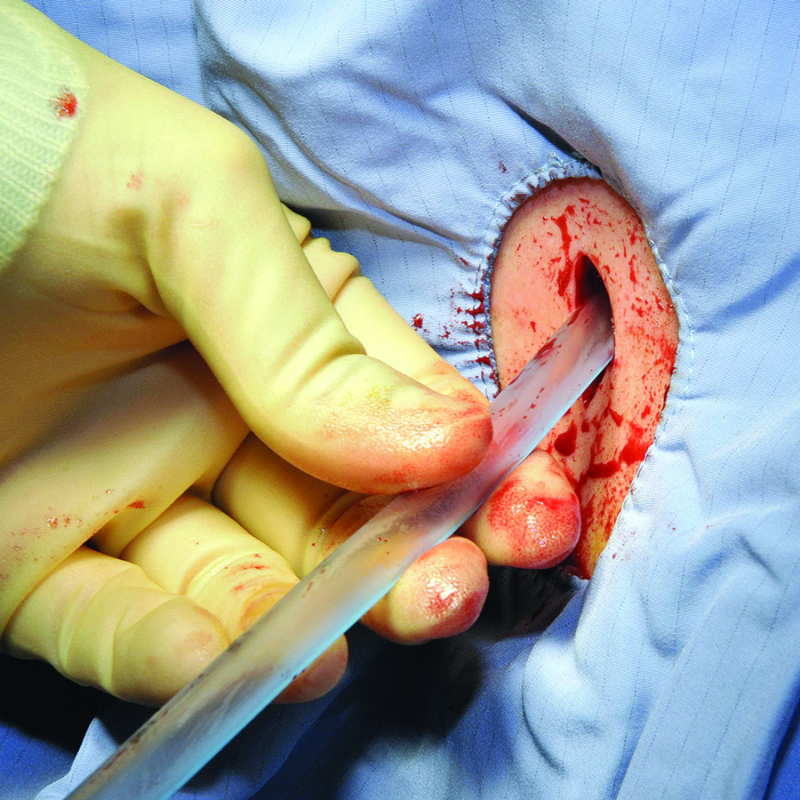
Chest tubes. Chest tubes are placed in the pleural space to evacuate air or fluid. They can be as thin as 20 French or as thick as 40 French (for adults). Chest tubes are typically placed between the fourth and fifth intercostal spaces in the anterior axillary or mid-axillary line; however, the location may vary according to the indication for placement. The tubes can be straight or angled.
The tubes are connected to a collecting system with a three-way chamber. The water chamber holds a column of water, which prevents air from being sucked into the pleural space with inhalation. The suction chamber can be attached to continuous wall suction to remove air or fluid, or it can be placed on “water seal” with no active suction mechanism. The third chamber is the collection chamber for fluid drainage.
Indications for a chest tube include pneumothorax, hemothorax, or a persistent or large pleural effusion. Pneumothorax and hemothorax usually require immediate chest tube placement. Chest tubes are also commonly placed at the end of thoracic surgeries to allow for appropriate re-expansion of the lung tissue.
A chest X-ray should be obtained after any chest tube insertion to ensure appropriate placement. Chest tubes are equipped with a radiopaque line along the longitudinal axis, which should be visible on X-ray. Respiratory variation in the fluid in the collecting tube, called “tidling,” should also be seen in a correctly placed chest tube, and should be monitored at the bedside to reassure continued appropriate location. The interventional radiologist or surgeon who placed the tube should determine the subsequent frequency of serial chest X-rays required to monitor the location of the chest tube.
If the patient has a pneumothorax, air bubbles will be visible in the water chamber; called an air leak, these are often more apparent when the patient coughs. The chest tube should initially be set to continuous suction at -20 mmHg to evacuate the air. Once the air leak has stopped, the chest tube should be placed on water seal to confirm resolution of the pneumothorax (water seal mimics normal physiology). If, after the transition from suction to water seal, resumption of the air leak is noted, it may indicate recurrence of the patient’s pneumothorax. A stat chest X-ray should be obtained, and the chest tube should be placed back on continuous suction. In general, a chest X-ray should be obtained any time the chest tube is changed from suction to water seal or vice versa.
If the patient experiences ongoing or worsening pain, fever, or inadequate drainage, a chest computed tomographic (CT) scan may be warranted to identify inappropriate positioning or other complications, such as occlusion or effusion of the tube. Blood or other debris might clog chest tubes; the surgical team may be able to evacuate the tube with suction tubing at the bedside. If unsuccessful, the tube may need to be removed and reinserted.
The team that placed the tube should help the hospitalist determine the timing of the chest tube removal. If the patient has a pleural effusion, the chest tube can usually be removed when the output is less than 100-200 mL per day and the lung is expanded. The tube should usually be taken off suction and placed on water seal to rule out pneumothorax prior to tube removal.
Penrose drains. Penrose drains are often used to drain fluid or to keep a space open for drainage. Surgeons may use sutures to anchor Penrose drains to skin. Common indications include:
- Ventral hernia repair;
- Debridement of infected pancreatitis; and
- Drainage of superficial abscess cavities.
Penrose drains are simple, flexible tubes that are open at both ends; in contrast to closed drains, they permit ingress as well as egress, facilitating colonization.
Closed suction drains. Closed suction drains with a plastic bulb attachment (i.e., Jackson-Pratt, Blake, Hemovac) are used to collect fluid from a postoperative cavity. Common indications include:
- Post-mastectomy to drain subcutaneous fluid;
- Abdominal surgery;
- Plastic surgery to prevent seroma formation and promote tissue apposition;
- Cholecystectomy if there is concern for damage to ducts of Luschka or other source of bile leak;
- Inadvertent postoperative leakage following a difficult rectal anastomosis; and
- Post-pancreatic surgery.
The quality and quantity of fluid drained should always be carefully noted and recorded. Changes in the fluid can imply development of bleed, leak, or other complications. The surgical team should be contacted immediately if changes are noted.
Typically, closed suction drains will be left in place until the drainage is less than 20 mL per day. These drains can be left in for weeks if necessary and will often be removed during the patient’s scheduled surgical follow-up. Rare complications include erosion into surrounding tissues and inadvertent suturing of the drain in place, such that reexploration is required to remove it. If a closed suction drain becomes occluded, contact the team that placed the drain for further recommendations on adjustment, replacement, or removal.
Nasogastric and duodenal tubes. Nasogastric tubes (NGTs) are often used in the nonoperative management of small bowel obstruction or ileus. They should be placed in the most dependent portion of the gastric lumen and confirmed by chest or abdominal X-ray. NGTs are sump pumps and have a double lumen, which includes an air port to assure flow. The air port should be patent for optimal functioning. The tube may be connected to continuous wall suction or intermittent suction, set to low (less than 60 mmHg) to avoid mucosal avulsion.
NGT output should decrease during the resolution of obstruction or ileus, and symptoms of nausea, vomiting, and abdominal distention should concomitantly improve. Persistently high output in a patient with other indicators of bowel function (flatus, for example) may suggest postpyloric placement (and placement should be checked by X-ray). The timing of NGT removal depends on resumption of bowel function.
Gastrostomy and jejunostomy tubes. Gastrostomy tubes are most commonly used for feeding but may also be used for decompression of functional or anatomic gastric outlet obstruction. They are indicated when patients need prolonged enteral access, such as those with prolonged mechanical ventilation or head and neck pathology that prohibits oral feeding. They are also rarely used for gastropexy to tack an atonic or patulous stomach to the abdominal wall or to prevent recurrence of paraesophageal hernias. These tubes can be placed percutaneously by interventional radiologists, endoscopically by surgeons and gastroenterologists, or laparoscopically or laparotomally by surgeons. This last option is often reserved for patients with difficult anatomy or those who are having laparotomy for another reason.
Because of the stomach’s generous lumen, gastrostomy tubes rarely clog. In the event that they do get clogged, carbonated liquids, meat tenderizer, or enzymes can help dissolve the obstruction. If a gastrostomy tube is left to drain, the patient may experience significant fluid and electrolyte losses, so these need to be carefully monitored.
Jejunostomy tubes are used exclusively for feeding and are usually placed 10-20 cm distal to the ligament of Treitz. These tubes are indicated in patients who require distal feedings due to gastric dysfunction or in those who have undergone a surgery in which a proximal anastomosis requires time to heal. These tubes are more apt to clog and can be more difficult to manage because the lumen of the small bowel is smaller than the stomach. Some prefer not to put pills down the tube to mitigate this risk. Routine flushes with water or saline (30 mL every four to six hours) are also helpful in mitigating the risk of clogging. In the event that they do get clogged, they may be treated like gastrostomy tubes, using carbonated liquids, meat tenderizer, or enzymes to help dissolve the obstruction.
Percutaneous tube sites should be examined frequently for signs of infection. Though gastrostomy and jejunostomy tubes are typically well secured intraabdominally, they can become dislodged. If a gastrostomy or jejunostomy tube has been in place for more than two weeks, it can easily be replaced at the bedside with a tube of comparable caliber by a member of the surgical team or by an experienced hospitalist. If the tube has been in place less than two weeks, it requires replacement with radiographic guidance, as the risk of creating a false lumen is high. Over time, tubes can become loose and fall out. If they need replacement, the preceding guidelines apply.
Back to the Case
A potential major complication of cholecystectomy is severance of the common bile duct, which necessitates significant further surgery. Less severe complications include injuries to the cholecystohepatic ducts (otherwise known as the ducts of Luschka), which can result in leakage of bile into the peritoneal cavity. A bile leak can lead to abscess and systemic infection if left undrained.
Surgeons who are concerned for such a complication intraoperatively may opt to leave a closed suction drain in the gallbladder fossa, such as a Blake drain, for monitoring and subsequent drainage. The drain will remain in place at least until the patient’s diet has been advanced fully, because digestion promotes the secretion of bile and may elucidate a leak. Bilious fluid in the Blake drain is suspicious for a leak.
The surgeon should be notified, and imaging should be obtained to find the nature of the injury to the biliary tree (CT scan with IV contrast, hepatobiliary iminodiacetic acid scan, or endoscopic retrograde cholangiopancreatography). If injury to major biliary structures (the cystic duct stump, the hepatic ducts, or the common bile duct) is diagnosed, a stent may be placed in order to restore ductile continuity.
Minor leaks, with damage to the cystic duct stump, hepatic ducts, and common bile duct ruled out, more often resolve on their own over time, and thus the patient’s closed suction drain will be left in place until biliary drainage ceases, without further initial intervention.
Bottom Line
Surgical tubes and drains have several placement indications. Alterations in quality and quantity of output can indicate changes in clinical status, and hospitalists should be able to handle initial troubleshooting. TH
Dr. Columbus is a general surgery resident at Brigham and Women’s Hospital in Boston. Dr. Havens is an instructor for the department of surgery at Brigham and Women’s Hospital. Dr. Peetz is an instructor for the department of surgery at University Hospital Case Medical Center in Cleveland.
Case
A 45-year-old woman was admitted with choledocholithiasis. Two days prior, following endoscopic retrograde cholangiopancreatography (ERCP), she had gone to the OR for cholecystectomy. The procedure was completed laparoscopically, though the surgeon reported a difficult dissection. The surgeon left a Blake drain in the gallbladder fossa, which initially contained punch-colored fluid. Today, there is bilious fluid in the drain.
Overview
Surgical drains are used to monitor for postoperative leaks or abscesses, to collect normal physiologic fluid, or to minimize dead space. A hospitalist caring for surgical patients may be the first provider to note when something changes in the color or volume of surgical drains. Table 1 lists various types of drains with their indications for use.
Surgical Tubes and Drains
Chest tubes. Chest tubes are placed in the pleural space to evacuate air or fluid. They can be as thin as 20 French or as thick as 40 French (for adults). Chest tubes are typically placed between the fourth and fifth intercostal spaces in the anterior axillary or mid-axillary line; however, the location may vary according to the indication for placement. The tubes can be straight or angled.
The tubes are connected to a collecting system with a three-way chamber. The water chamber holds a column of water, which prevents air from being sucked into the pleural space with inhalation. The suction chamber can be attached to continuous wall suction to remove air or fluid, or it can be placed on “water seal” with no active suction mechanism. The third chamber is the collection chamber for fluid drainage.
Indications for a chest tube include pneumothorax, hemothorax, or a persistent or large pleural effusion. Pneumothorax and hemothorax usually require immediate chest tube placement. Chest tubes are also commonly placed at the end of thoracic surgeries to allow for appropriate re-expansion of the lung tissue.
A chest X-ray should be obtained after any chest tube insertion to ensure appropriate placement. Chest tubes are equipped with a radiopaque line along the longitudinal axis, which should be visible on X-ray. Respiratory variation in the fluid in the collecting tube, called “tidling,” should also be seen in a correctly placed chest tube, and should be monitored at the bedside to reassure continued appropriate location. The interventional radiologist or surgeon who placed the tube should determine the subsequent frequency of serial chest X-rays required to monitor the location of the chest tube.
If the patient has a pneumothorax, air bubbles will be visible in the water chamber; called an air leak, these are often more apparent when the patient coughs. The chest tube should initially be set to continuous suction at -20 mmHg to evacuate the air. Once the air leak has stopped, the chest tube should be placed on water seal to confirm resolution of the pneumothorax (water seal mimics normal physiology). If, after the transition from suction to water seal, resumption of the air leak is noted, it may indicate recurrence of the patient’s pneumothorax. A stat chest X-ray should be obtained, and the chest tube should be placed back on continuous suction. In general, a chest X-ray should be obtained any time the chest tube is changed from suction to water seal or vice versa.
If the patient experiences ongoing or worsening pain, fever, or inadequate drainage, a chest computed tomographic (CT) scan may be warranted to identify inappropriate positioning or other complications, such as occlusion or effusion of the tube. Blood or other debris might clog chest tubes; the surgical team may be able to evacuate the tube with suction tubing at the bedside. If unsuccessful, the tube may need to be removed and reinserted.
The team that placed the tube should help the hospitalist determine the timing of the chest tube removal. If the patient has a pleural effusion, the chest tube can usually be removed when the output is less than 100-200 mL per day and the lung is expanded. The tube should usually be taken off suction and placed on water seal to rule out pneumothorax prior to tube removal.
Penrose drains. Penrose drains are often used to drain fluid or to keep a space open for drainage. Surgeons may use sutures to anchor Penrose drains to skin. Common indications include:
- Ventral hernia repair;
- Debridement of infected pancreatitis; and
- Drainage of superficial abscess cavities.
Penrose drains are simple, flexible tubes that are open at both ends; in contrast to closed drains, they permit ingress as well as egress, facilitating colonization.
Closed suction drains. Closed suction drains with a plastic bulb attachment (i.e., Jackson-Pratt, Blake, Hemovac) are used to collect fluid from a postoperative cavity. Common indications include:
- Post-mastectomy to drain subcutaneous fluid;
- Abdominal surgery;
- Plastic surgery to prevent seroma formation and promote tissue apposition;
- Cholecystectomy if there is concern for damage to ducts of Luschka or other source of bile leak;
- Inadvertent postoperative leakage following a difficult rectal anastomosis; and
- Post-pancreatic surgery.
The quality and quantity of fluid drained should always be carefully noted and recorded. Changes in the fluid can imply development of bleed, leak, or other complications. The surgical team should be contacted immediately if changes are noted.
Typically, closed suction drains will be left in place until the drainage is less than 20 mL per day. These drains can be left in for weeks if necessary and will often be removed during the patient’s scheduled surgical follow-up. Rare complications include erosion into surrounding tissues and inadvertent suturing of the drain in place, such that reexploration is required to remove it. If a closed suction drain becomes occluded, contact the team that placed the drain for further recommendations on adjustment, replacement, or removal.
Nasogastric and duodenal tubes. Nasogastric tubes (NGTs) are often used in the nonoperative management of small bowel obstruction or ileus. They should be placed in the most dependent portion of the gastric lumen and confirmed by chest or abdominal X-ray. NGTs are sump pumps and have a double lumen, which includes an air port to assure flow. The air port should be patent for optimal functioning. The tube may be connected to continuous wall suction or intermittent suction, set to low (less than 60 mmHg) to avoid mucosal avulsion.
NGT output should decrease during the resolution of obstruction or ileus, and symptoms of nausea, vomiting, and abdominal distention should concomitantly improve. Persistently high output in a patient with other indicators of bowel function (flatus, for example) may suggest postpyloric placement (and placement should be checked by X-ray). The timing of NGT removal depends on resumption of bowel function.
Gastrostomy and jejunostomy tubes. Gastrostomy tubes are most commonly used for feeding but may also be used for decompression of functional or anatomic gastric outlet obstruction. They are indicated when patients need prolonged enteral access, such as those with prolonged mechanical ventilation or head and neck pathology that prohibits oral feeding. They are also rarely used for gastropexy to tack an atonic or patulous stomach to the abdominal wall or to prevent recurrence of paraesophageal hernias. These tubes can be placed percutaneously by interventional radiologists, endoscopically by surgeons and gastroenterologists, or laparoscopically or laparotomally by surgeons. This last option is often reserved for patients with difficult anatomy or those who are having laparotomy for another reason.
Because of the stomach’s generous lumen, gastrostomy tubes rarely clog. In the event that they do get clogged, carbonated liquids, meat tenderizer, or enzymes can help dissolve the obstruction. If a gastrostomy tube is left to drain, the patient may experience significant fluid and electrolyte losses, so these need to be carefully monitored.
Jejunostomy tubes are used exclusively for feeding and are usually placed 10-20 cm distal to the ligament of Treitz. These tubes are indicated in patients who require distal feedings due to gastric dysfunction or in those who have undergone a surgery in which a proximal anastomosis requires time to heal. These tubes are more apt to clog and can be more difficult to manage because the lumen of the small bowel is smaller than the stomach. Some prefer not to put pills down the tube to mitigate this risk. Routine flushes with water or saline (30 mL every four to six hours) are also helpful in mitigating the risk of clogging. In the event that they do get clogged, they may be treated like gastrostomy tubes, using carbonated liquids, meat tenderizer, or enzymes to help dissolve the obstruction.
Percutaneous tube sites should be examined frequently for signs of infection. Though gastrostomy and jejunostomy tubes are typically well secured intraabdominally, they can become dislodged. If a gastrostomy or jejunostomy tube has been in place for more than two weeks, it can easily be replaced at the bedside with a tube of comparable caliber by a member of the surgical team or by an experienced hospitalist. If the tube has been in place less than two weeks, it requires replacement with radiographic guidance, as the risk of creating a false lumen is high. Over time, tubes can become loose and fall out. If they need replacement, the preceding guidelines apply.
Back to the Case
A potential major complication of cholecystectomy is severance of the common bile duct, which necessitates significant further surgery. Less severe complications include injuries to the cholecystohepatic ducts (otherwise known as the ducts of Luschka), which can result in leakage of bile into the peritoneal cavity. A bile leak can lead to abscess and systemic infection if left undrained.
Surgeons who are concerned for such a complication intraoperatively may opt to leave a closed suction drain in the gallbladder fossa, such as a Blake drain, for monitoring and subsequent drainage. The drain will remain in place at least until the patient’s diet has been advanced fully, because digestion promotes the secretion of bile and may elucidate a leak. Bilious fluid in the Blake drain is suspicious for a leak.
The surgeon should be notified, and imaging should be obtained to find the nature of the injury to the biliary tree (CT scan with IV contrast, hepatobiliary iminodiacetic acid scan, or endoscopic retrograde cholangiopancreatography). If injury to major biliary structures (the cystic duct stump, the hepatic ducts, or the common bile duct) is diagnosed, a stent may be placed in order to restore ductile continuity.
Minor leaks, with damage to the cystic duct stump, hepatic ducts, and common bile duct ruled out, more often resolve on their own over time, and thus the patient’s closed suction drain will be left in place until biliary drainage ceases, without further initial intervention.
Bottom Line
Surgical tubes and drains have several placement indications. Alterations in quality and quantity of output can indicate changes in clinical status, and hospitalists should be able to handle initial troubleshooting. TH
Dr. Columbus is a general surgery resident at Brigham and Women’s Hospital in Boston. Dr. Havens is an instructor for the department of surgery at Brigham and Women’s Hospital. Dr. Peetz is an instructor for the department of surgery at University Hospital Case Medical Center in Cleveland.
Case
A 45-year-old woman was admitted with choledocholithiasis. Two days prior, following endoscopic retrograde cholangiopancreatography (ERCP), she had gone to the OR for cholecystectomy. The procedure was completed laparoscopically, though the surgeon reported a difficult dissection. The surgeon left a Blake drain in the gallbladder fossa, which initially contained punch-colored fluid. Today, there is bilious fluid in the drain.
Overview
Surgical drains are used to monitor for postoperative leaks or abscesses, to collect normal physiologic fluid, or to minimize dead space. A hospitalist caring for surgical patients may be the first provider to note when something changes in the color or volume of surgical drains. Table 1 lists various types of drains with their indications for use.
Surgical Tubes and Drains
Chest tubes. Chest tubes are placed in the pleural space to evacuate air or fluid. They can be as thin as 20 French or as thick as 40 French (for adults). Chest tubes are typically placed between the fourth and fifth intercostal spaces in the anterior axillary or mid-axillary line; however, the location may vary according to the indication for placement. The tubes can be straight or angled.
The tubes are connected to a collecting system with a three-way chamber. The water chamber holds a column of water, which prevents air from being sucked into the pleural space with inhalation. The suction chamber can be attached to continuous wall suction to remove air or fluid, or it can be placed on “water seal” with no active suction mechanism. The third chamber is the collection chamber for fluid drainage.
Indications for a chest tube include pneumothorax, hemothorax, or a persistent or large pleural effusion. Pneumothorax and hemothorax usually require immediate chest tube placement. Chest tubes are also commonly placed at the end of thoracic surgeries to allow for appropriate re-expansion of the lung tissue.
A chest X-ray should be obtained after any chest tube insertion to ensure appropriate placement. Chest tubes are equipped with a radiopaque line along the longitudinal axis, which should be visible on X-ray. Respiratory variation in the fluid in the collecting tube, called “tidling,” should also be seen in a correctly placed chest tube, and should be monitored at the bedside to reassure continued appropriate location. The interventional radiologist or surgeon who placed the tube should determine the subsequent frequency of serial chest X-rays required to monitor the location of the chest tube.
If the patient has a pneumothorax, air bubbles will be visible in the water chamber; called an air leak, these are often more apparent when the patient coughs. The chest tube should initially be set to continuous suction at -20 mmHg to evacuate the air. Once the air leak has stopped, the chest tube should be placed on water seal to confirm resolution of the pneumothorax (water seal mimics normal physiology). If, after the transition from suction to water seal, resumption of the air leak is noted, it may indicate recurrence of the patient’s pneumothorax. A stat chest X-ray should be obtained, and the chest tube should be placed back on continuous suction. In general, a chest X-ray should be obtained any time the chest tube is changed from suction to water seal or vice versa.
If the patient experiences ongoing or worsening pain, fever, or inadequate drainage, a chest computed tomographic (CT) scan may be warranted to identify inappropriate positioning or other complications, such as occlusion or effusion of the tube. Blood or other debris might clog chest tubes; the surgical team may be able to evacuate the tube with suction tubing at the bedside. If unsuccessful, the tube may need to be removed and reinserted.
The team that placed the tube should help the hospitalist determine the timing of the chest tube removal. If the patient has a pleural effusion, the chest tube can usually be removed when the output is less than 100-200 mL per day and the lung is expanded. The tube should usually be taken off suction and placed on water seal to rule out pneumothorax prior to tube removal.
Penrose drains. Penrose drains are often used to drain fluid or to keep a space open for drainage. Surgeons may use sutures to anchor Penrose drains to skin. Common indications include:
- Ventral hernia repair;
- Debridement of infected pancreatitis; and
- Drainage of superficial abscess cavities.
Penrose drains are simple, flexible tubes that are open at both ends; in contrast to closed drains, they permit ingress as well as egress, facilitating colonization.
Closed suction drains. Closed suction drains with a plastic bulb attachment (i.e., Jackson-Pratt, Blake, Hemovac) are used to collect fluid from a postoperative cavity. Common indications include:
- Post-mastectomy to drain subcutaneous fluid;
- Abdominal surgery;
- Plastic surgery to prevent seroma formation and promote tissue apposition;
- Cholecystectomy if there is concern for damage to ducts of Luschka or other source of bile leak;
- Inadvertent postoperative leakage following a difficult rectal anastomosis; and
- Post-pancreatic surgery.
The quality and quantity of fluid drained should always be carefully noted and recorded. Changes in the fluid can imply development of bleed, leak, or other complications. The surgical team should be contacted immediately if changes are noted.
Typically, closed suction drains will be left in place until the drainage is less than 20 mL per day. These drains can be left in for weeks if necessary and will often be removed during the patient’s scheduled surgical follow-up. Rare complications include erosion into surrounding tissues and inadvertent suturing of the drain in place, such that reexploration is required to remove it. If a closed suction drain becomes occluded, contact the team that placed the drain for further recommendations on adjustment, replacement, or removal.
Nasogastric and duodenal tubes. Nasogastric tubes (NGTs) are often used in the nonoperative management of small bowel obstruction or ileus. They should be placed in the most dependent portion of the gastric lumen and confirmed by chest or abdominal X-ray. NGTs are sump pumps and have a double lumen, which includes an air port to assure flow. The air port should be patent for optimal functioning. The tube may be connected to continuous wall suction or intermittent suction, set to low (less than 60 mmHg) to avoid mucosal avulsion.
NGT output should decrease during the resolution of obstruction or ileus, and symptoms of nausea, vomiting, and abdominal distention should concomitantly improve. Persistently high output in a patient with other indicators of bowel function (flatus, for example) may suggest postpyloric placement (and placement should be checked by X-ray). The timing of NGT removal depends on resumption of bowel function.
Gastrostomy and jejunostomy tubes. Gastrostomy tubes are most commonly used for feeding but may also be used for decompression of functional or anatomic gastric outlet obstruction. They are indicated when patients need prolonged enteral access, such as those with prolonged mechanical ventilation or head and neck pathology that prohibits oral feeding. They are also rarely used for gastropexy to tack an atonic or patulous stomach to the abdominal wall or to prevent recurrence of paraesophageal hernias. These tubes can be placed percutaneously by interventional radiologists, endoscopically by surgeons and gastroenterologists, or laparoscopically or laparotomally by surgeons. This last option is often reserved for patients with difficult anatomy or those who are having laparotomy for another reason.
Because of the stomach’s generous lumen, gastrostomy tubes rarely clog. In the event that they do get clogged, carbonated liquids, meat tenderizer, or enzymes can help dissolve the obstruction. If a gastrostomy tube is left to drain, the patient may experience significant fluid and electrolyte losses, so these need to be carefully monitored.
Jejunostomy tubes are used exclusively for feeding and are usually placed 10-20 cm distal to the ligament of Treitz. These tubes are indicated in patients who require distal feedings due to gastric dysfunction or in those who have undergone a surgery in which a proximal anastomosis requires time to heal. These tubes are more apt to clog and can be more difficult to manage because the lumen of the small bowel is smaller than the stomach. Some prefer not to put pills down the tube to mitigate this risk. Routine flushes with water or saline (30 mL every four to six hours) are also helpful in mitigating the risk of clogging. In the event that they do get clogged, they may be treated like gastrostomy tubes, using carbonated liquids, meat tenderizer, or enzymes to help dissolve the obstruction.
Percutaneous tube sites should be examined frequently for signs of infection. Though gastrostomy and jejunostomy tubes are typically well secured intraabdominally, they can become dislodged. If a gastrostomy or jejunostomy tube has been in place for more than two weeks, it can easily be replaced at the bedside with a tube of comparable caliber by a member of the surgical team or by an experienced hospitalist. If the tube has been in place less than two weeks, it requires replacement with radiographic guidance, as the risk of creating a false lumen is high. Over time, tubes can become loose and fall out. If they need replacement, the preceding guidelines apply.
Back to the Case
A potential major complication of cholecystectomy is severance of the common bile duct, which necessitates significant further surgery. Less severe complications include injuries to the cholecystohepatic ducts (otherwise known as the ducts of Luschka), which can result in leakage of bile into the peritoneal cavity. A bile leak can lead to abscess and systemic infection if left undrained.
Surgeons who are concerned for such a complication intraoperatively may opt to leave a closed suction drain in the gallbladder fossa, such as a Blake drain, for monitoring and subsequent drainage. The drain will remain in place at least until the patient’s diet has been advanced fully, because digestion promotes the secretion of bile and may elucidate a leak. Bilious fluid in the Blake drain is suspicious for a leak.
The surgeon should be notified, and imaging should be obtained to find the nature of the injury to the biliary tree (CT scan with IV contrast, hepatobiliary iminodiacetic acid scan, or endoscopic retrograde cholangiopancreatography). If injury to major biliary structures (the cystic duct stump, the hepatic ducts, or the common bile duct) is diagnosed, a stent may be placed in order to restore ductile continuity.
Minor leaks, with damage to the cystic duct stump, hepatic ducts, and common bile duct ruled out, more often resolve on their own over time, and thus the patient’s closed suction drain will be left in place until biliary drainage ceases, without further initial intervention.
Bottom Line
Surgical tubes and drains have several placement indications. Alterations in quality and quantity of output can indicate changes in clinical status, and hospitalists should be able to handle initial troubleshooting. TH
Dr. Columbus is a general surgery resident at Brigham and Women’s Hospital in Boston. Dr. Havens is an instructor for the department of surgery at Brigham and Women’s Hospital. Dr. Peetz is an instructor for the department of surgery at University Hospital Case Medical Center in Cleveland.
Private Insurers to Reap Bulk of Spending on Hospitalized Patient Care
Spending on care of hospitalized patients is expected to pass $1 trillion in 2015, a new high. Thomas Selden, PhD, of the Agency for Healthcare Research and Quality recently asked where that money is likely to go. The answer: private insurers.
Dr. Selden and his colleagues report in Health Affairs this month that in 2012, private insurers’ payment rates for inpatient hospital stays were approximately 75% greater than Medicare’s payment rates, a sharp increase from the differential of approximately 10% percent during the period of 1996 to 2001. “We need to understand who’s paying what,” Dr. Selden says. “It’s the first step to a better understanding of public policy.”
The report found that “the predicted percentage difference between the rates of private insurers and those of Medicare has increased substantially over time.” In 1996, private insurers paid 106.1% of Medicare payment rates, a payment rate difference of 6.1% (95% CI: -3.2, 15.5). The difference climbed to 64.1% (95% CI: 48.3, 80.0) in 2011 and 75.3% (95% CI: 52.0, 98.6) in 2012. Medicaid payment rates averaged approximately 90% of Medicare payment rates throughout the study period.
Dr. Selden is hopeful that stakeholders will use the data his team collected to determine the impetus for the widening gap. He also plans to research whether payment differences affect quality metrics.
“Anytime you’re talking about a trillion dollars, it’s really important when a payment difference opens up of this magnitude,” he adds. “The difference is real … what the policy implications are is for the policy makers to decide.” TH
Visit our website for more information on healthcare payment models.
Spending on care of hospitalized patients is expected to pass $1 trillion in 2015, a new high. Thomas Selden, PhD, of the Agency for Healthcare Research and Quality recently asked where that money is likely to go. The answer: private insurers.
Dr. Selden and his colleagues report in Health Affairs this month that in 2012, private insurers’ payment rates for inpatient hospital stays were approximately 75% greater than Medicare’s payment rates, a sharp increase from the differential of approximately 10% percent during the period of 1996 to 2001. “We need to understand who’s paying what,” Dr. Selden says. “It’s the first step to a better understanding of public policy.”
The report found that “the predicted percentage difference between the rates of private insurers and those of Medicare has increased substantially over time.” In 1996, private insurers paid 106.1% of Medicare payment rates, a payment rate difference of 6.1% (95% CI: -3.2, 15.5). The difference climbed to 64.1% (95% CI: 48.3, 80.0) in 2011 and 75.3% (95% CI: 52.0, 98.6) in 2012. Medicaid payment rates averaged approximately 90% of Medicare payment rates throughout the study period.
Dr. Selden is hopeful that stakeholders will use the data his team collected to determine the impetus for the widening gap. He also plans to research whether payment differences affect quality metrics.
“Anytime you’re talking about a trillion dollars, it’s really important when a payment difference opens up of this magnitude,” he adds. “The difference is real … what the policy implications are is for the policy makers to decide.” TH
Visit our website for more information on healthcare payment models.
Spending on care of hospitalized patients is expected to pass $1 trillion in 2015, a new high. Thomas Selden, PhD, of the Agency for Healthcare Research and Quality recently asked where that money is likely to go. The answer: private insurers.
Dr. Selden and his colleagues report in Health Affairs this month that in 2012, private insurers’ payment rates for inpatient hospital stays were approximately 75% greater than Medicare’s payment rates, a sharp increase from the differential of approximately 10% percent during the period of 1996 to 2001. “We need to understand who’s paying what,” Dr. Selden says. “It’s the first step to a better understanding of public policy.”
The report found that “the predicted percentage difference between the rates of private insurers and those of Medicare has increased substantially over time.” In 1996, private insurers paid 106.1% of Medicare payment rates, a payment rate difference of 6.1% (95% CI: -3.2, 15.5). The difference climbed to 64.1% (95% CI: 48.3, 80.0) in 2011 and 75.3% (95% CI: 52.0, 98.6) in 2012. Medicaid payment rates averaged approximately 90% of Medicare payment rates throughout the study period.
Dr. Selden is hopeful that stakeholders will use the data his team collected to determine the impetus for the widening gap. He also plans to research whether payment differences affect quality metrics.
“Anytime you’re talking about a trillion dollars, it’s really important when a payment difference opens up of this magnitude,” he adds. “The difference is real … what the policy implications are is for the policy makers to decide.” TH
Visit our website for more information on healthcare payment models.
SHM Members Can Share Their Success Stories on Social Media
From care transitions to antibiotic stewardship

What does that mean? When you experience a success related to being a member, attend an SHM event, or use an SHM resource or program, tweet about it using the hashtag #SHeMpowered and mention SHM, @SHMLive. We’ll retweet and share your fantastic work with our thousands of followers.

What kinds of things can you tweet about? Pretty much anything! Attended Leadership Academy? Finished a course in the Learning Portal? Implemented a checklist from Project BOOST in your hospital? Tweet about it! Just remember to use the hashtag #SHeMpowered to be a part of the larger conversation on social media, and follow the hashtag to see how others are using their SHM membership to enhance patient care.

For more information, visit www.hospitalmedicine.org/getinvolved, and be sure to follow SHM on Twitter @SHMLive. While you’re online, check out our Facebook page, YouTube channel, and LinkedIn group, and get #SHeMpowered!
From care transitions to antibiotic stewardship
What does that mean? When you experience a success related to being a member, attend an SHM event, or use an SHM resource or program, tweet about it using the hashtag #SHeMpowered and mention SHM, @SHMLive. We’ll retweet and share your fantastic work with our thousands of followers.
What kinds of things can you tweet about? Pretty much anything! Attended Leadership Academy? Finished a course in the Learning Portal? Implemented a checklist from Project BOOST in your hospital? Tweet about it! Just remember to use the hashtag #SHeMpowered to be a part of the larger conversation on social media, and follow the hashtag to see how others are using their SHM membership to enhance patient care.
For more information, visit www.hospitalmedicine.org/getinvolved, and be sure to follow SHM on Twitter @SHMLive. While you’re online, check out our Facebook page, YouTube channel, and LinkedIn group, and get #SHeMpowered!
From care transitions to antibiotic stewardship
What does that mean? When you experience a success related to being a member, attend an SHM event, or use an SHM resource or program, tweet about it using the hashtag #SHeMpowered and mention SHM, @SHMLive. We’ll retweet and share your fantastic work with our thousands of followers.
What kinds of things can you tweet about? Pretty much anything! Attended Leadership Academy? Finished a course in the Learning Portal? Implemented a checklist from Project BOOST in your hospital? Tweet about it! Just remember to use the hashtag #SHeMpowered to be a part of the larger conversation on social media, and follow the hashtag to see how others are using their SHM membership to enhance patient care.
For more information, visit www.hospitalmedicine.org/getinvolved, and be sure to follow SHM on Twitter @SHMLive. While you’re online, check out our Facebook page, YouTube channel, and LinkedIn group, and get #SHeMpowered!
Value of Ultra-Brief Cognitive Assessments in Predicting Negative Hospital Outcomes
Clinical question: What is the value of ultra-brief cognitive assessments in predicting hospital outcomes?

Background: Cognitive assessment tools can be used to predict patient outcomes in the hospital setting. Physician time constraints limit use of longer traditional cognitive testing, and little is known about the effectiveness of ultra-brief (less than one minute) assessments and their predictive value.
Study design: Secondary data analysis of a quality improvement project.
Setting: Tertiary, Veterans Administration hospital.
Synopsis: Using data from a prior inpatient database, 3,232 patients over the age of 60 were screened on admission using the modified Richmond Agitation and Sedation Scale (mRASS) for arousal and the months of the year backwards (MOTYB) for attention. Abnormal mRASS and incorrect MOTYB predicted negative hospital outcomes: increased length of stay (incident rate ratio 1.23, 95% CI 1.17-1.3); increased restraint use (risk ratio 5.05, 95% CI); increased hospital mortality (RR 3.46, 95% CI 1.24-9.63); and decreased rates of being discharged home (RR 2.97, 95% CI: 2.42-3.64).
This study highlights the value of two ultra-brief cognitive assessment tools in the prediction of potential poor outcomes during inpatient admission. Hospitalists need to identify high-risk patients, and these tools allow for rapid assessment at the time of admission, without a significant time constraint for the busy hospitalist.
Bottom Line: The use of ultra-brief cognitive assessment tools in patients over age 60 can predict negative inpatient outcomes.
Citation: Yevchak AM, Doherty K, Archambault EG, Kelly B, Fonda JR, Rudolph JL. The association between an ultra-brief cognitive screening in older adults and hospital outcomes. J Hosp Med. 2015;10(10):651-657.
Clinical question: What is the value of ultra-brief cognitive assessments in predicting hospital outcomes?
Background: Cognitive assessment tools can be used to predict patient outcomes in the hospital setting. Physician time constraints limit use of longer traditional cognitive testing, and little is known about the effectiveness of ultra-brief (less than one minute) assessments and their predictive value.
Study design: Secondary data analysis of a quality improvement project.
Setting: Tertiary, Veterans Administration hospital.
Synopsis: Using data from a prior inpatient database, 3,232 patients over the age of 60 were screened on admission using the modified Richmond Agitation and Sedation Scale (mRASS) for arousal and the months of the year backwards (MOTYB) for attention. Abnormal mRASS and incorrect MOTYB predicted negative hospital outcomes: increased length of stay (incident rate ratio 1.23, 95% CI 1.17-1.3); increased restraint use (risk ratio 5.05, 95% CI); increased hospital mortality (RR 3.46, 95% CI 1.24-9.63); and decreased rates of being discharged home (RR 2.97, 95% CI: 2.42-3.64).
This study highlights the value of two ultra-brief cognitive assessment tools in the prediction of potential poor outcomes during inpatient admission. Hospitalists need to identify high-risk patients, and these tools allow for rapid assessment at the time of admission, without a significant time constraint for the busy hospitalist.
Bottom Line: The use of ultra-brief cognitive assessment tools in patients over age 60 can predict negative inpatient outcomes.
Citation: Yevchak AM, Doherty K, Archambault EG, Kelly B, Fonda JR, Rudolph JL. The association between an ultra-brief cognitive screening in older adults and hospital outcomes. J Hosp Med. 2015;10(10):651-657.
Clinical question: What is the value of ultra-brief cognitive assessments in predicting hospital outcomes?
Background: Cognitive assessment tools can be used to predict patient outcomes in the hospital setting. Physician time constraints limit use of longer traditional cognitive testing, and little is known about the effectiveness of ultra-brief (less than one minute) assessments and their predictive value.
Study design: Secondary data analysis of a quality improvement project.
Setting: Tertiary, Veterans Administration hospital.
Synopsis: Using data from a prior inpatient database, 3,232 patients over the age of 60 were screened on admission using the modified Richmond Agitation and Sedation Scale (mRASS) for arousal and the months of the year backwards (MOTYB) for attention. Abnormal mRASS and incorrect MOTYB predicted negative hospital outcomes: increased length of stay (incident rate ratio 1.23, 95% CI 1.17-1.3); increased restraint use (risk ratio 5.05, 95% CI); increased hospital mortality (RR 3.46, 95% CI 1.24-9.63); and decreased rates of being discharged home (RR 2.97, 95% CI: 2.42-3.64).
This study highlights the value of two ultra-brief cognitive assessment tools in the prediction of potential poor outcomes during inpatient admission. Hospitalists need to identify high-risk patients, and these tools allow for rapid assessment at the time of admission, without a significant time constraint for the busy hospitalist.
Bottom Line: The use of ultra-brief cognitive assessment tools in patients over age 60 can predict negative inpatient outcomes.
Citation: Yevchak AM, Doherty K, Archambault EG, Kelly B, Fonda JR, Rudolph JL. The association between an ultra-brief cognitive screening in older adults and hospital outcomes. J Hosp Med. 2015;10(10):651-657.

Criteria for Appropriate Use of Peripherally Inserted Central Catheters
Clinical question: What are criteria for appropriate and inappropriate use of PICCs?
Background: PICCs are commonly used in medical care in a variety of clinical contexts; however, criteria defining the appropriate use of PICCs and practices related to PICC placement have not been previously established.
Study design: A multispecialty panel classified indications for PICC use as appropriate or inappropriate using the RAND/UCLA Appropriateness Method.
Synopsis: Selected appropriate PICC uses include:
- Infusion of peripherally compatible infusates, intermittent infusions, or infrequent phlebotomy in patients with poor or difficult venous access when the expected duration of use is at least six days;
- Phlebotomy at least every eight hours when the expected duration of use is at least six days; and
- Invasive hemodynamic monitoring in a critically ill patient only if the duration of use is expected to exceed 15 days.
Selected appropriate PICC-related practices:
- Verify PICC tip position using a chest radiograph only after non-ECG or non-fluoroscopically guided PICC insertion;
- Provide an interval without a PICC to allow resolution of bacteremia when managing PICC-related bloodstream infections; and
- For PICC-related DVT, provide at least three months of systemic anticoagulation if not otherwise contraindicated.
Selected inappropriate PICC-related practices:
- Adjustment of PICC tips that reside in the lower third of the superior vena cava, cavoatrial junction, or right atrium; and
- Removal or replacement of PICCs that are clinically necessary, well positioned, and functional in the setting of PICC-related DVT or without evidence of catheter-associated bloodstream infection.
Bottom line: A multispecialty expert panel provides guidance for appropriate use of PICCs and PICC-related practices.
Citation: Chopra V, Flanders SA, Saint S, et al. The Michigan appropriateness guide for intravenous catheters (MAGIC): results from a multispecialty panel using the RAND/UCLA appropriateness method. Ann Intern Med. 2015;163(6):S1-S40.
Clinical question: What are criteria for appropriate and inappropriate use of PICCs?
Background: PICCs are commonly used in medical care in a variety of clinical contexts; however, criteria defining the appropriate use of PICCs and practices related to PICC placement have not been previously established.
Study design: A multispecialty panel classified indications for PICC use as appropriate or inappropriate using the RAND/UCLA Appropriateness Method.
Synopsis: Selected appropriate PICC uses include:
- Infusion of peripherally compatible infusates, intermittent infusions, or infrequent phlebotomy in patients with poor or difficult venous access when the expected duration of use is at least six days;
- Phlebotomy at least every eight hours when the expected duration of use is at least six days; and
- Invasive hemodynamic monitoring in a critically ill patient only if the duration of use is expected to exceed 15 days.
Selected appropriate PICC-related practices:
- Verify PICC tip position using a chest radiograph only after non-ECG or non-fluoroscopically guided PICC insertion;
- Provide an interval without a PICC to allow resolution of bacteremia when managing PICC-related bloodstream infections; and
- For PICC-related DVT, provide at least three months of systemic anticoagulation if not otherwise contraindicated.
Selected inappropriate PICC-related practices:
- Adjustment of PICC tips that reside in the lower third of the superior vena cava, cavoatrial junction, or right atrium; and
- Removal or replacement of PICCs that are clinically necessary, well positioned, and functional in the setting of PICC-related DVT or without evidence of catheter-associated bloodstream infection.
Bottom line: A multispecialty expert panel provides guidance for appropriate use of PICCs and PICC-related practices.
Citation: Chopra V, Flanders SA, Saint S, et al. The Michigan appropriateness guide for intravenous catheters (MAGIC): results from a multispecialty panel using the RAND/UCLA appropriateness method. Ann Intern Med. 2015;163(6):S1-S40.
Clinical question: What are criteria for appropriate and inappropriate use of PICCs?
Background: PICCs are commonly used in medical care in a variety of clinical contexts; however, criteria defining the appropriate use of PICCs and practices related to PICC placement have not been previously established.
Study design: A multispecialty panel classified indications for PICC use as appropriate or inappropriate using the RAND/UCLA Appropriateness Method.
Synopsis: Selected appropriate PICC uses include:
- Infusion of peripherally compatible infusates, intermittent infusions, or infrequent phlebotomy in patients with poor or difficult venous access when the expected duration of use is at least six days;
- Phlebotomy at least every eight hours when the expected duration of use is at least six days; and
- Invasive hemodynamic monitoring in a critically ill patient only if the duration of use is expected to exceed 15 days.
Selected appropriate PICC-related practices:
- Verify PICC tip position using a chest radiograph only after non-ECG or non-fluoroscopically guided PICC insertion;
- Provide an interval without a PICC to allow resolution of bacteremia when managing PICC-related bloodstream infections; and
- For PICC-related DVT, provide at least three months of systemic anticoagulation if not otherwise contraindicated.
Selected inappropriate PICC-related practices:
- Adjustment of PICC tips that reside in the lower third of the superior vena cava, cavoatrial junction, or right atrium; and
- Removal or replacement of PICCs that are clinically necessary, well positioned, and functional in the setting of PICC-related DVT or without evidence of catheter-associated bloodstream infection.
Bottom line: A multispecialty expert panel provides guidance for appropriate use of PICCs and PICC-related practices.
Citation: Chopra V, Flanders SA, Saint S, et al. The Michigan appropriateness guide for intravenous catheters (MAGIC): results from a multispecialty panel using the RAND/UCLA appropriateness method. Ann Intern Med. 2015;163(6):S1-S40.
ACP Guidelines for Evaluation of Suspected Pulmonary Embolism
Clinical question: What are best practices for evaluating patients with suspected acute pulmonary embolism (PE)?
Background: Use of CT in the evaluation of PE has increased across all clinical settings without improving mortality. Contrast CT carries the risks of radiation exposure, contrast-induced nephropathy, and incidental findings that require further investigation. The authors highlight evidence-based strategies for evaluation of PE, focusing on delivering high-value care.
Study design: Clinical guideline.
Setting: Literature review of studies across all adult clinical settings.
Synopsis: The clinical guidelines committee of the American College of Physicians conducted a literature search surrounding evaluation of suspected acute PE. From their review, they concluded:
- Pretest probability should initially be determined based on validated prediction tools (Wells score, Revised Geneva);
- In patients found to have low pretest probability and meeting the pulmonary embolism rule-out criteria (PERC), clinicians can forego d-dimer testing;
- In those with intermediate pretest probability or those with low pre-test probability who do not pass PERC, d-dimer measurement should be obtained;
- The d-dimer threshold should be age adjusted and imaging should not be pursued in patients whose d-dimer level falls below this cutoff, while those with positive d-dimers should receive CT pulmonary angiography (CTPA); and
- Patients with high pretest probability should undergo CTPA (or V/Q scan if CTPA is contraindicated) without d-dimer testing.
Bottom line: In suspected acute PE, first determine pretest probability using Wells and Revised Geneva, and then use this probability in conjunction with the PERC and d-dimer (as indicated) to guide decisions about imaging.
Citation: Raja AS, Greenberg JO, Qaseem A, Denberg TD, Fitterman N, Schuur JD. Evaluation of patients with suspected acute pulmonary embolism: best practice advice from the clinical guidelines committee of the American College of Physicians. Ann Intern Med. 2015;163(9):701-711.
Clinical question: What are best practices for evaluating patients with suspected acute pulmonary embolism (PE)?
Background: Use of CT in the evaluation of PE has increased across all clinical settings without improving mortality. Contrast CT carries the risks of radiation exposure, contrast-induced nephropathy, and incidental findings that require further investigation. The authors highlight evidence-based strategies for evaluation of PE, focusing on delivering high-value care.
Study design: Clinical guideline.
Setting: Literature review of studies across all adult clinical settings.
Synopsis: The clinical guidelines committee of the American College of Physicians conducted a literature search surrounding evaluation of suspected acute PE. From their review, they concluded:
- Pretest probability should initially be determined based on validated prediction tools (Wells score, Revised Geneva);
- In patients found to have low pretest probability and meeting the pulmonary embolism rule-out criteria (PERC), clinicians can forego d-dimer testing;
- In those with intermediate pretest probability or those with low pre-test probability who do not pass PERC, d-dimer measurement should be obtained;
- The d-dimer threshold should be age adjusted and imaging should not be pursued in patients whose d-dimer level falls below this cutoff, while those with positive d-dimers should receive CT pulmonary angiography (CTPA); and
- Patients with high pretest probability should undergo CTPA (or V/Q scan if CTPA is contraindicated) without d-dimer testing.
Bottom line: In suspected acute PE, first determine pretest probability using Wells and Revised Geneva, and then use this probability in conjunction with the PERC and d-dimer (as indicated) to guide decisions about imaging.
Citation: Raja AS, Greenberg JO, Qaseem A, Denberg TD, Fitterman N, Schuur JD. Evaluation of patients with suspected acute pulmonary embolism: best practice advice from the clinical guidelines committee of the American College of Physicians. Ann Intern Med. 2015;163(9):701-711.
Clinical question: What are best practices for evaluating patients with suspected acute pulmonary embolism (PE)?
Background: Use of CT in the evaluation of PE has increased across all clinical settings without improving mortality. Contrast CT carries the risks of radiation exposure, contrast-induced nephropathy, and incidental findings that require further investigation. The authors highlight evidence-based strategies for evaluation of PE, focusing on delivering high-value care.
Study design: Clinical guideline.
Setting: Literature review of studies across all adult clinical settings.
Synopsis: The clinical guidelines committee of the American College of Physicians conducted a literature search surrounding evaluation of suspected acute PE. From their review, they concluded:
- Pretest probability should initially be determined based on validated prediction tools (Wells score, Revised Geneva);
- In patients found to have low pretest probability and meeting the pulmonary embolism rule-out criteria (PERC), clinicians can forego d-dimer testing;
- In those with intermediate pretest probability or those with low pre-test probability who do not pass PERC, d-dimer measurement should be obtained;
- The d-dimer threshold should be age adjusted and imaging should not be pursued in patients whose d-dimer level falls below this cutoff, while those with positive d-dimers should receive CT pulmonary angiography (CTPA); and
- Patients with high pretest probability should undergo CTPA (or V/Q scan if CTPA is contraindicated) without d-dimer testing.
Bottom line: In suspected acute PE, first determine pretest probability using Wells and Revised Geneva, and then use this probability in conjunction with the PERC and d-dimer (as indicated) to guide decisions about imaging.
Citation: Raja AS, Greenberg JO, Qaseem A, Denberg TD, Fitterman N, Schuur JD. Evaluation of patients with suspected acute pulmonary embolism: best practice advice from the clinical guidelines committee of the American College of Physicians. Ann Intern Med. 2015;163(9):701-711.