User login
Nyaz Didehbani, PhD


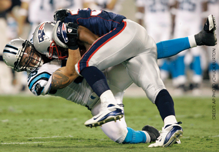
Concussion May Lead to High Depression Rate in Retired NFL Players
SAN DIEGO—Among older retired players from the National Football League (NFL) who have a history of concussion, about 40% have mild to moderate symptoms of depression, a rate nearly three times higher than that of the general population, according to research presented at the 65th Annual Meeting of the American Academy of Neurology.
Concussions in the former football players were specifically related to cognitive symptoms of depression, such as sadness, feelings of worthlessness or self-criticism, and suicidal thoughts, reported Nyaz Didehbani, PhD, and colleagues from the University of Texas at Dallas and University of Texas Southwestern Medical Center. In addition, somatic complaints were more prominent in the athletes compared with other items on the Beck Depression Inventory–II (BDI–II), even though the symptoms were not significantly correlated with concussions.
“Overall, results indicated that individuals having sustained concussions in early adulthood may be at a higher risk for developing depression as they age, compared to the general population,” stated Dr. Didehbani, a research psychologist at the Center for BrainHealth, University of Texas at Dallas. “More specifically, concussions were related to cognitive symptoms of depression, and it is possible that the high endorsement of somatic symptoms may be related to pain or other factors.”
Postconcussion Effects in Former NFL Players
Previous research has shown that concussions can lead to cognitive and mood disturbances, though few studies had explored the effects that may occur as players age. Dr. Didehbani’s group compared 30 retired NFL veterans who had had a concussion with 29 age-, education-, and IQ-matched healthy controls without a history of concussion. In both groups, ages ranged from 41 to 77, with a mean of 59 in the athletes and a mean of 60 in controls.
The investigators performed detailed neuropsychologic examinations in all participants to rule out cognitive impairment. All subjects completed the BDI–II, and participants’ scores were assessed according to a three-factor model of depressive symptoms (cognitive, affective, and somatic). Data regarding concussions were retrospectively obtained with use of the American Academy of Neurology Practice Parameter for grading concussions.
A Significant Correlation Between Concussion and Depression
The researchers found a significant correlation between the number of lifetime concussions in the former players and total scores on the BDI–II. In addition, the number of concussions was significantly correlated with cognitive symptoms on the BDI–II (r = 0.56). BDI-II scores were not associated with age, education, IQ, or the number of years played. Furthermore, the former athletes had significantly higher scores regarding cognitive, affective, and somatic symptoms.
Twelve of the 30 players (40%) had mild to moderate symptoms of depression, compared with the national average rate of 15% in older adults. The athletes had more symptoms on every BDI–II item, and on 13 of the 21 items the rate of symptoms was at least 20% higher, compared with controls. The largest differences between athletes and controls were observed in reports of problems with concentration (53% vs 19%, respectively), changes in appetite (47% vs 13%), loss of energy (60% vs 29%), changes in sleep (57% vs 26%), and decreased interest in sex (43% vs 16%).
Overall, the findings “highlight the need to educate individuals and families about somatic and psychologic symptoms associated with depression and to thoroughly assess depressive symptoms throughout the lifespan in professional athletes,” concluded Dr. Didehbani.
—Colby Stong
Editor
Suggested Reading
Didehbani N, Munro Cullum C, Mansinghani S, et al. Depressive symptoms and concussions in aging retired NFL players. Arch Clin Neuropsychol. 2013 May 3 [Epub ahead of print].
Hart J Jr, Kraut MA, Womack KB, et al. Neuroimaging of cognitive dysfunction and depression in aging retired National Football League players: a cross-sectional study. JAMA Neurol. 2013;70(3):326-335.
[To listen to an audiocast with Nyaz Didehbani, click here.]
DTI May Detect Axonal Injury After Sports-Related Concussion
SAN DIEGO—Diffusion tensor imaging (DTI) may help neurologists detect axonal injury before any symptoms appear in an athlete who has had a concussion, said Jeffrey J. Bazarian, MD, MPH. Neurologists can maximize the technique’s sensitivity by comparing it with a DTI scan taken at baseline, he noted at the 65th Annual Meeting of the American Academy of Neurology.
A pattern of decreased fractional anisotropy and elevated mean diffusivity on DTI suggests axonal loss. The opposite pattern, which suggests axonal edema, may occur at the same time. “We still don’t know the clinical significance of these changes,” said Dr. Bazarian, Associate Professor of Emergency Medicine at the University of Rochester Medical Center in New York. The relationship of these changes to short-term cognitive outcome and neurologic function is uncertain. Similarly unclear is the relationship of these changes to the development of chronic traumatic encephalopathy.
Water Movement Can Reveal Axonal Swelling
Axonal injury is the primary pathophysiologic process that occurs after brain injury. After concussion, the brain rotates and creates shear forces along the length of the axon. “If the axon gets stretched at the right threshold of stretch force, little pores form along the axon, and things start to fall apart,” said Dr. Bazarian. The axon swells, stops functioning, and may disconnect itself from the neuron over time.
CT and MRI scans cannot show axonal injury, but DTI can by illustrating the movement of water in the brain. A DTI scan measures fractional anisotropy, which indicates the degree to which water’s movement in the brain is straight, and mean diffusivity, which reveals the amount of water movement in the brain. “If axons swell up, then we would guess that the straightness of water motion would go up and the overall diffusivity would go down,” said Dr. Bazarian. “Conversely, if the axon degenerated, just the opposite would occur. This might be a nice way to indirectly see axonal swelling or axonal loss.”
Concussion Correlated With Changes on DTI
Dr. Bazarian and his colleagues examined seven high school football players to test whether DTI detects axonal injury after concussion. Each player underwent a DTI scan at the beginning of the football season and another scan at the end of the season. One player had a concussion, and six players served as controls. Players took cognitive exams before and after the football season. During the season, players used a diary to record the number of times they had been hit in the helmet. The investigators used wild bootstrapping to look for statistically significant differences between voxels before and after injury.
The player with concussion was cognitively worse than the other players, said Dr. Bazarian. The investigators found no cognitive difference between the other six players and nonathletes. All of the players were hit in the helmet between 50 and 400 times during the season.
Compared with the other players, the player with concussion had the greatest proportion (3%) of significant changes in fractional anisotropy and mean diffusivity in his brain after the season ended. The controls had nearly as much white-matter change as the player with concussion. The amount of change in fractional anisotropy and mean diffusivity in the players correlated with the number of times they reported being hit in the head and to the increases in their postconcussion symptom score at the end of the season. For the player with concussion, any part of the brain with post-season changes in fractional anisotropy also had changes in mean diffusivity, which suggested that DTI was detecting axonal injury, said Dr. Bazarian.
DTI May Indicate a Threshold for White Matter Damage
In a follow-up study, Dr. Bazarian and colleagues gave 10 college football players helmets with sensors that recorded blows stronger than 10 g. Players wore the helmets during practices and games and underwent a DTI scan at the beginning of the season, at the end of the season, and after six months of rest. Players also underwent cognitive and physical exams at these same three time points. Investigators used wild bootstrapping to compare each player’s three scans.
All players had increases and decreases in fractional anisotropy and mean diffusivity at the end of the season. The percentage of the brain with decreased fractional anisotropy correlated well with almost all of the helmet readings and with some neurologic outcomes. “That may be the important metric, in terms of axonal injury,” said Dr. Bazarian. Three players had a greater proportion of brain with decreased fractional anisotropy than the other players and the controls. Postseason changes correlated well with balance and some aspects of cognition, said Dr. Bazarian.
After six months’ rest, two additional players had increased proportions of brain with decreased fractional anisotropy. One player who had had decreased fractional anisotropy at the end of the season returned to normal after six months’ rest. Another player with decreased fractional anisotropy at the end of the season was unchanged after six months’ rest. A third player seemed to be recovering after six months’ rest, but was still not back to baseline.
“These data allow us to draw some thresholds above which you start to see white matter damage,” said Dr. Bazarian. “The one that stood out was players who had had head hits resulting in rotational acceleration greater than 4,500 radians/s2. Once you got above 40 or 45 hits, the amount of your white matter that has changes went up above what controls’ levels were. What those changes mean over the long term is not clear.”
—Erik Greb
Senior Associate Editor
Suggested Reading
Bazarian JJ, Zhu T, Blyth B, et al. Subject-specific changes in brain white matter on diffusion tensor imaging after sports-related concussion. Magn Reson Imaging. 2012;30(2):171-180.
Henry LC, Tremblay J, Tremblay S, et al. Acute and chronic changes in diffusivity measures after sports concussion. J Neurotrauma. 2011;28(10):2049-2059.
Marchi N, Bazarian JJ, Puvenna V, et al. Consequences of repeated blood-brain barrier disruption in football players. PLoS One. 2013;8(3):e56805
Charles W. Hoge, MD

An Atlas of Stats; Coordinating Traumatic Brain Injury Services; Voices of the Native American Experience
Psychogenic Nonepileptic Seizures Are Associated With Mild TBI
SAN DIEGO—Neurologists evaluating patients with a history of traumatic brain injury (TBI) and seizures should consider a diagnosis of psychogenic seizures, according to research presented at the 66th Annual Meeting of the American Epilepsy Society. The diagnosis may be suspected in patients with seizures related to mild TBI and in patients with a history of post-traumatic stress disorder (PTSD), said Martin Salinsky, MD, Director of the Veterans Affairs Medical Center in Portland.
"Just because a patient has a history of TBI and subsequently developed seizures does not necessarily mean that he or she has epilepsy," said Dr. Salinsky. About 25% of all patients evaluated in epilepsy monitoring units are discharged with a diagnosis of psychogenic seizures, which are alterations in behavior that resemble epileptic seizures, but do not result from paroxysmal neuronal discharges or any other physiologic abnormality. The exact prevalence of psychogenic nonepileptic seizures is unknown, however. In previous population-based studies of post-traumatic epilepsy, which were based on chart reviews or administrative databases, cases of epilepsy were rarely confirmed by EEG monitoring.
Psychogenic nonepileptic seizures are often mistaken for epilepsy and treated unsuccessfully with antiepileptic drugs. Patients with psychogenic seizures often are disabled, experience the side effects of treatment with antiepileptic drugs, and feel the psychosocial and economic effects associated with poorly controlled seizures.
Mild TBI Is Weakly Associated With Epilepsy
The first two studies to provide evidence for an association between head injuries and psychogenic seizures were published in Epilepsia in 1998, said Dr. Salinsky. Both studies were retrospective reviews of patients who had been diagnosed with psychogenic seizures on video EEG monitoring. Between one-quarter and one-third of the patients in both studies reported an antecedent head injury as the likely cause of the seizure.
Of the patients who suspected previous head injury as the cause of their seizures, about 80% had had a mild head injury, approximately 10% had had a moderate head injury, and about 10% had had a severe head injury. "This is not the distribution you would expect to see with post-traumatic epilepsy," which is more commonly associated with moderate to severe head injury than with mild head injury, said Dr. Salinsky.
Mild TBI is strongly associated with psychogenic seizures, but weakly associated with epilepsy. In a recent study, more than 80% of US veterans who entered an epilepsy monitoring unit with a history of mild TBI as a cause of their seizures were diagnosed with psychogenic seizures, said Dr. Salinksy. In contrast, approximately 90% of veterans with a history of severe TBI were diagnosed with epilepsy.
Post-Traumatic Stress Disorder May Contribute to Psychogenic Seizures
Psychogenic seizures are just as common in veterans as they are in civilians. But a new study showed that more than twice as many veterans (56%) as civilians (26%) had suspected a preceding head injury as the cause of the seizures. For veterans and civilians alike, most of the injuries were mild TBI, according to Dr. Salinksy.
In a recent analysis, veterans diagnosed with psychogenic seizures tended to have greater psychopathology (ie, more Axis I diagnoses) than veterans diagnosed with epileptic seizures. In particular, veterans diagnosed with psychogenic seizures were significantly more likely to have PTSD than veterans diagnosed with epileptic seizures. "Other than PTSD, differences between these two groups were relatively small and were not statistically significant," said Dr. Salinksy.
A multivariate analysis showed PTSD to be the only predictive factor for psychogenic seizures. "The odds ratio was fairly high, and the p value highly significant," said Dr. Salinsky. This finding mainly resulted from patients with a history of mild TBI, he added.
"In our veteran population, we are beginning to see a model develop whereby the development of psychogenic seizures in patients who have mild TBI may be mediated through the mechanism of PTSD," said Dr. Salinsky. Several studies suggest that health problems in veterans who have had mild TBI generally are mediated through PTSD. Perhaps not all psychogenic seizures in patients with TBI are mediated through PTSD, "but this does appear to be one valid mechanism in veterans, and perhaps in civilians as well," concluded Dr. Salinsky.
Senior Associate Editor
Suggested Reading
Annegers JF, Coan SP. The risks of epilepsy after traumatic brain injury. Seizure. 2000;9(7):453-457.
Holmes MD, Dodrill CB. What is the significance of subjective events recorded during long-term EEG video monitoring? Epilepsia. 1998;39(8):857-862.
Sigurdardottir KR, Olafsson E. Incidence of psychogenic seizures in adults: a population-based study in Iceland. Epilepsia. 1998;39(7):749-752.
SAN DIEGO—Neurologists evaluating patients with a history of traumatic brain injury (TBI) and seizures should consider a diagnosis of psychogenic seizures, according to research presented at the 66th Annual Meeting of the American Epilepsy Society. The diagnosis may be suspected in patients with seizures related to mild TBI and in patients with a history of post-traumatic stress disorder (PTSD), said Martin Salinsky, MD, Director of the Veterans Affairs Medical Center in Portland.
"Just because a patient has a history of TBI and subsequently developed seizures does not necessarily mean that he or she has epilepsy," said Dr. Salinsky. About 25% of all patients evaluated in epilepsy monitoring units are discharged with a diagnosis of psychogenic seizures, which are alterations in behavior that resemble epileptic seizures, but do not result from paroxysmal neuronal discharges or any other physiologic abnormality. The exact prevalence of psychogenic nonepileptic seizures is unknown, however. In previous population-based studies of post-traumatic epilepsy, which were based on chart reviews or administrative databases, cases of epilepsy were rarely confirmed by EEG monitoring.
Psychogenic nonepileptic seizures are often mistaken for epilepsy and treated unsuccessfully with antiepileptic drugs. Patients with psychogenic seizures often are disabled, experience the side effects of treatment with antiepileptic drugs, and feel the psychosocial and economic effects associated with poorly controlled seizures.
Mild TBI Is Weakly Associated With Epilepsy
The first two studies to provide evidence for an association between head injuries and psychogenic seizures were published in Epilepsia in 1998, said Dr. Salinsky. Both studies were retrospective reviews of patients who had been diagnosed with psychogenic seizures on video EEG monitoring. Between one-quarter and one-third of the patients in both studies reported an antecedent head injury as the likely cause of the seizure.
Of the patients who suspected previous head injury as the cause of their seizures, about 80% had had a mild head injury, approximately 10% had had a moderate head injury, and about 10% had had a severe head injury. "This is not the distribution you would expect to see with post-traumatic epilepsy," which is more commonly associated with moderate to severe head injury than with mild head injury, said Dr. Salinsky.
Mild TBI is strongly associated with psychogenic seizures, but weakly associated with epilepsy. In a recent study, more than 80% of US veterans who entered an epilepsy monitoring unit with a history of mild TBI as a cause of their seizures were diagnosed with psychogenic seizures, said Dr. Salinksy. In contrast, approximately 90% of veterans with a history of severe TBI were diagnosed with epilepsy.
Post-Traumatic Stress Disorder May Contribute to Psychogenic Seizures
Psychogenic seizures are just as common in veterans as they are in civilians. But a new study showed that more than twice as many veterans (56%) as civilians (26%) had suspected a preceding head injury as the cause of the seizures. For veterans and civilians alike, most of the injuries were mild TBI, according to Dr. Salinksy.
In a recent analysis, veterans diagnosed with psychogenic seizures tended to have greater psychopathology (ie, more Axis I diagnoses) than veterans diagnosed with epileptic seizures. In particular, veterans diagnosed with psychogenic seizures were significantly more likely to have PTSD than veterans diagnosed with epileptic seizures. "Other than PTSD, differences between these two groups were relatively small and were not statistically significant," said Dr. Salinksy.
A multivariate analysis showed PTSD to be the only predictive factor for psychogenic seizures. "The odds ratio was fairly high, and the p value highly significant," said Dr. Salinsky. This finding mainly resulted from patients with a history of mild TBI, he added.
"In our veteran population, we are beginning to see a model develop whereby the development of psychogenic seizures in patients who have mild TBI may be mediated through the mechanism of PTSD," said Dr. Salinsky. Several studies suggest that health problems in veterans who have had mild TBI generally are mediated through PTSD. Perhaps not all psychogenic seizures in patients with TBI are mediated through PTSD, "but this does appear to be one valid mechanism in veterans, and perhaps in civilians as well," concluded Dr. Salinsky.
Senior Associate Editor
Suggested Reading
Annegers JF, Coan SP. The risks of epilepsy after traumatic brain injury. Seizure. 2000;9(7):453-457.
Holmes MD, Dodrill CB. What is the significance of subjective events recorded during long-term EEG video monitoring? Epilepsia. 1998;39(8):857-862.
Sigurdardottir KR, Olafsson E. Incidence of psychogenic seizures in adults: a population-based study in Iceland. Epilepsia. 1998;39(7):749-752.
AAN Releases Updated Sports Concussion Guideline
SAN DIEGO—The American Academy of Neurology (AAN) has released an evidence-based guideline for the assessment and management of athletes with concussion. This new guideline replaces the 1997 AAN guideline on the same topic. The new guideline was announced at a press conference at the 65th Annual Meeting of the AAN and was published in the March 18, 2013, online issue of Neurology.
"With over a million athletes experiencing sports-related concussion per year in the United States, the AAN is pleased to release this evidence-based guideline," said lead author Christopher C. Giza, MD, from the David Geffen School of Medicine and Mattel Children's Hospital at UCLA.
The guideline was developed using an evidence-based process and was multidisciplinary in nature, including neurologists and a panel of professionals from other specialties who are involved in the care and assessment of athletes with sports concussions. "These recommendations are the best summation of available science regarding the assessment and management of sports concussion," Dr. Giza said.
"One of the most important recommendations from this guideline is that an athlete who sustained or is suspected of sustaining a concussion should be removed from play that day and then be assessed by a licensed health care provider with expertise in management of concussion," Dr. Giza said.
Another important component that came out of the evidence was the effect of concussion on youth. "Evidence suggests that high school-age individuals with concussion take longer to recover than their older professional counterparts, and so they should be managed more conservatively." The guideline committee also found a dearth of evidence on concussion under the age of high school and this, they said, was an area for future investigation.
"Individuals with suspected injury should be removed from contact risk activity after concussion," Dr. Giza said. "The evidence is really sparse for complete cognitive restriction after concussion—complete cognitive rest. The guideline recommends that athletes be removed from sports activity that would put them back at contact risk. An important distinction of this guideline from the 1997 guideline is that it moves away from the grading system of concussion. We don't find that the evidence supports trying to rate the concussion at the moment of impact and determine how severe it was or how long a return to play would be. In this case the assessment of concussion would be done individually with multiple diagnostic tools, and the management plans are individualized because there are different factors that contribute to how long an athlete might recover from concussion."
Among the sports in the studies evaluated, risk of concussion is greatest in football and rugby, followed by hockey and soccer. The risk of concussion for young women and girls is greatest in soccer and basketball.
According to the guideline:
• An athlete who has a history of one or more concussions is at greater risk of being diagnosed with another concussion.
• The first 10 days after a concussion appear to be the period of greatest risk of being diagnosed with another concussion.
• There is no clear evidence that one type of football helmet can better protect against concussion compared with another kind of helmet. Helmets should fit properly and be well maintained.
• Licensed health care professionals trained in treating concussion should look for ongoing symptoms (especially headache and fogginess), history of concussions, and younger age in the athlete. Each of these factors has been linked to a longer recovery after a concussion.
• Risk factors linked to chronic neurobehavioral impairment in professional athletes include prior concussion, longer exposure to the sport, and having the APOE ε4 gene.
• Concussion is a clinical diagnosis. Symptom checklists, the Standardized Assessment of Concussion (SAC), neuropsychologic testing (paper-and-pencil and computerized), and the Balance Error Scoring System may be helpful tools in diagnosing and managing concussions but should not be used alone for making a diagnosis.
• Signs and symptoms of a concussion include headache and sensitivity to light and sound; changes to reaction time, balance, and coordination; changes in memory, judgment, speech, and sleep; loss of consciousness or a "blackout" (occurring in less than 10% of cases).
"If in doubt, sit it out," said Jeffrey S. Kutcher, MD, from the University of Michigan Medical School in Ann Arbor and a coauthor of the guideline. "Being seen by a trained professional is extremely important after a concussion. If headaches or other symptoms return with the start of exercise, stop the activity and consult a doctor."
The new AAN guideline has been endorsed by the National Football League Players Association, the Child Neurology Society, the National Association of Emergency Medical Service Physicians, the National Association of School Psychologists, the National Athletic Trainers Association, the American Football Coaches Association, the National Academy of Neuropsychology, and the Neurocritical Care Society.
Vice President/Group Editor
Suggested Reading
Giza C, Kutcher JS, Ashwal S, et al. Summary of evidence-based guideline update: evaluation and management of concussion in sports. Report of the Guideline Development Subcommittee of the American Academy of Neurology. Neurology. 2013 March 18 [Epub ahead of print].
SAN DIEGO—The American Academy of Neurology (AAN) has released an evidence-based guideline for the assessment and management of athletes with concussion. This new guideline replaces the 1997 AAN guideline on the same topic. The new guideline was announced at a press conference at the 65th Annual Meeting of the AAN and was published in the March 18, 2013, online issue of Neurology.
"With over a million athletes experiencing sports-related concussion per year in the United States, the AAN is pleased to release this evidence-based guideline," said lead author Christopher C. Giza, MD, from the David Geffen School of Medicine and Mattel Children's Hospital at UCLA.
The guideline was developed using an evidence-based process and was multidisciplinary in nature, including neurologists and a panel of professionals from other specialties who are involved in the care and assessment of athletes with sports concussions. "These recommendations are the best summation of available science regarding the assessment and management of sports concussion," Dr. Giza said.
"One of the most important recommendations from this guideline is that an athlete who sustained or is suspected of sustaining a concussion should be removed from play that day and then be assessed by a licensed health care provider with expertise in management of concussion," Dr. Giza said.
Another important component that came out of the evidence was the effect of concussion on youth. "Evidence suggests that high school-age individuals with concussion take longer to recover than their older professional counterparts, and so they should be managed more conservatively." The guideline committee also found a dearth of evidence on concussion under the age of high school and this, they said, was an area for future investigation.
"Individuals with suspected injury should be removed from contact risk activity after concussion," Dr. Giza said. "The evidence is really sparse for complete cognitive restriction after concussion—complete cognitive rest. The guideline recommends that athletes be removed from sports activity that would put them back at contact risk. An important distinction of this guideline from the 1997 guideline is that it moves away from the grading system of concussion. We don't find that the evidence supports trying to rate the concussion at the moment of impact and determine how severe it was or how long a return to play would be. In this case the assessment of concussion would be done individually with multiple diagnostic tools, and the management plans are individualized because there are different factors that contribute to how long an athlete might recover from concussion."
Among the sports in the studies evaluated, risk of concussion is greatest in football and rugby, followed by hockey and soccer. The risk of concussion for young women and girls is greatest in soccer and basketball.
According to the guideline:
• An athlete who has a history of one or more concussions is at greater risk of being diagnosed with another concussion.
• The first 10 days after a concussion appear to be the period of greatest risk of being diagnosed with another concussion.
• There is no clear evidence that one type of football helmet can better protect against concussion compared with another kind of helmet. Helmets should fit properly and be well maintained.
• Licensed health care professionals trained in treating concussion should look for ongoing symptoms (especially headache and fogginess), history of concussions, and younger age in the athlete. Each of these factors has been linked to a longer recovery after a concussion.
• Risk factors linked to chronic neurobehavioral impairment in professional athletes include prior concussion, longer exposure to the sport, and having the APOE ε4 gene.
• Concussion is a clinical diagnosis. Symptom checklists, the Standardized Assessment of Concussion (SAC), neuropsychologic testing (paper-and-pencil and computerized), and the Balance Error Scoring System may be helpful tools in diagnosing and managing concussions but should not be used alone for making a diagnosis.
• Signs and symptoms of a concussion include headache and sensitivity to light and sound; changes to reaction time, balance, and coordination; changes in memory, judgment, speech, and sleep; loss of consciousness or a "blackout" (occurring in less than 10% of cases).
"If in doubt, sit it out," said Jeffrey S. Kutcher, MD, from the University of Michigan Medical School in Ann Arbor and a coauthor of the guideline. "Being seen by a trained professional is extremely important after a concussion. If headaches or other symptoms return with the start of exercise, stop the activity and consult a doctor."
The new AAN guideline has been endorsed by the National Football League Players Association, the Child Neurology Society, the National Association of Emergency Medical Service Physicians, the National Association of School Psychologists, the National Athletic Trainers Association, the American Football Coaches Association, the National Academy of Neuropsychology, and the Neurocritical Care Society.
Vice President/Group Editor
Suggested Reading
Giza C, Kutcher JS, Ashwal S, et al. Summary of evidence-based guideline update: evaluation and management of concussion in sports. Report of the Guideline Development Subcommittee of the American Academy of Neurology. Neurology. 2013 March 18 [Epub ahead of print].
Suggested Reading
Giza C, Kutcher JS, Ashwal S, et al. Summary of evidence-based guideline update: evaluation and management of concussion in sports. Report of the Guideline Development Subcommittee of the American Academy of Neurology. Neurology. 2013 March 18 [Epub ahead of print].
Kristen Sahler, MD, Talks With Alan Rapoport, MD, About Post-Traumatic Headache

Hemodynamic Complications Are Common in Veterans After TBI
HONOLULU—Cerebral vasospasm and intracranial hypertension may be frequent secondary insults following wartime traumatic brain injury (TBI), according to research presented at the 2013 International Stroke Conference.
In a retrospective study of 122 consecutive veterans, transcranial Doppler (TCD) ultrasonography signs of mild, moderate, and severe vasospasm involving anterior circulation vessels were observed in 71%, 42%, and 16% of the veterans with TBI, respectively. TCD signs involving posterior circulation vessels were observed in 57%, 32%, and 14%, respectively.
Intracranial hypertension was recorded in 43% of patients, lead author Alexander Razumovsky, PhD, reported. “Cerebral blood flow velocity measurements permit detection of early signs of cerebral vasospasm and allow physicians to better define management strategies,” he said.
TCD Commonly Used by US Army
TCD monitoring is routinely used by the US Army, but it has not been fully embraced elsewhere because many physicians are unaware of how it can be used within the continuum of TBI care to help reverse outcomes, said study coauthor Col. Rocco Armonda, MC, USA.
“There’s this lack of drive to change outcomes, because people haven’t seen how you can take someone who is in a comatose state and eventually gain functional independence,” said Dr. Armonda, who is the Director of Cerebrovascular Surgery and Interventional Neuroradiology at the National Naval Medical Center in Bethesda, Maryland.
A recent study from the Walter Reed National Military Medical Center, however, reported that 63% of veterans with a penetrating TBI and a mean discharge Glasgow Coma Scale (GCS) score of 6 to 8 progressed to functional independence, a number twice as high as would be expected, Dr. Armonda commented.
Dr. Razumovsky said that simple, noninvasive TCD monitoring should be performed in the daily management of hospitalized TBI patients to complement other monitoring strategies, and it can be used alone in emergency situations when intracranial pressure monitoring is contraindicated or not readily available.
TCD monitoring is not necessary during outpatient follow-up of patients with TBI, with the exception of the subset who have undergone hemicraniectomy, Dr. Armonda said.
“In those patients who’ve had hemicraniectomy, some of them suffer prolonged duration from atmospheric pressure of the cranial defect, and TCD has actually been helpful to look at the cerebral blood flow dynamics,” he said at a press briefing on the study.
The results confirm earlier data that TBI is associated with a high incidence of cerebral vasospasm, particularly in patients with severe TBI, and are applicable to civilian TBI patients, because a significant number will experience post-traumatic bleeding that will lead to vasospasm and intracranial hypertension, said Dr. Razumovsky, Director of Sentient NeuroCare Services in Hunt Valley, Maryland, which provides services to the National Naval Medical Hospital.
Detecting Vasospasm in TBI Patients
In the current study, patients were admitted to Walter Reed a mean of 6.7 days after injury from October 1, 2008, to November 30, 2012. Mean cerebral blood flow velocity (CBFV) of 100 to 139 cm/s was used to define mild vasospasm, 140 to 199 cm/s for moderate vasospasm, and more than 200 cm/s severe for vasospasm.
In all, 88 patients had penetrating head injury, of whom 45 (51%) had a secondary diagnosis of blast injury; and 34 patients had a closed head injury, of whom 15 (44%) had a secondary diagnosis of blast injury. Their mean age was 26 years.
In the mild vasospasm group, the mean anterior circulation CBFV was 122.5 cm/s and mean posterior CBFV was 61 cm/s. Their average GCS score changed from 8 at baseline to 6.1 at discharge, Dr. Razumovsky said.
In the moderate vasospasm group, the mean anterior circulation CBFV was 156.6 cm/s and mean posterior CBFV was 77.5 cm/s. Their average GCS score changed from 5.9 at baseline to 5.3 at discharge.
Patients with severe vasospasm had an average anterior circulation CBFV of 241.9 cm/s and a mean posterior CBFV of 101.8 cm/s. Their average GCS score was 4.8 at both time periods. Eight patients (7%) underwent transluminal angioplasty for post-traumatic symptomatic vasospasm, he said.
Larry Goldstein, MD, Director of the Duke Stroke Center in Durham, North Carolina, said in an interview that the detection of vasospasm by TCD was potentially important, because there is an increased risk of vasospasm after subarachnoid hemorrhage that can lead to delayed ischemic neurologic deficits. Although it was unclear from the data how many patients had traumatic subarachnoid hemorrhage associated with their brain injury, he noted that the risk of vasospasm seems to be similar for traumatic and nontraumatic subarachnoid hemorrhage.
“In many centers, both groups of patients are routinely monitored with transcranial Dopplers over the first couple of weeks or so, which is when the peak period of vasospasm develops,” Dr. Goldstein said. “Showing that monitoring leads to interventions that then change outcome, that’s not so straightforward.”
IMNG Medical News
Can Prophylaxis Prevent Epilepsy in Patients With Traumatic Brain Injury?
SAN DIEGO—Prophylactic treatment with established antiepileptic drugs (AEDs) has not prevented epilepsy in trials involving patients with traumatic brain injury (TBI), said investigators at the 66th Annual Meeting of the American Epilepsy Society. Newer AEDs have not been tested in randomized trials, however, and other prophylactic measures have shown promise in animal models, neurologists reported.
In its report titled "Epilepsy Across the Spectrum," the Institute of Medicine called for the development and evaluation of prevention efforts that focus on established risk factors for epilepsy, such as TBI. The prevalence of TBI among returning soldiers and among National Football League players has drawn attention to this risk factor. Preventing epilepsy after known risks, such as TBI is "one of the major issues in epilepsy at the present time," according to Marc A. Dichter, MD, PhD, Professor of Neurology at the University of Pennsylvania in Philadelphia.
Established AEDs Did Not Prevent Post-Traumatic Epilepsy
Almost all clinical trials of prophylactic AED treatment have defined post-traumatic epilepsy as unprovoked seizures that arise more than seven days after an acute brain trauma, said Patrick Kwan, MD, PhD, Professor of Neurology at the University of Melbourne. Seizures that occur within seven days of an acute brain trauma are classified as acute symptomatic or provoked seizures.
Observational studies conducted in the late 1970s and early 1980s suggested that treating patients prophylactically with the established AEDs (eg, phenytoin, phenobarbital, and valproate) would help prevent post-traumatic epilepsy. But a seminal double-blind, randomized trial performed by Nancy Temkin, PhD, Professor of Neurosurgery and Biostatistics at the University of Washington in Seattle, found no significant difference between patients who received phenytoin and controls in the development of late seizures. Patients who received phenytoin had a significantly reduced risk of developing early seizures, however.
In a second trial, Dr. Temkin randomized patients with severe TBI to receive phenytoin for one week or valproate for one month or six months. Neither drug prevented late seizures, and investigators observed a trend toward higher incidence of post-traumatic epilepsy in patients receiving valproate. Furthermore, patients who received valproate for six months had a trend toward high mortality, compared with untreated patients.
The results of the American Academy of Neurology's systematic review of prospective, controlled trials of AED prophylaxis for post-traumatic epilepsy were consistent with Dr. Temkin's findings. The pooled analysis of class 1 studies concluded that giving phenytoin prophylactically reduced early post-traumatic seizures. But a pooled analysis of class 1 and class 2 studies found that prophylactic phenytoin did not prevent late post-traumatic seizures.
Newer AEDs Have Not Been Tested in Randomized Trials
A recent nonrandomized phase II study compared levetiracetam prophylaxis with no treatment for the prevention of post-traumatic epilepsy. Levetiracetam was associated with a reduced risk of developing late seizures, compared with no treatment, but all patients were also given phenytoin for the first week of the trial to prevent early post-traumatic seizures, said Dr. Kwan. In addition, patients who presented within eight hours of a severe head injury received levetiracetam, but patients who presented between eight and 24 hours received no treatment.
Patients were followed for 24 months, and 12% of patients stopped taking levetiracetam. "Although it was not statistically significant, there was a numerically higher mortality rate in people who were treated, compared with the untreated group," observed Dr. Kwan. "Now, perhaps this was because those who were treated [had] presented early, and they tended to have a more severe head injury. These patients did have a lower Glasgow Coma Scale score, so it just shows that the study was not really looking at efficacy, but rather at feasibility and safety."
"We really need to examine the new AEDs to figure this out," said Dr. Dichter. "We need more basic research, including testing with the new AEDs," he added. "We need more clinical research to learn how best to perform clinical trials in this area, as well as to find effective therapies. AEDs may prove useful, but other strategies involving drugs that do not directly suppress seizures, or maybe devices, are likely to be useful as well. If we do not begin clinical trials now, once we do find effective antiepileptogenic therapies in animal models, the wait to bring them to the clinic will be much longer," he concluded.
Brain Cooling Prevented Post-Traumatic Epilepsy in Rats
Researchers have successfully prevented post-traumatic epilepsy in animals without using AEDs. Raimondo D'Ambrosio, PhD, Associate Professor of Neurosurgery at the University of Washington, administered frontal fluid percussion injuries to rats and subsequently observed, by invasive EEG, small, localized neocortical seizures that he called grade 1 seizures. The seizures eventually spread to the rest of the cortex, and Dr. D'Ambrosio called these spreading seizures grade 2 seizures. "If you record from the scalp during grade 1 events, you don't see anything at all," said Dr. Dichter, "but you will see the spreading grade 2 seizures."
When Dr. D'Ambrosio cooled the rats' brains by 2 °C, they did not develop post-traumatic epilepsy. Cooling eliminated short, medium-sized, and long seizures in treated animals. The amount of cooling performed in the study is "much less than we do in the ICU now for people who have had cardiac arrest," observed Dr. Dichter. So far, no studies have investigated whether cooling can prevent post-traumatic epilepsy in humans.
Suggested Reading
Klein P, Herr D, Pearl PL, et al. Results of phase II pharmacokinetic study of levetiracetam for prevention of post-traumatic epilepsy. Epilepsy Behav. 2012;24(4):457-461.
Temkin NR. Preventing and treating posttraumatic seizures: the human experience. Epilepsia. 2009;50(Suppl 2):10-13.
Temkin NR, Dikmen SS, Wilensky AJ, et al. A randomized, double-blind study of phenytoin for the prevention of post-traumatic seizures. N Engl J Med. 1990;323(8):497-502.
SAN DIEGO—Prophylactic treatment with established antiepileptic drugs (AEDs) has not prevented epilepsy in trials involving patients with traumatic brain injury (TBI), said investigators at the 66th Annual Meeting of the American Epilepsy Society. Newer AEDs have not been tested in randomized trials, however, and other prophylactic measures have shown promise in animal models, neurologists reported.
In its report titled "Epilepsy Across the Spectrum," the Institute of Medicine called for the development and evaluation of prevention efforts that focus on established risk factors for epilepsy, such as TBI. The prevalence of TBI among returning soldiers and among National Football League players has drawn attention to this risk factor. Preventing epilepsy after known risks, such as TBI is "one of the major issues in epilepsy at the present time," according to Marc A. Dichter, MD, PhD, Professor of Neurology at the University of Pennsylvania in Philadelphia.
Established AEDs Did Not Prevent Post-Traumatic Epilepsy
Almost all clinical trials of prophylactic AED treatment have defined post-traumatic epilepsy as unprovoked seizures that arise more than seven days after an acute brain trauma, said Patrick Kwan, MD, PhD, Professor of Neurology at the University of Melbourne. Seizures that occur within seven days of an acute brain trauma are classified as acute symptomatic or provoked seizures.
Observational studies conducted in the late 1970s and early 1980s suggested that treating patients prophylactically with the established AEDs (eg, phenytoin, phenobarbital, and valproate) would help prevent post-traumatic epilepsy. But a seminal double-blind, randomized trial performed by Nancy Temkin, PhD, Professor of Neurosurgery and Biostatistics at the University of Washington in Seattle, found no significant difference between patients who received phenytoin and controls in the development of late seizures. Patients who received phenytoin had a significantly reduced risk of developing early seizures, however.
In a second trial, Dr. Temkin randomized patients with severe TBI to receive phenytoin for one week or valproate for one month or six months. Neither drug prevented late seizures, and investigators observed a trend toward higher incidence of post-traumatic epilepsy in patients receiving valproate. Furthermore, patients who received valproate for six months had a trend toward high mortality, compared with untreated patients.
The results of the American Academy of Neurology's systematic review of prospective, controlled trials of AED prophylaxis for post-traumatic epilepsy were consistent with Dr. Temkin's findings. The pooled analysis of class 1 studies concluded that giving phenytoin prophylactically reduced early post-traumatic seizures. But a pooled analysis of class 1 and class 2 studies found that prophylactic phenytoin did not prevent late post-traumatic seizures.
Newer AEDs Have Not Been Tested in Randomized Trials
A recent nonrandomized phase II study compared levetiracetam prophylaxis with no treatment for the prevention of post-traumatic epilepsy. Levetiracetam was associated with a reduced risk of developing late seizures, compared with no treatment, but all patients were also given phenytoin for the first week of the trial to prevent early post-traumatic seizures, said Dr. Kwan. In addition, patients who presented within eight hours of a severe head injury received levetiracetam, but patients who presented between eight and 24 hours received no treatment.
Patients were followed for 24 months, and 12% of patients stopped taking levetiracetam. "Although it was not statistically significant, there was a numerically higher mortality rate in people who were treated, compared with the untreated group," observed Dr. Kwan. "Now, perhaps this was because those who were treated [had] presented early, and they tended to have a more severe head injury. These patients did have a lower Glasgow Coma Scale score, so it just shows that the study was not really looking at efficacy, but rather at feasibility and safety."
"We really need to examine the new AEDs to figure this out," said Dr. Dichter. "We need more basic research, including testing with the new AEDs," he added. "We need more clinical research to learn how best to perform clinical trials in this area, as well as to find effective therapies. AEDs may prove useful, but other strategies involving drugs that do not directly suppress seizures, or maybe devices, are likely to be useful as well. If we do not begin clinical trials now, once we do find effective antiepileptogenic therapies in animal models, the wait to bring them to the clinic will be much longer," he concluded.
Brain Cooling Prevented Post-Traumatic Epilepsy in Rats
Researchers have successfully prevented post-traumatic epilepsy in animals without using AEDs. Raimondo D'Ambrosio, PhD, Associate Professor of Neurosurgery at the University of Washington, administered frontal fluid percussion injuries to rats and subsequently observed, by invasive EEG, small, localized neocortical seizures that he called grade 1 seizures. The seizures eventually spread to the rest of the cortex, and Dr. D'Ambrosio called these spreading seizures grade 2 seizures. "If you record from the scalp during grade 1 events, you don't see anything at all," said Dr. Dichter, "but you will see the spreading grade 2 seizures."
When Dr. D'Ambrosio cooled the rats' brains by 2 °C, they did not develop post-traumatic epilepsy. Cooling eliminated short, medium-sized, and long seizures in treated animals. The amount of cooling performed in the study is "much less than we do in the ICU now for people who have had cardiac arrest," observed Dr. Dichter. So far, no studies have investigated whether cooling can prevent post-traumatic epilepsy in humans.
Suggested Reading
Klein P, Herr D, Pearl PL, et al. Results of phase II pharmacokinetic study of levetiracetam for prevention of post-traumatic epilepsy. Epilepsy Behav. 2012;24(4):457-461.
Temkin NR. Preventing and treating posttraumatic seizures: the human experience. Epilepsia. 2009;50(Suppl 2):10-13.
Temkin NR, Dikmen SS, Wilensky AJ, et al. A randomized, double-blind study of phenytoin for the prevention of post-traumatic seizures. N Engl J Med. 1990;323(8):497-502.