User login
Managing asthma in children: Pets don’t always have to go
SAN ANTONIO – It may not always be necessary to tell parents of children with asthma to get rid of the household pet, a recent study suggests.

Children with had significant improvements in a variety of asthma measures, regardless of whether parents reported pets or smoking at home, according to results of the 4-year, 471-patient prospective study.
Those results suggest that clinicians should be working to make sure the guidelines are being closely followed before, for example, telling parents they need to consider getting rid of the family pet, said Shahid Sheikh, MD, FCCP, of Nationwide Children’s Hospital, Columbus, Ohio.
“As the guidelines still work, we need to focus and develop the connections with the family to make sure the patients are on the right treatment, and that they’re getting the medications,” Dr. Sheikh said in an interview at the annual meeting of the American College of Chest Physicians.
The prospective cohort study by Dr. Sheikh and his colleagues, presented in a poster session, included children referred to a pediatric asthma center with the diagnosis of uncontrolled asthma. All patients received asthma care according to National Asthma Education and Prevention Program Expert Panel Report 3 guidelines.
Medications were changed as needed, and the asthma action plan was revised accordingly and reviewed with the family at each visit, Dr. Sheikh reported. After a baseline evaluation, clinic visits for the study occurred at 3 months, 6 months, and then at 1, 2, 3, and 4 years.
Out of 471 patients, 258 had pets, and 125 were in homes where smoking took place, according to parent reports.
Asthma control test scores were 15.1 at baseline for children in no-pet households, and 16.5 for those with pets; by the 3-month visit, scores increased to 20.1 and 20.3 for the no-pet and pet groups, and at 4 years, those scores had edged up to 22.2 and 22.7 (P = .371), Dr. Sheikh reported.
Similarly, after care was started, there was no significant difference between the no-pet and pet groups in mean percent of predicted forced expiratory volume in 1 second (FEV1), wheezing, nighttime cough, albuterol use, and other factors over the 4 years of follow-up, he said.
Likewise, looking at the data by nonsmoking vs. smoking households, asthma control test scores at baseline were 16.1 and 15.1, respectively, and at 4 years they were 22.2 and 22.3 (P = .078), with a similar lack of difference in predicted FEV1, wheezing, and all other factors evaluated.
Getting rid of the family pet may need to be a consideration for some families, but based on these data, that might not be necessary for the majority of families, Dr. Sheikh said in the interview.
“On the other hand, we are not saying that if you are smoking, you should continue to smoke,” he added.
“What we are saying is that smoking is bad, but if your child is not getting better, I don’t want to blame your smoking for it. There may be something else which may be more important than smoking which we are missing – the child may not be getting the medicine, or may not be on the right medicine, or may have other comorbidities.”
Dr. Sheikh and his coinvestigators disclosed that they had no relationships relevant to the study.
SOURCE: Sheikh S et al. CHEST 2018. doi: 10.1016/j.chest.2018.08.666.
SAN ANTONIO – It may not always be necessary to tell parents of children with asthma to get rid of the household pet, a recent study suggests.
Children with had significant improvements in a variety of asthma measures, regardless of whether parents reported pets or smoking at home, according to results of the 4-year, 471-patient prospective study.
Those results suggest that clinicians should be working to make sure the guidelines are being closely followed before, for example, telling parents they need to consider getting rid of the family pet, said Shahid Sheikh, MD, FCCP, of Nationwide Children’s Hospital, Columbus, Ohio.
“As the guidelines still work, we need to focus and develop the connections with the family to make sure the patients are on the right treatment, and that they’re getting the medications,” Dr. Sheikh said in an interview at the annual meeting of the American College of Chest Physicians.
The prospective cohort study by Dr. Sheikh and his colleagues, presented in a poster session, included children referred to a pediatric asthma center with the diagnosis of uncontrolled asthma. All patients received asthma care according to National Asthma Education and Prevention Program Expert Panel Report 3 guidelines.
Medications were changed as needed, and the asthma action plan was revised accordingly and reviewed with the family at each visit, Dr. Sheikh reported. After a baseline evaluation, clinic visits for the study occurred at 3 months, 6 months, and then at 1, 2, 3, and 4 years.
Out of 471 patients, 258 had pets, and 125 were in homes where smoking took place, according to parent reports.
Asthma control test scores were 15.1 at baseline for children in no-pet households, and 16.5 for those with pets; by the 3-month visit, scores increased to 20.1 and 20.3 for the no-pet and pet groups, and at 4 years, those scores had edged up to 22.2 and 22.7 (P = .371), Dr. Sheikh reported.
Similarly, after care was started, there was no significant difference between the no-pet and pet groups in mean percent of predicted forced expiratory volume in 1 second (FEV1), wheezing, nighttime cough, albuterol use, and other factors over the 4 years of follow-up, he said.
Likewise, looking at the data by nonsmoking vs. smoking households, asthma control test scores at baseline were 16.1 and 15.1, respectively, and at 4 years they were 22.2 and 22.3 (P = .078), with a similar lack of difference in predicted FEV1, wheezing, and all other factors evaluated.
Getting rid of the family pet may need to be a consideration for some families, but based on these data, that might not be necessary for the majority of families, Dr. Sheikh said in the interview.
“On the other hand, we are not saying that if you are smoking, you should continue to smoke,” he added.
“What we are saying is that smoking is bad, but if your child is not getting better, I don’t want to blame your smoking for it. There may be something else which may be more important than smoking which we are missing – the child may not be getting the medicine, or may not be on the right medicine, or may have other comorbidities.”
Dr. Sheikh and his coinvestigators disclosed that they had no relationships relevant to the study.
SOURCE: Sheikh S et al. CHEST 2018. doi: 10.1016/j.chest.2018.08.666.
REPORTING FROM CHEST 2018
Key clinical point: Exposure to pets and tobacco smoke may have very little effect on the improvement of asthma in children who are being managed according to guidelines.
Major finding: ACT scores were 15.1 and 16.5 at baseline for children in no-pet and pet households, respectively, and were 22.2 and 22.7 at the 4-year evaluation.
Study details: A 4-year prospective cohort study of 471 children with uncontrolled asthma seen in a pediatric asthma center.
Disclosures: The study authors had no disclosures.
Source: Sheikh S et al. CHEST 2018. doi: 10.1016/j.chest.2018.08.666.
A Ticking Noise From the Chest: Recognition of the Hamman Sign
The authors describe a case of a 21-year-old woman who presented with shortness of breath and exhibited a Hamman sign, an uncommon clinical finding.
Traditionally, a physician develops a differential diagnosis based primarily (>70%) on the history and the physical examination of a patient.1 While modern medicine has developed with new technological devices and a growing number of diagnostic tests, one must not forget the value of a thorough physical examination.
Case
A 21-year-old woman, in previous good health, presented to the ED with the chief complaint of shortness of breath. She stated that she woke up with acute dyspnea and a stabbing pain on the left side of her thorax, related to her breathing. The patient looked distressed upon presentation.
Her vital signs at presentation were: blood pressure, 150/85 mm Hg; heart rate, 120 beats/min; respiratory
During physical examination, a loud ticking noise was heard originating from the thorax, even without a stethoscope (an example of the sound can be heard at https://www.youtube.com/watch?v=mXJHtJeL1mM). During auscultation, the ticking noise was prominent in early systole and audible over all parts of the thorax. The sound was only heard when the patient was in the supine position and disappeared when she sat up. It persisted when the patient was holding her breath. Breath sounds were equal and clear bilaterally. There was no subcutaneous emphysema palpable over the thorax or neck region.
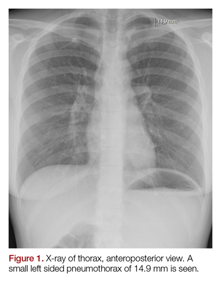
The electrocardiogram and blood results, including D-dimer, were normal. The chest X-ray showed an apical pneumothorax of 1.5 cm on the left side (Figures 1 and 2). There was no evidence of pneumomediastinum or pneumopericardium. The patient received acetaminophen and ibuprofen tablets for pain, and she was discharged home. At the follow-up 2 weeks later, she had no remaining symptoms and the ticking sound had disappeared.

Discussion
These loud intermittent noises originating from the thorax were described for the first time at the beginning of the 19th century.2,3 However, it was Louis Virgil Hamman whose name would be linked to this physical examination finding. In 1937 he described typical clicking, crackling, and popping sounds over the precordium, synchronized with the heartbeat. This was usually in combination with subcutaneous emphysema in the neck region. Hamman presumed that the symptoms were due to mediastinal air caused by rupture alveoli or bronchioles, resulting in interstitial emphysema of the lung parenchyma. In addition, air could leak into the pleural space, causing a pneumothorax. He concluded that the clinical findings were pathognomonic for spontaneous mediastinal emphysema, and this physical examination finding became known as the “Hamman sign”.4-11
However, in the following years it was demonstrated that the appearance of loud, systolic clicking noises over the precordium could also be present in patients with a small spontaneous left-sided pneumothorax.5-9 It was assumed the pneumothorax caused a small amount of air to accumulate in the pleural space in the major fissure inferiorly, which shifted with the cardiac contraction. This results in the noise being present while in the supine position. In the sitting position, the air moves cranially above the heart, meaning it is not influenced by the cardiac contractions and the noise disappears.5-8 The Hamman sign is absent in right-sided pneumothorax, presumably because of the smaller contact surface between the lung pleura and the mediastinal pleura overlying the heart in comparison to the left side. Also, the contractions of the right side of the heart are much weaker and generate less pressure in comparison to the left atrium and ventricle.8,11Only a small amount of air, approximately 25 mL, is enough to produce the typical sound. In larger pneumothorax’s, with more than 125 mL of intrapleural air, these sounds are absent, because the contractions of the heart cannot create enough pressure to cause the accumulated air to shift in the pleural space.5,8-9Sound analysis in left-sided pneumothorax by Roelandt et al7 showed multiple murmurs which can be present in both systole and diastole. In contrast to pulmonic noises, the Hamman sign persists when the patient is holding their breath and disappears with sitting or standing.3,6-10 Furthermore, it must not be confused with extra heart sounds, which present as a “gallop rhythm”, with a strong resemblance in quality to the first normal heart sound (S1). In addition, extra heart sounds are uncommon in healthy patients and do not appear suddenly or temporarily.5,8
The Hamman sign is a rare physical examination finding, only identified in less than 1% of all patients with a pneumothorax.9 However, its presence is so specific that it is strong evidence for an underlying pneumothorax or pneumomediastinum, even if radiographic imaging is normal.10 As previously stated, since the Hamman sign is mostly commonly associated with a pneumothorax consisting of less than 125 mL of air, these can usually be treated conservatively, without the necessity of placing a chest tube or aspiration. However, when a patient experiences significant shortness of breath, the emergency physician should consider ordering additional imaging, in the form of an ultrasound or a computed tomography scan to identify the underlying cause of the Hamman sign and place a chest tube when clinically indicated.
Conclusion
The Hamman sign is a rare clinical examination finding in left-sided pneumothorax or pneumomediastinum, in which a ticking or crackling noise is heard over the thorax. This is mostly synchronous with the heartbeat and not related to respiration. It is caused by a small amount of accumulated air in the pleural space, which is being displaced by cardiac contractions during the cardiac cycle. Although typically small, pneumothoraces have a good prognosis. Recognition of the Hamman sign is important, and physicians must realize that even a normal chest X-ray does not rule out the diagnosis.
1. Sandler G. Costs of unnecessary tests. Br Med J. 1979;2(6181):21-24.
2. Laennec RTH, Andral G, Laennec M. Traité De L’auscultation Médiate, Et Des Maladies Des Poumons Et Du Coeur. Paris, France: Paris, J.S.Chaudé, 1826; 1837.
3. Lister WA, Camb MB, Lond MRCP. A case of pericardial knock associated with spontaneous pneumothorax. Lancet. 1928;211(5468):1225-1226.
4. Hamman L. Spontaneous interstitial emphysema of the lungs. Tr A Am Physicians. 1937;52:311-319.
5. Scadding JG, Lond MRCP, Wood P. Systolic clicks due to left-sided pneumothorax. Lancet. 1939;234(6067):1208-1211.
6. Scott JT. Mediastinal emphysema and left pneumothorax. Dist Chest. 1957;32(4):421-434.
7. Roelandt J, Willems J, van der Hauwaert LG, de Geest H. Clicks and sounds (whoops) in left-sided pneumothorax. Clinical and phonocardiographic study. Dis Chest. 1969;56(1):31-36.
8. Smit FW, van Embden Andresen GH, Ubbens R. “Hammans’s sign”, pneumomediastinum and pneumothorax. Ned Tijdschr Geneeskd. 1974;118(22):828-833.
9. Baumann MH, Sahn SA. Hamman’s sign revisited. Pneumothorax or pneumomediastinum? Chest. 1992;102(4):1281-1282.
10. Remmelts HH, Banga JD. Popping pneumothorax. Neth J Med. 2010;68(4):187.
11. Jaiganesh T, Wright K, Sadana A. Mobile diagnosis: Hamman’s crunch in a primary spontaneous pneumothorax. Emerg Med J. 2010;27(6):482-483. doi:10.1136/emj.2009.079681.
The authors describe a case of a 21-year-old woman who presented with shortness of breath and exhibited a Hamman sign, an uncommon clinical finding.
The authors describe a case of a 21-year-old woman who presented with shortness of breath and exhibited a Hamman sign, an uncommon clinical finding.
Traditionally, a physician develops a differential diagnosis based primarily (>70%) on the history and the physical examination of a patient.1 While modern medicine has developed with new technological devices and a growing number of diagnostic tests, one must not forget the value of a thorough physical examination.
Case
A 21-year-old woman, in previous good health, presented to the ED with the chief complaint of shortness of breath. She stated that she woke up with acute dyspnea and a stabbing pain on the left side of her thorax, related to her breathing. The patient looked distressed upon presentation.
Her vital signs at presentation were: blood pressure, 150/85 mm Hg; heart rate, 120 beats/min; respiratory
During physical examination, a loud ticking noise was heard originating from the thorax, even without a stethoscope (an example of the sound can be heard at https://www.youtube.com/watch?v=mXJHtJeL1mM). During auscultation, the ticking noise was prominent in early systole and audible over all parts of the thorax. The sound was only heard when the patient was in the supine position and disappeared when she sat up. It persisted when the patient was holding her breath. Breath sounds were equal and clear bilaterally. There was no subcutaneous emphysema palpable over the thorax or neck region.
The electrocardiogram and blood results, including D-dimer, were normal. The chest X-ray showed an apical pneumothorax of 1.5 cm on the left side (Figures 1 and 2). There was no evidence of pneumomediastinum or pneumopericardium. The patient received acetaminophen and ibuprofen tablets for pain, and she was discharged home. At the follow-up 2 weeks later, she had no remaining symptoms and the ticking sound had disappeared.
Discussion
These loud intermittent noises originating from the thorax were described for the first time at the beginning of the 19th century.2,3 However, it was Louis Virgil Hamman whose name would be linked to this physical examination finding. In 1937 he described typical clicking, crackling, and popping sounds over the precordium, synchronized with the heartbeat. This was usually in combination with subcutaneous emphysema in the neck region. Hamman presumed that the symptoms were due to mediastinal air caused by rupture alveoli or bronchioles, resulting in interstitial emphysema of the lung parenchyma. In addition, air could leak into the pleural space, causing a pneumothorax. He concluded that the clinical findings were pathognomonic for spontaneous mediastinal emphysema, and this physical examination finding became known as the “Hamman sign”.4-11
However, in the following years it was demonstrated that the appearance of loud, systolic clicking noises over the precordium could also be present in patients with a small spontaneous left-sided pneumothorax.5-9 It was assumed the pneumothorax caused a small amount of air to accumulate in the pleural space in the major fissure inferiorly, which shifted with the cardiac contraction. This results in the noise being present while in the supine position. In the sitting position, the air moves cranially above the heart, meaning it is not influenced by the cardiac contractions and the noise disappears.5-8 The Hamman sign is absent in right-sided pneumothorax, presumably because of the smaller contact surface between the lung pleura and the mediastinal pleura overlying the heart in comparison to the left side. Also, the contractions of the right side of the heart are much weaker and generate less pressure in comparison to the left atrium and ventricle.8,11Only a small amount of air, approximately 25 mL, is enough to produce the typical sound. In larger pneumothorax’s, with more than 125 mL of intrapleural air, these sounds are absent, because the contractions of the heart cannot create enough pressure to cause the accumulated air to shift in the pleural space.5,8-9Sound analysis in left-sided pneumothorax by Roelandt et al7 showed multiple murmurs which can be present in both systole and diastole. In contrast to pulmonic noises, the Hamman sign persists when the patient is holding their breath and disappears with sitting or standing.3,6-10 Furthermore, it must not be confused with extra heart sounds, which present as a “gallop rhythm”, with a strong resemblance in quality to the first normal heart sound (S1). In addition, extra heart sounds are uncommon in healthy patients and do not appear suddenly or temporarily.5,8
The Hamman sign is a rare physical examination finding, only identified in less than 1% of all patients with a pneumothorax.9 However, its presence is so specific that it is strong evidence for an underlying pneumothorax or pneumomediastinum, even if radiographic imaging is normal.10 As previously stated, since the Hamman sign is mostly commonly associated with a pneumothorax consisting of less than 125 mL of air, these can usually be treated conservatively, without the necessity of placing a chest tube or aspiration. However, when a patient experiences significant shortness of breath, the emergency physician should consider ordering additional imaging, in the form of an ultrasound or a computed tomography scan to identify the underlying cause of the Hamman sign and place a chest tube when clinically indicated.
Conclusion
The Hamman sign is a rare clinical examination finding in left-sided pneumothorax or pneumomediastinum, in which a ticking or crackling noise is heard over the thorax. This is mostly synchronous with the heartbeat and not related to respiration. It is caused by a small amount of accumulated air in the pleural space, which is being displaced by cardiac contractions during the cardiac cycle. Although typically small, pneumothoraces have a good prognosis. Recognition of the Hamman sign is important, and physicians must realize that even a normal chest X-ray does not rule out the diagnosis.
1. Sandler G. Costs of unnecessary tests. Br Med J. 1979;2(6181):21-24.
2. Laennec RTH, Andral G, Laennec M. Traité De L’auscultation Médiate, Et Des Maladies Des Poumons Et Du Coeur. Paris, France: Paris, J.S.Chaudé, 1826; 1837.
3. Lister WA, Camb MB, Lond MRCP. A case of pericardial knock associated with spontaneous pneumothorax. Lancet. 1928;211(5468):1225-1226.
4. Hamman L. Spontaneous interstitial emphysema of the lungs. Tr A Am Physicians. 1937;52:311-319.
5. Scadding JG, Lond MRCP, Wood P. Systolic clicks due to left-sided pneumothorax. Lancet. 1939;234(6067):1208-1211.
6. Scott JT. Mediastinal emphysema and left pneumothorax. Dist Chest. 1957;32(4):421-434.
7. Roelandt J, Willems J, van der Hauwaert LG, de Geest H. Clicks and sounds (whoops) in left-sided pneumothorax. Clinical and phonocardiographic study. Dis Chest. 1969;56(1):31-36.
8. Smit FW, van Embden Andresen GH, Ubbens R. “Hammans’s sign”, pneumomediastinum and pneumothorax. Ned Tijdschr Geneeskd. 1974;118(22):828-833.
9. Baumann MH, Sahn SA. Hamman’s sign revisited. Pneumothorax or pneumomediastinum? Chest. 1992;102(4):1281-1282.
10. Remmelts HH, Banga JD. Popping pneumothorax. Neth J Med. 2010;68(4):187.
11. Jaiganesh T, Wright K, Sadana A. Mobile diagnosis: Hamman’s crunch in a primary spontaneous pneumothorax. Emerg Med J. 2010;27(6):482-483. doi:10.1136/emj.2009.079681.
Adjuvanted flu vaccine reduces hospitalizations in oldest old
SAN FRANCISCO – presented at an annual scientific meeting on infectious diseases.
“It’s one thing to say you have a more immunogenic vaccine, it’s another thing to be able to say it offers clinical benefit, especially in the oldest old and the frailest frail,” says Stefan Gravenstein, MD, professor of medicine and health services, policy and practice at the Brown University School of Public Health, Providence, R.I. Dr. Gravenstein presented a poster outlying a randomized, clinical trial of the Fluad vaccine in nursing homes.
The study randomized the nursing homes so that some facilities would offer Fluad as part of their standard of care. The design helped address the problem of consent. Any clinical trial that requires individual consent would likely exclude many of the frailest patients, leading to an unrepresentative sample. “So if you want to have a generalizable result, you’d like to have it applied to the population the way you would in the real world, so randomizing the nursing homes rather than the people makes a lot of sense,” said Dr. Gravenstein.
Dr. Gravenstein chose to test the vaccine in nursing home residents, hoping to see a signal in a population in which flu complications are more common. “If you can get a difference in a nursing home population, that’s clinically important, that gives you hope that you can see it in all the other populations, too,” he said.
SOURCE: Gravenstein S et al. IDWeek 2018, Abstract 996.
REPORTING FROM ID WEEK 2018

Laryngeal Breathing Tubes Improve Survival
Switching breathing tubes may save more lives. A study funded by the National Heart, Lung, and Blood Institute shows that when a laryngeal tube instead of an endotracheal tube is used to open and access the airway in someone who has suffered cardiac arrest, the patient is more likely to survive.
The Pragmatic Airway Resuscitation Trial, a multicenter study conducted by the Resuscitation Outcomes Consortium, compared survival rates among 3,000 adults treated for cardiac arrest by paramedic crews from 27 emergency medical service (EMS) agencies. In half the cases, the EMS team used the newer laryngeal tube, and the other half used traditional endotracheal intubation.
Outcomes were significantly better in the laryngeal group: 18.3% of patients survived 3 days in the hospital compared with 15.4% of the endotracheal group. Moreover, 10.8% of the laryngeal group survived to discharge compared with 8.1% of the other group. The proportion of patients surviving with good brain function was also higher in the laryngeal group.
Obesity paradox extends to PE patients
SAN ANTONIO – compared with those who are not obese, according to results of a retrospective analysis covering 13 years and nearly 2 million PE discharges.

The obese patients in the analysis had a lower mortality risk, despite receiving more thrombolytics and mechanical intubation, said investigator Zubair Khan, MD, an internal medicine resident at the University of Toledo (Ohio) Medical Center.
“Surprisingly, the mortality of PE was significantly less in obese patients,” Dr. Khan said in a podium presentation at the annual meeting of the American College of Chest Physicians. “When we initiated the study, we did not expect this result.”
The association between obesity and lower mortality, sometimes called the “obesity paradox,” has been observed in studies of other chronic health conditions including stable heart failure, coronary artery disease, unstable angina, MI, and also in some PE studies, Dr. Khan said.
The study by Dr. Khan and his colleagues, based on the National Inpatient Sample (NIS) database, included adults with a primary discharge diagnosis of PE between 2002 and 2014. They included 1,959,018 PE discharges, of which 312,770 (16%) had an underlying obesity diagnosis.
Obese PE patients had more risk factors and more severe disease but had an overall mortality of 2.2%, compared with 3.7% in PE patients without obesity (P less than .001), Dr. Khan reported.
Hypertension was significantly more prevalent in the obese PE patients (65% vs. 50.5%; P less than .001), as was chronic lung disease and chronic liver disease, he noted in his presentation.
Obese patients more often received thrombolytics (3.6% vs. 1.9%; P less than .001) and mechanical ventilation (5.8% vs. 4%; P less than .001), and more frequently had cardiogenic shock (0.65% vs. 0.45%; P less than .001), he said.
The obese PE patients were more often female, black, and younger than 65 years of age, it was reported.
Notably, the prevalence of obesity in PE patients more than doubled over the course of the study period, from 10.2% in 2002 to 22.6% in 2014, Dr. Khan added.
The paradoxically lower mortality in obese patients might be explained by increased levels of endocannabinoids, which have shown protective effects in rat and mouse studies, Dr. Khan told attendees at the meeting.
“I think it’s a rich area for more and further research, especially in basic science,” Dr. Khan said.
Dr. Khan and his coauthors disclosed that they had no relationships relevant to the study.
SOURCE: Khan Z et al. CHEST. 2018 Oct. doi: 10.1016/j.chest.2018.08.919.
SAN ANTONIO – compared with those who are not obese, according to results of a retrospective analysis covering 13 years and nearly 2 million PE discharges.
The obese patients in the analysis had a lower mortality risk, despite receiving more thrombolytics and mechanical intubation, said investigator Zubair Khan, MD, an internal medicine resident at the University of Toledo (Ohio) Medical Center.
“Surprisingly, the mortality of PE was significantly less in obese patients,” Dr. Khan said in a podium presentation at the annual meeting of the American College of Chest Physicians. “When we initiated the study, we did not expect this result.”
The association between obesity and lower mortality, sometimes called the “obesity paradox,” has been observed in studies of other chronic health conditions including stable heart failure, coronary artery disease, unstable angina, MI, and also in some PE studies, Dr. Khan said.
The study by Dr. Khan and his colleagues, based on the National Inpatient Sample (NIS) database, included adults with a primary discharge diagnosis of PE between 2002 and 2014. They included 1,959,018 PE discharges, of which 312,770 (16%) had an underlying obesity diagnosis.
Obese PE patients had more risk factors and more severe disease but had an overall mortality of 2.2%, compared with 3.7% in PE patients without obesity (P less than .001), Dr. Khan reported.
Hypertension was significantly more prevalent in the obese PE patients (65% vs. 50.5%; P less than .001), as was chronic lung disease and chronic liver disease, he noted in his presentation.
Obese patients more often received thrombolytics (3.6% vs. 1.9%; P less than .001) and mechanical ventilation (5.8% vs. 4%; P less than .001), and more frequently had cardiogenic shock (0.65% vs. 0.45%; P less than .001), he said.
The obese PE patients were more often female, black, and younger than 65 years of age, it was reported.
Notably, the prevalence of obesity in PE patients more than doubled over the course of the study period, from 10.2% in 2002 to 22.6% in 2014, Dr. Khan added.
The paradoxically lower mortality in obese patients might be explained by increased levels of endocannabinoids, which have shown protective effects in rat and mouse studies, Dr. Khan told attendees at the meeting.
“I think it’s a rich area for more and further research, especially in basic science,” Dr. Khan said.
Dr. Khan and his coauthors disclosed that they had no relationships relevant to the study.
SOURCE: Khan Z et al. CHEST. 2018 Oct. doi: 10.1016/j.chest.2018.08.919.
REPORTING FROM CHEST 2018
Key clinical point: The obesity paradox observed in other chronic conditions held true in this study of patients with pulmonary embolism (PE).
Major finding: Obese PE patients had more risk factors and more severe disease, but an overall mortality of 2.2% vs 3.7% in nonobese PE patients.
Study details: Retrospective analysis of the National Inpatient Sample (NIS) database including almost 2 million individuals with a primary discharge diagnosis of PE.
Disclosures: Study authors had no disclosures.
Source: Khan Z et al. CHEST. 2018 Oct. doi: 10.1016/j.chest.2018.08.919.
Planning for ventilator-dependent patients during natural disasters

SAN ANTONIO – For patients with neuromuscular disorders, the stress and danger from natural disasters such Hurricane Harvey are best avoided by leaving the area as soon as possible, according to Venessa A. Holland, MD, FCCP, of Houston Methodist Hospital.
While none of Dr. Holland’s patients died during this catastrophic hurricane, there were considerable challenges, particularly for those trapped by the many trillion gallons of water fell on Texas and Louisiana in August 2017. Houston was flooded, and hospitals and other medical facilities were hit hard. The vulnerability of ventilator-dependent and incapacitated patients was of particular concern.
In one case, a ventilator-dependent patient trapped by flood waters at home became diaphoretic and hypotensive. The patient was treated with electrolyte-replacement sports drink administered via percutaneous endoscopic gastrostomy (PEG) tube, Dr. Holland told attendees at the annual meeting of the American College of Chest Physicians.
Dr. Holland spoke in a video interview about how neuromuscular disorder patients fared during Hurricane Harvey and her recommendations for the next natural disaster.
Dr. Holland disclosed that she previously served as a consultant to Hill-Rom.
REPORTING FROM CHEST 2018

Latest clinical trials advance COPD management

SAN ANTONIO – Recent studies have shown that the use of a long-acting beta2-agonist/long-acting muscarinic antagonist (LABA/LAMA) combination is superior to LAMA alone in endpoints including exacerbation, Nicola A. Hanania, MD, FCCP, said in a panel discussion session at the annual meeting of the American College of Chest Physicians.
Other recent evidence has shown that the use of LABA/LAMA has cardiovascular benefits in hyperinflated patients with COPD, according to Dr. Hanania, director of the Airways Clinical Research Center at Baylor College of Medicine, Houston.
Meanwhile, emerging data in patients with advanced COPD have demonstrated the benefits of single-inhaler triple therapy with inhaled corticosteroid (ICS)/LABA/LAMA versus LABA/LAMA or ICS/LABA combinations, Dr. Hanania said in an interview.
The past year also has brought news that ICS de-escalation is possible in patients with moderate COPD with no exacerbation risk, though it may not be possible in patients with high baseline blood eosinophils, he added.
Recent developments have not all been about drug therapy. The Zephyr endobronchial valve improved outcomes in patients with little to no collateral ventilation in target lobes, Dr. Hanania said. However, the therapy comes with a potential risk of pneumothorax, so patients need to be monitored in the hospital.
Dr. Hanania provided disclosures related to Roche (Genentech), AstraZeneca, Boehringer Ingelheim, Novartis, GlaxoSmithKline, and Sanofi/Regeneron, as well as institutional research grant support from the National Heart, Lung, and Blood Institute and the American Lung Association.
REPORTING FROM CHEST 2018

Precision medicine poised to improve IPF management
SAN ANTONIO – Although the research is in early phases, (IPF), Justin Oldham, MD, said in a plenary presentation at the annual meeting of the American College of Chest Physicians.

“IPF is a highly variable disease,” said Dr. Oldham, director of the Interstitial Lung Disease Program at the University of California, Davis. “Some patients have more rapidly progressive disease than others, and so using precision medicine to identify those who are most likely to have that progressive phenotype is very important, not only for risk stratification but also treatment considerations.”
Some retrospective studies suggest certain subgroups of patients with IPF are genetically predisposed to respond to certain types of therapy, he said in a video interview. Now, researchers are in the process of submitting grants to better study that in a prospective fashion.
Dr. Oldham reported receiving funding from the National Institutes of Health, the American Lung Association, and the ACCP, as well as consulting and speaker fees from Genentech and Boehringer Ingelheim.
SAN ANTONIO – Although the research is in early phases, (IPF), Justin Oldham, MD, said in a plenary presentation at the annual meeting of the American College of Chest Physicians.
“IPF is a highly variable disease,” said Dr. Oldham, director of the Interstitial Lung Disease Program at the University of California, Davis. “Some patients have more rapidly progressive disease than others, and so using precision medicine to identify those who are most likely to have that progressive phenotype is very important, not only for risk stratification but also treatment considerations.”
Some retrospective studies suggest certain subgroups of patients with IPF are genetically predisposed to respond to certain types of therapy, he said in a video interview. Now, researchers are in the process of submitting grants to better study that in a prospective fashion.
Dr. Oldham reported receiving funding from the National Institutes of Health, the American Lung Association, and the ACCP, as well as consulting and speaker fees from Genentech and Boehringer Ingelheim.
EXPERT ANALYSIS FROM CHEST 2018

Pulmonary NP ensures care continuity, reduces readmissions
SAN ANTONIO – Unplanned whose discharge process involved a pulmonary nurse practitioner to coordinate continuity of care, a study of more than 70 patients has found.

Despite an increase over time in the rate of discharges, readmissions fell, Sarah Barry, CRNP, of Children’s Hospital of Philadelphia (CHOP), said at the annual meeting of the American College of Chest Physicians.
“The technology-dependent pediatric population who is going home with tracheostomy and ventilator dependence is at risk for hospital readmission, and having an advanced practice provider in a continuity role promotes adherence to our standards of practice and improves transition to home,” Ms. Barry said in an interview.
She noted previous research showing that 40% of 109 home mechanical ventilation patients discharged between 2003 and 2009 had unplanned readmissions, 28% of which occurred within the first month after discharge.
Nearly two thirds (64%) of those readmissions were related to a pulmonary and/or tracheostomy problem. That study also found that changes in condition management 1 week before discharge, such as medications, ventilator settings, or feeding regimens, was associated with unplanned readmission.
That research “makes us ask ourselves if our readmissions are avoidable and what can we do to get these kids home safe and to keep them home,” Ms. Barry told attendees, adding that CHOP was unhappy with their readmission rates.
“Kids were often not making it to their first pulmonary appointment, and it was a burden for these families,” she said. “We questioned whether or not having a nurse practitioner in a role to promote adherence to our standards would have a positive impact on our unplanned route.”
They evaluated the effect of such an NP on unplanned readmissions among tracheostomy/ventilator-supported children. The NP’s role was to track patients, mostly from the progressive care unit, who required a tracheostomy and ventilator and were expected to be discharged home or to a long-term care facility. The NP provided continuity for medical management and coordinated care at discharge.
“We also do not make changes for 2 weeks before discharge so that we can focus on all the other coordination that goes into getting these kids home,” Ms. Barry said.
She reviewed the patients’ electronic charts to record time to scheduled follow-up visit, days until hospital readmission, admitting diagnosis at readmission, and length of stay after readmission. With consideration for the time needed for transition into this new process, the population studied was assessed within three cohorts.
The first cohort comprised the 22 children discharged between April 2016 and March 2017, the full year before a pulmonary NP began coordinating the discharge process. These patients averaged 1.8 discharges per month with an initial follow-up of 2-12 weeks.
Just over a quarter (27%) of the first cohort were readmitted before their scheduled follow-up, ranging from 2 to 25 days after discharge. Five percent were readmitted within a week of discharge, and 27% were readmitted within a month; their average length of stay was 13 days after readmission. Most (83%) of these discharges were respiratory related while the other 17% were gastrointestinal related.
The second cohort involved the 11 patients discharged between April 2017 and August 2017, the first 5 months after a pulmonary NP began overseeing the discharge readiness process.
“We chose 5 months because it took about 5months for me to develop my own protocols and standards of practice,” Ms. Barry explained.
An average 2.2 discharges occurred monthly with 2-8 weeks of initial postdischarge follow-up. Though nearly half these children (45%) were readmitted before their scheduled follow-up, their length of stay was shorter, an average of 11 days.
Readmission within a week after discharge occurred among 27% of the children, and 45% of them were readmitted within a month of discharge. Sixty percent of these patients were readmitted for respiratory issues, compared with 40% with GI issues.
The third cohort included all 38 patients discharged from September 2017 to August 2018, the year after a pulmonary NP had become fully established in the continuity role, with an average 3.2 discharges occurred per month. Readmission rates were considerably lower: Eighteen percent of patients were readmitted before their scheduled follow-up appointment, which ranged from 1 to 13 weeks after discharge.
Five percent were readmitted within a week of discharge, and 24% were readmitted within a month, ranging from 1 to 26 days post discharge. But length of stay was shorter still at an average of 9 days.
The reasons for readmission varied more in this cohort: While 56% were respiratory related, 22% were related to fever, and 11% were related to neurodevelopment concerns or social reasons, such as necessary involvement of social services.
Ms. Barry’s colleague, Howard B. Panitch, MD, also on the staff of CHOP, noted during the discussion that the NP’s role is invaluable in “keeping the inpatient teams honest.
“She reminds her colleagues in critical care that you can’t make that ventilator change when on your way out the door or very close to discharge.”
Ms. Barry had no disclosures. No external funding was noted.
SOURCE: Barry S et al. CHEST 2018 Oct. doi: 10.1016/j.chest.2018.08.743.
SAN ANTONIO – Unplanned whose discharge process involved a pulmonary nurse practitioner to coordinate continuity of care, a study of more than 70 patients has found.
Despite an increase over time in the rate of discharges, readmissions fell, Sarah Barry, CRNP, of Children’s Hospital of Philadelphia (CHOP), said at the annual meeting of the American College of Chest Physicians.
“The technology-dependent pediatric population who is going home with tracheostomy and ventilator dependence is at risk for hospital readmission, and having an advanced practice provider in a continuity role promotes adherence to our standards of practice and improves transition to home,” Ms. Barry said in an interview.
She noted previous research showing that 40% of 109 home mechanical ventilation patients discharged between 2003 and 2009 had unplanned readmissions, 28% of which occurred within the first month after discharge.
Nearly two thirds (64%) of those readmissions were related to a pulmonary and/or tracheostomy problem. That study also found that changes in condition management 1 week before discharge, such as medications, ventilator settings, or feeding regimens, was associated with unplanned readmission.
That research “makes us ask ourselves if our readmissions are avoidable and what can we do to get these kids home safe and to keep them home,” Ms. Barry told attendees, adding that CHOP was unhappy with their readmission rates.
“Kids were often not making it to their first pulmonary appointment, and it was a burden for these families,” she said. “We questioned whether or not having a nurse practitioner in a role to promote adherence to our standards would have a positive impact on our unplanned route.”
They evaluated the effect of such an NP on unplanned readmissions among tracheostomy/ventilator-supported children. The NP’s role was to track patients, mostly from the progressive care unit, who required a tracheostomy and ventilator and were expected to be discharged home or to a long-term care facility. The NP provided continuity for medical management and coordinated care at discharge.
“We also do not make changes for 2 weeks before discharge so that we can focus on all the other coordination that goes into getting these kids home,” Ms. Barry said.
She reviewed the patients’ electronic charts to record time to scheduled follow-up visit, days until hospital readmission, admitting diagnosis at readmission, and length of stay after readmission. With consideration for the time needed for transition into this new process, the population studied was assessed within three cohorts.
The first cohort comprised the 22 children discharged between April 2016 and March 2017, the full year before a pulmonary NP began coordinating the discharge process. These patients averaged 1.8 discharges per month with an initial follow-up of 2-12 weeks.
Just over a quarter (27%) of the first cohort were readmitted before their scheduled follow-up, ranging from 2 to 25 days after discharge. Five percent were readmitted within a week of discharge, and 27% were readmitted within a month; their average length of stay was 13 days after readmission. Most (83%) of these discharges were respiratory related while the other 17% were gastrointestinal related.
The second cohort involved the 11 patients discharged between April 2017 and August 2017, the first 5 months after a pulmonary NP began overseeing the discharge readiness process.
“We chose 5 months because it took about 5months for me to develop my own protocols and standards of practice,” Ms. Barry explained.
An average 2.2 discharges occurred monthly with 2-8 weeks of initial postdischarge follow-up. Though nearly half these children (45%) were readmitted before their scheduled follow-up, their length of stay was shorter, an average of 11 days.
Readmission within a week after discharge occurred among 27% of the children, and 45% of them were readmitted within a month of discharge. Sixty percent of these patients were readmitted for respiratory issues, compared with 40% with GI issues.
The third cohort included all 38 patients discharged from September 2017 to August 2018, the year after a pulmonary NP had become fully established in the continuity role, with an average 3.2 discharges occurred per month. Readmission rates were considerably lower: Eighteen percent of patients were readmitted before their scheduled follow-up appointment, which ranged from 1 to 13 weeks after discharge.
Five percent were readmitted within a week of discharge, and 24% were readmitted within a month, ranging from 1 to 26 days post discharge. But length of stay was shorter still at an average of 9 days.
The reasons for readmission varied more in this cohort: While 56% were respiratory related, 22% were related to fever, and 11% were related to neurodevelopment concerns or social reasons, such as necessary involvement of social services.
Ms. Barry’s colleague, Howard B. Panitch, MD, also on the staff of CHOP, noted during the discussion that the NP’s role is invaluable in “keeping the inpatient teams honest.
“She reminds her colleagues in critical care that you can’t make that ventilator change when on your way out the door or very close to discharge.”
Ms. Barry had no disclosures. No external funding was noted.
SOURCE: Barry S et al. CHEST 2018 Oct. doi: 10.1016/j.chest.2018.08.743.
REPORTING FROM CHEST 2018
Key clinical point: Use of pulmonary NP for continuity care decreases unplanned readmissions among pediatric tracheostomy/ventilator patients.
Major finding: Unplanned readmission rates declined from 27% to 18% before the patient’s first follow-up appointment.
Study details: A retrospective electronic chart review of 71 tracheostomy/ventilator-dependent children discharged between April 2016 and August 2018 at Children’s Hospital of Philadelphia.
Disclosures: Ms. Barry had no disclosures. No external funding was noted.
Source: Barry S et al. CHEST 2018 Oct. doi: 10.1016/j.chest.2018.08.743.
Pulmonary circulation disorders predict noninvasive vent failure
SAN ANTONIO – after noninvasive ventilation (NIV) failed for acute exacerbations, found a new study.

Patients with fluid and electrolyte abnormalities or alcohol abuse also had a greater risk of escalating beyond NIV for exacerbations, according to the findings.
“Patients with these underlying conditions should be monitored closely, especially individuals with existing pulmonary disorders as they are at highest risk,” Di Pan, DO, of Mount Sinai Hospital, New York, reported at annual meeting of the American College of Chest Physicians.
The researchers used the 2012-2014 Nationwide Inpatient Sample database to retrospectively analyze data from 73,480 patients, average age 67.8 years, who had a primary diagnosis of COPD exacerbation and who had received initial treatment with NIV in their first 24 hours after hospitalization. The report is in CHEST® Journal(2018 Oct. doi: 10.1016/j.chest.2018.08.340).
The researchers examined associations between NIV failure and 29 Elixhauser comorbidity measures to identify what clinical characteristics might predict the need for invasive ventilation. They defined NIV failure as requiring intubation at any time within 30 days of admission.
Pulmonary circulation disorders emerged as the strongest predictor of the need for intubation, with a fourfold increase in relative risk (hazard ratio [HR]: 4.19, P less than .001). Alcohol abuse (HR: 1.85, P = .01) and fluid and electrolyte abnormalities (HR: 1.3, P less than .001) followed as additional factors associated with NIV failure. The latter included irregularities in potassium or sodium, acid-base disorders, hypervolemia and hypovolemia.
Among the 3,740 patients with alcohol abuse, additional statistically significant associations with intubation included a slightly higher mean age, female sex, and the mean Charlson comorbidity index. Mean age of those requiring intubation in this group was 62.28 years, compared 61.47 years among those in whom NIV was adequate (P = .03). Among those intubated, 30.2% of the patients were female, compared with 26.3% female patients in the nonintubated group.
Among the 26,150 patients with fluid, electrolyte and acid-base disturbances, younger patients were more likely to require intubation: The average age of those needing intubation was 67.23 years, compared with 69.3 years for those non-intubated (P less than .001). While a higher Charlson index (2.83 vs. 2.53) was again correlated with greater risk of needing intubation (P less than .001), males were now more likely to require intubation: 58.1% of those without intubation were female, compared with 53.9% of those needing intubation (P less than .001).
Within the 890 patients with pulmonary circulation disorders, mean age was 68.03 years for intubation and 70.77 years for nonintubation (P less than .001). In this group, 56.4% of the patients requiring intubation were female, compared to 47.9% of patients not intubated. The average Charlson index was lower (3.11) among those requiring intubation than among those not needing it (3.57, P less than .001).
The findings were limited by the lack of disease severity stratification and use of now-outdated ICD-9 coding. The researchers also lacked detailed clinical data, such as lab values, imaging results, and vital signs, and Dr. Pan acknowledged the broad variation within the diagnoses of the also-broad Elixhauser comorbidity index.
“For the next steps, we can do a stratified analysis” to identify which specific pulmonary circulation diseases primarily account for the association with intubation, Dr. Pan said.
No external funding was noted. The authors reported having no disclosures.
SOURCE: Pan D. et al. CHEST 2018. https://doi.org/10.1016/j.chest.2018.08.340.
SAN ANTONIO – after noninvasive ventilation (NIV) failed for acute exacerbations, found a new study.
Patients with fluid and electrolyte abnormalities or alcohol abuse also had a greater risk of escalating beyond NIV for exacerbations, according to the findings.
“Patients with these underlying conditions should be monitored closely, especially individuals with existing pulmonary disorders as they are at highest risk,” Di Pan, DO, of Mount Sinai Hospital, New York, reported at annual meeting of the American College of Chest Physicians.
The researchers used the 2012-2014 Nationwide Inpatient Sample database to retrospectively analyze data from 73,480 patients, average age 67.8 years, who had a primary diagnosis of COPD exacerbation and who had received initial treatment with NIV in their first 24 hours after hospitalization. The report is in CHEST® Journal(2018 Oct. doi: 10.1016/j.chest.2018.08.340).
The researchers examined associations between NIV failure and 29 Elixhauser comorbidity measures to identify what clinical characteristics might predict the need for invasive ventilation. They defined NIV failure as requiring intubation at any time within 30 days of admission.
Pulmonary circulation disorders emerged as the strongest predictor of the need for intubation, with a fourfold increase in relative risk (hazard ratio [HR]: 4.19, P less than .001). Alcohol abuse (HR: 1.85, P = .01) and fluid and electrolyte abnormalities (HR: 1.3, P less than .001) followed as additional factors associated with NIV failure. The latter included irregularities in potassium or sodium, acid-base disorders, hypervolemia and hypovolemia.
Among the 3,740 patients with alcohol abuse, additional statistically significant associations with intubation included a slightly higher mean age, female sex, and the mean Charlson comorbidity index. Mean age of those requiring intubation in this group was 62.28 years, compared 61.47 years among those in whom NIV was adequate (P = .03). Among those intubated, 30.2% of the patients were female, compared with 26.3% female patients in the nonintubated group.
Among the 26,150 patients with fluid, electrolyte and acid-base disturbances, younger patients were more likely to require intubation: The average age of those needing intubation was 67.23 years, compared with 69.3 years for those non-intubated (P less than .001). While a higher Charlson index (2.83 vs. 2.53) was again correlated with greater risk of needing intubation (P less than .001), males were now more likely to require intubation: 58.1% of those without intubation were female, compared with 53.9% of those needing intubation (P less than .001).
Within the 890 patients with pulmonary circulation disorders, mean age was 68.03 years for intubation and 70.77 years for nonintubation (P less than .001). In this group, 56.4% of the patients requiring intubation were female, compared to 47.9% of patients not intubated. The average Charlson index was lower (3.11) among those requiring intubation than among those not needing it (3.57, P less than .001).
The findings were limited by the lack of disease severity stratification and use of now-outdated ICD-9 coding. The researchers also lacked detailed clinical data, such as lab values, imaging results, and vital signs, and Dr. Pan acknowledged the broad variation within the diagnoses of the also-broad Elixhauser comorbidity index.
“For the next steps, we can do a stratified analysis” to identify which specific pulmonary circulation diseases primarily account for the association with intubation, Dr. Pan said.
No external funding was noted. The authors reported having no disclosures.
SOURCE: Pan D. et al. CHEST 2018. https://doi.org/10.1016/j.chest.2018.08.340.
REPORTING FROM CHEST 2018
Key clinical point: Invasive ventilation is more likely in COPD patients with pulmonary circulation disorders, alcohol abuse, and fluid/electrolyte abnormalities.
Major finding: Patients with COPD exacerbations were 4.19 times more likely to need invasive ventilation if they had a pulmonary circulation disorder (HR 4.19, P less than .001).
Study details: The findings are based on a retrospective analysis of comorbidity and outcomes data from 73,480 COPD patients in the 2012-2014 Nationwide Inpatient Sample database.
Disclosures: No external funding was noted. The authors reported having no disclosures.
Source: Pan D et al. CHEST 2018.