User login
Enzyme plays key role in MM, group finds

Credit: Darren Baker
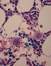
Researchers have identified an enzyme that appears key to prognosis and progression in multiple myeloma (MM).
The group found the enzyme, ST3GAL6, is overexpressed in MM cell lines and patients with disease.
This overexpression increases glycosylation, which escalates the interaction between MM cells and selectins.
And this encourages the circulation and spread of MM cells, as well as their retention in the bone marrow.
The researchers recounted this series of events in Blood.
“[W]e focused on alterations in [glycosylation] because of its role in cell-cell interactions and the spread of cancer cells in the blood,” said study author Michael O’Dwyer, MD, of National University of Ireland, Galway.
Using gene set enrichment analysis, he and his colleagues confirmed the overexpression of glycosylation-related signatures in MM. They also discovered the sialyltransferase ST3GAL6 was “one of the most significantly increased genes” in MM patients, when compared to healthy donors.
The team observed increased ST3GAL6 levels in both relapsed and newly diagnosed MM. And they found that higher ST3GAL6 levels were associated with decreased survival.
To expand upon these findings, the researchers went on to test 5 MM cell lines—MM1S, MM1R, U266, RPMI-8226, and H929. They found significantly higher levels of ST3GAL6 mRNA and protein in the cell lines compared to healthy CD138+ cells.
Knocking down ST3GAL6 in 2 of the cell lines—MM1S and RPMI-8226—significantly reduced the amount of alpha 2,3 sialic acid at the cell surface, which suggests that ST3GAL6 contributes to the synthesis of this glycan.
In addition, knocking down ST3GAL6 significantly reduced MM cells’ adhesion to bone marrow stem cells, human umbilical vein endothelial cells, and fibronectin. It also reduced MM cells’ transendothelial migration, attenuated Src activation in MM cells, and reduced the cells’ ability to roll on p-selectin.
Likewise, in mouse models, knockdown of ST3GAL6 reduced MM cell homing and engraftment. It also significantly decreased tumor burden and increased survival in the mice.
The researchers concluded that these findings highlight the importance of altered glycosylation, particularly sialylation, in MM cell adhesion and migration.
“Our research is crucial because it sheds new light on the biology of [MM], which could lead to new strategies to overcome resistance to treatment,” Dr O’Dwyer said. “Our aim now is to prevent these interactions that cause the spread [of MM cells] using specific enzyme and selectin inhibitors.” ![]()
Credit: Darren Baker
Researchers have identified an enzyme that appears key to prognosis and progression in multiple myeloma (MM).
The group found the enzyme, ST3GAL6, is overexpressed in MM cell lines and patients with disease.
This overexpression increases glycosylation, which escalates the interaction between MM cells and selectins.
And this encourages the circulation and spread of MM cells, as well as their retention in the bone marrow.
The researchers recounted this series of events in Blood.
“[W]e focused on alterations in [glycosylation] because of its role in cell-cell interactions and the spread of cancer cells in the blood,” said study author Michael O’Dwyer, MD, of National University of Ireland, Galway.
Using gene set enrichment analysis, he and his colleagues confirmed the overexpression of glycosylation-related signatures in MM. They also discovered the sialyltransferase ST3GAL6 was “one of the most significantly increased genes” in MM patients, when compared to healthy donors.
The team observed increased ST3GAL6 levels in both relapsed and newly diagnosed MM. And they found that higher ST3GAL6 levels were associated with decreased survival.
To expand upon these findings, the researchers went on to test 5 MM cell lines—MM1S, MM1R, U266, RPMI-8226, and H929. They found significantly higher levels of ST3GAL6 mRNA and protein in the cell lines compared to healthy CD138+ cells.
Knocking down ST3GAL6 in 2 of the cell lines—MM1S and RPMI-8226—significantly reduced the amount of alpha 2,3 sialic acid at the cell surface, which suggests that ST3GAL6 contributes to the synthesis of this glycan.
In addition, knocking down ST3GAL6 significantly reduced MM cells’ adhesion to bone marrow stem cells, human umbilical vein endothelial cells, and fibronectin. It also reduced MM cells’ transendothelial migration, attenuated Src activation in MM cells, and reduced the cells’ ability to roll on p-selectin.
Likewise, in mouse models, knockdown of ST3GAL6 reduced MM cell homing and engraftment. It also significantly decreased tumor burden and increased survival in the mice.
The researchers concluded that these findings highlight the importance of altered glycosylation, particularly sialylation, in MM cell adhesion and migration.
“Our research is crucial because it sheds new light on the biology of [MM], which could lead to new strategies to overcome resistance to treatment,” Dr O’Dwyer said. “Our aim now is to prevent these interactions that cause the spread [of MM cells] using specific enzyme and selectin inhibitors.” ![]()
Study reveals potential targets for MYC-dependent cancers

Credit: Juha Klefstrom
New research suggests the MYC protein drives cell growth by inhibiting a handful of genes involved in DNA packaging and cell death.
The study showed that MYC works through a microRNA to suppress the genes’ expression.
This marks the first time that a subset of MYC-controlled genes have been identified as critical players in the protein’s cancer-causing function, and it points to new therapeutic targets for MYC-dependent cancers.
“This is a different way of thinking about the roles of microRNA and chromatin packaging in cancer,” said Dean Felsher, MD, PhD, of the Stanford University School of Medicine in California.
“We were very surprised to learn that the overexpression of one microRNA can mimic the cancerous effect of MYC.”
Dr Felsher and his colleagues reported this discovery in Cancer Cell.
The team noted that MYC overexpression has been known to prompt an increase in the levels of a microRNA family called miR-17-92.
“People have known for several years that MYC regulates the expression of microRNAs,” Dr Felsher said. “But it wasn’t clear how this was related to MYC’s oncogenic function.”
To gain some insight, Dr Felsher and his colleagues analyzed MYC-dependent cancer cells in vitro and in vivo.
The cells in which miR-17-92 expression was locked in the “on” position kept dividing even when MYC expression was blocked. This suggested that MYC works through the microRNA family to exert its cancer-causing effects.
The researchers then looked for an overlap among genes affected by MYC overexpression and those affected by miR-17-92. There were about 401 genes whose expression was either increased or suppressed by both MYC and miR-17-92.
The team chose to focus on genes that were suppressed because these genes exhibited, on average, many more binding sites for the microRNAs. They further narrowed their panel down to 15 genes regulated by more than one miR-17-92 binding site.
Of these genes, 5 stood out. Four of them—Sin3b, Hbp1, Suv420h1, and Btg1—encode proteins known to regulate chromatin packaging.
These 4 proteins affect cell proliferation and senescence by regulating gene accessibility within the chromatin. They had never before been identified as MYC or miR-17-92 targets.
The fifth gene encodes the apoptotic protein Bim. Previous research suggested that Bim expression is affected by miR-17-92.
All 5 of the proteins are known to affect either cellular proliferation, entry into senescence, or apoptosis, in part by granting or prohibiting access to genes in tightly packaged stretches of DNA in the chromatin.
“MYC is still a general amplifier of gene transcription and expression,” Dr Felsher said. “But our study shows that the maintenance of the cancerous state relies on a more focused mechanism.”
Lastly, the researchers showed that suppressing the expression of the 5 target genes, effectively mimicking MYC overexpression, partially mitigates the effect of MYC deactivation.
Up to 30% of MYC-dependent cancer cells in culture continued to grow—compared to 11% of control cells—in the absence of MYC expression. And tumors in mice either failed to regress or recurred within a few weeks.
“One of the biggest unanswered questions in oncology is how oncogenes cause cancer, and whether you can replace an oncogene with another gene product,” Dr Felsher said.
“These experiments begin to reveal how MYC affects the self-renewal decisions of cells. They may also help us target those aspects of MYC overexpression that contribute to the cancer phenotype.” ![]()
FDA approves bortezomib retreatment in MM
The US Food and Drug Administration (FDA) has approved bortezomib (Velcade) for the retreatment of adults with multiple myeloma (MM) who previously responded to bortezomib and relapsed at least 6 months after that treatment.
Bortezomib’s label has been updated to include dosing guidelines and safety and efficacy findings for single-agent bortezomib and bortezomib in combination with dexamethasone in patients previously treated with bortezomib.
Bortezomib retreatment may be started at the last dose the patient tolerated.
The FDA approved bortezomib retreatment based on results of the phase 2 RETRIEVE study and other supportive data.
The single-arm RETRIEVE trial included 130 MM patients aged 18 years and older who had previously responded to bortezomib-based therapy and relapsed at least 6 months after the treatment. The patients had received a median of 2 prior therapies (range, 1 to 7).
In this study, 94 of the patients received bortezomib in combination with dexamethasone.
One patient achieved complete response to treatment, and 49 achieved partial responses, for an overall response rate of 38.5%. The median duration of response was 6.5 months (range, 0.6 to 19.3 months).
The safety profile with bortezomib retreatment was consistent with the known safety profile of intravenous bortezomib in relapsed MM. Researchers did not observer cumulative toxicities upon retreatment.
The most common adverse event was thrombocytopenia, which occurred in 52% of patients. The incidence of grade 3 or higher thrombocytopenia was 24%.
Peripheral neuropathy was also common, occurring in 28% of patients. Grade 3 or higher peripheral neuropathy occurred in 6% of patients.
The rate of serious adverse events was 12.3%. The most commonly reported serious adverse events were thrombocytopenia (3.8%), diarrhea (2.3%), herpes zoster (1.5%), and pneumonia (1.5%). Adverse events leading to discontinuation occurred in 13% of patients.
Bortezomib is co-developed by Millennium/Takeda and Janssen Pharmaceutical Companies. Millennium is responsible for commercialization of bortezomib in the US. Janssen Pharmaceutical Companies are responsible for commercialization in Europe and the rest of the world.
Takeda Pharmaceutical Company Limited and Janssen Pharmaceutical K.K. co-promote bortezomib in Japan. Bortezomib is approved in more than 90 countries.
Method could speed up cancer diagnosis

Credit: NIGMS
A new technique could enable faster diagnosis of cancer and various prenatal conditions, according to a paper published in Proceedings of the National Academy of Sciences.
The method, known as convex lens-induced confinement (CLIC), allows researchers to load long strands of DNA into a tunable, nanoscale imaging chamber in ways that maintain their structural identity and under conditions that are similar to those in the human body.
CLIC lets researchers map large genomes rapidly and identify specific gene sequences from single cells with single-molecule resolution, a process that is critical to diagnosing diseases like cancer.
“Current practices of genomic analysis typically require tens of thousands of cells worth of genomic material to obtain the information we need, but this new approach works with single cells,” said study author Rob Sladek, MD, of McGill University in Montreal, Canada.
“CLIC will allow researchers to avoid having to spend time stitching together maps of entire genomes, as we do under current techniques, and promises to make genomic analysis a much simpler and more efficient process.”
The CLIC imaging chamber can sit on top of a standard inverted fluorescence microscope, and strands of DNA can be loaded into the chamber from above, which allows the strands to maintain their integrity.
Existing tools used for genomic analysis rely on side-loading DNA under pressure into nanochannels in the imaging chamber. This breaks the DNA molecules into small pieces, making it a challenge to reconstruct the genome.
CLIC, on the other hand, is “like squeezing many soft spaghetti noodles into long, narrow tubes without breaking them,” according to study author Sabrina Leslie, PhD, also of McGill University.
“Once these long strands of DNA are gently squeezed down into nanochannels from a nanoscale bath above, they become effectively rigid, which means that we can map positions along uniformly stretched strands of DNA, while holding them still,” she said.
“This means diagnostics can be performed quickly, one cell at a time, which is critical for diagnosing many prenatal conditions and the onset of cancer.” ![]()
Interim data appear positive for MM drug
Interim results of the phase 3 ASPIRE trial suggest carfilzomib can improve progression-free survival (PFS) in patients with relapsed multiple myeloma (MM).
Patients who received carfilzomib, lenalidomide, and dexamethasone lived 8.7 months longer without progression than patients who received only lenalidomide and dexamethasone.
The companies developing carfilzomib, Amgen and its subsidiary, Onyx Pharmaceuticals, Inc., recently shared these results.
They said additional results will be submitted for presentation at the 56th Annual ASH Meeting in December.
The companies also said data from the ASPIRE trial will form the basis for regulatory submissions for carfilzomib throughout the world.
In the US, the data may support the conversion of accelerated approval to full approval and expand the current indication for carfilzomib.
The ASPIRE trial includes 792 patients with relapsed MM who were randomized to treatment at sites in North America, Europe, and Israel. Prior to study treatment, the patients had received 1 to 3 therapeutic regimens.
The patients were randomized to receive carfilzomib (20 mg/m2 on days 1 and 2 of cycle 1 only, then 27 mg/m2), in addition to a standard dosing schedule of lenalidomide (25 mg per day for 21 days on, 7 days off) and low-dose dexamethasone (40 mg per week in 4-week cycles), or lenalidomide and low-dose dexamethasone alone.
The primary endpoint was PFS, and secondary endpoints included overall survival, overall response rate, duration of response, disease control rate, health-related quality of life, and safety.
At a planned interim analysis, patients in the carfilzomib arm had a significantly longer median PFS than patients in the other arm—26.3 months and 17.6 months, respectively (P<0.0001).
The data for overall survival are not yet mature, but the analysis showed a trend in favor of carfilzomib that did not reach statistical significance, according to Amgen and Onyx.
The companies said the safety profile in this study is consistent with previous studies, including the rate of cardiac events.
Treatment discontinuation due to adverse events and on-study deaths were comparable between the 2 arms, and researchers did not identify any new safety signals. ![]()
Drug could prevent thrombocytopenia in MM
in the bone marrow
Researchers say they’ve identified a previously unknown but crucial component of the platelet production process.
And this discovery could help spare multiple myeloma (MM) patients from thrombocytopenia induced by the proteasome inhibitor bortezomib.
The researchers found that proteasome inhibition blocked platelet production in vitro and in vivo.
But fasudil, a Rho kinase inhibitor that is approved for use outside the US, restored platelet counts.
The researchers believe these findings, published in The Journal of Clinical Investigation, could translate to MM patients.
“A low platelet count is a big issue for people who receive bortezomib for this cancer,” said study author Andrew S. Weyrich, PhD, of the University of Utah in Salt Lake City.
“When platelet levels drop too low, it can mean interrupting treatment to allow the platelet count to recover. Fasudil potentially could help keep platelet counts normal while multiple myeloma patients receive bortezomib.”
Dr Weyrich and his colleagues found that bortezomib-induced proteasome inhibition prevented the production of proplatelets in both human and mouse megakaryocytes.
Megakaryocytes isolated from mice lacking PSMC1, an essential subunit of the 26S proteasome, also failed to produce proplatelets.
Further study revealed that the megakaryocytes’ inability to generate platelets was caused by the hyperactivation of RhoA, a protein that helps megakaryocytes maintain the proper shape to produce platelets.
When the researchers inhibited RhoA or its downstream target, Rho-associated protein kinase, in vitro, they were able to restore megakaryocyte proplatelet formation in the setting of proteasome inhibition.
Likewise, the Rho kinase inhibitor fasudil restored platelet counts in adult mice that had thrombocytopenia induced by proteasome inhibition.
Fasudil is approved in Japan and elsewhere to treat cerebral vasospasms, or constricted arteries that arise as a complication of brain aneurysms.
The drug is under investigation in US clinical trials for treating high blood pressure, diabetic macular edema, and other health issues.
There are no trials investigating fasudil’s effects on thrombocytopenia, but Dr Weyrich and his colleagues hope their study might change that. And if clinical trials produce favorable results, fasudil might be made available for MM patients much faster than a new drug.
“If the Food and Drug Administration did approve fasudil for use by multiple myeloma patients, it could, in principle, be moved to the clinic relatively fast in the United States,” Dr Weyrich said. ![]()
Biosimilar can treat chemo-induced anemia

Credit: Rhoda Baer
A biosimilar of the erythropoiesis-stimulating agent epoetin alfa can elicit responses in patients with chemotherapy-induced anemia, according to a study published in BMC Cancer.
The agent, epoetin zeta (Retacrit), produced a hemoglobin (Hb) response in more than 80% of patients at 3- and 6-month time points.
Response rates were similar in patients with hematologic malignancies and those with solid tumors.
And the rate of clinically significant adverse events was low. This included thromboembolic events, bleeding, infection, local intolerability, and increased blood pressure.
Mauricette Michallet, MD, PhD, of Centre Hospitalier Lyon in France, and her colleagues conducted this study, known as ORHEO. It was sponsored by Hospira, the makers of epoetin zeta.
The researchers evaluated 2310 adult patients with chemotherapy-induced anemia (Hb<11 g/dL). Patients had solid tumors (n=1838), lymphomas (n=301), or multiple myeloma (n=171).
Patients were taking a number of treatments aside from epoetin zeta and chemotherapy. This included intravenous iron (10%), oral iron (16%), antithrombotic agents (12%), folates (7%), vitamin B (4%), and other vitamins (2%). An additional 17% of patients were reported as being on “other treatments.”
In all, 99.9% of patients received epoetin zeta. The primary endpoint was the rate of response.
Response was defined as an increase in Hb levels to at least 10 g/dL since enrollment, an increase in Hb levels of at least 1 g/dL since enrollment, or reaching target Hb levels set at the start of study, without any blood transfusions in the 3 weeks prior to measurement. In patients with baseline Hb levels of at least 10 g/dL, only those who reached their Hb target or had an increase greater than 1 g/dL were considered responders.
Eighty-two percent of patients achieved a response at 3 months, and 87% had a response at 6 months. The overall mean change in Hb level was 1.52 ± 1.61 at 3 months and 1.72 ± 1.61 g/dL at 6 months. The rate of transfusion was 9% at 3 months and 6% at 6 months.
Between enrollment and month 6, 1202 patients discontinued epoetin zeta. Forty percent stopped because Hb levels were met, 27% stopped because they were stopping or changing chemotherapy, 15% stopped for both of the aforementioned reasons, 11% stopped because epoetin zeta was ineffective, and 2% stopped due to adverse events.
Seventeen percent of patients experienced an adverse event, including thromboembolic events (4%), infection (5%), bleeding (2%), local intolerability (0.2%), increased blood pressure (2%), and “other” events (9%).
Epoetin zeta was approved in Europe in 2007. For epoetin biosimilars to gain approval in the European Union, companies must agree to conduct post-marketing studies. ![]()
Fasting can have beneficial effects in cancer setting

in the bone marrow
New research indicates that cycles of prolonged fasting may prevent chemotherapy-induced immunosuppressive toxicity and induce regeneration of the hematopoietic system.
Long periods of fasting reduced damage in bone marrow stem and progenitor cells and protected both mice and humans from chemotoxicity.
In mice, the fasting cycles “flipped a regenerative switch,” changing the signaling pathways for hematopoietic stem cells (HSCs).
Researchers reported these results in Cell Stem Cell.
“We could not predict that prolonged fasting would have such a remarkable effect in promoting stem cell-based regeneration of the hematopoietic system,” said study author Valter Longo, PhD, of the University of Southern California in Los Angeles.
“When you starve, the system tries to save energy, and one of the things it can do to save energy is to recycle a lot of the immune cells that are not needed, especially those that may be damaged. What we started noticing in both our human work and animal work is that the white blood cell count goes down with prolonged fasting. Then, when you re-feed, the blood cells come back. So we started thinking, well, where does it come from?”
The researchers found that prolonged fasting reduced the enzyme PKA, which regulates HSC self-renewal and pluripotency.
“PKA is the key gene that needs to shut down in order for these stem cells to switch into regenerative mode,” Dr Longo said. “It gives the ‘okay’ for stem cells to go ahead and begin proliferating and rebuild the entire system.”
Prolonged fasting also lowered levels of IGF-1, a growth-factor hormone that has been linked to aging, tumor progression, and cancer risk.
In addition to downregulating the IGF-1/PKA pathway in HSCs, prolonged fasting protected hematopoietic cells from chemotoxicity and promoted HSC self-renewal to reverse immunosuppression.
Experiments revealed that inhibiting IGF-1 or PKA signaling mimicked the effects of prolonged fasting.
The researchers also analyzed a small group of patients from a pilot study evaluating the effects of fasting before chemotherapy. Fasting for 72 hours, but not 24 hours, ensured that patients had normal lymphocyte counts and maintained a normal lineage balance in white blood cells after chemotherapy.
Dr Longo’s lab is now conducting further research on controlled dietary interventions and stem cell regeneration in both animal and clinical studies. ![]()
in the bone marrow
New research indicates that cycles of prolonged fasting may prevent chemotherapy-induced immunosuppressive toxicity and induce regeneration of the hematopoietic system.
Long periods of fasting reduced damage in bone marrow stem and progenitor cells and protected both mice and humans from chemotoxicity.
In mice, the fasting cycles “flipped a regenerative switch,” changing the signaling pathways for hematopoietic stem cells (HSCs).
Researchers reported these results in Cell Stem Cell.
“We could not predict that prolonged fasting would have such a remarkable effect in promoting stem cell-based regeneration of the hematopoietic system,” said study author Valter Longo, PhD, of the University of Southern California in Los Angeles.
“When you starve, the system tries to save energy, and one of the things it can do to save energy is to recycle a lot of the immune cells that are not needed, especially those that may be damaged. What we started noticing in both our human work and animal work is that the white blood cell count goes down with prolonged fasting. Then, when you re-feed, the blood cells come back. So we started thinking, well, where does it come from?”
The researchers found that prolonged fasting reduced the enzyme PKA, which regulates HSC self-renewal and pluripotency.
“PKA is the key gene that needs to shut down in order for these stem cells to switch into regenerative mode,” Dr Longo said. “It gives the ‘okay’ for stem cells to go ahead and begin proliferating and rebuild the entire system.”
Prolonged fasting also lowered levels of IGF-1, a growth-factor hormone that has been linked to aging, tumor progression, and cancer risk.
In addition to downregulating the IGF-1/PKA pathway in HSCs, prolonged fasting protected hematopoietic cells from chemotoxicity and promoted HSC self-renewal to reverse immunosuppression.
Experiments revealed that inhibiting IGF-1 or PKA signaling mimicked the effects of prolonged fasting.
The researchers also analyzed a small group of patients from a pilot study evaluating the effects of fasting before chemotherapy. Fasting for 72 hours, but not 24 hours, ensured that patients had normal lymphocyte counts and maintained a normal lineage balance in white blood cells after chemotherapy.
Dr Longo’s lab is now conducting further research on controlled dietary interventions and stem cell regeneration in both animal and clinical studies.
Protein map may point to new cancer treatments

the endoplasmic reticulum
in green, mitochondria in red,
and chromosomes in blue
Credit: Wellcome Images
Researchers say they’ve uncovered the structure of one of the most important protein complexes involved in cell division, and their finding has implications for cancer treatment.
The team mapped the anaphase-promoting complex (APC/C), which performs a wide range of tasks associated with mitosis.
The researchers believe that seeing APC/C in unprecedented detail could transform our understanding of how cells divide and reveal binding sites for future cancer drugs.
“It’s very rewarding to finally tie down the detailed structure of this important protein, which is both one of the most important and most complicated found in all of nature,” said David Barford, DPhil, of the Medical Research Council Laboratory of Molecular Biology in Cambridge, UK.
“We hope our discovery will open up whole new avenues of research that increase our understanding of the process of mitosis and ultimately lead to the discovery of new cancer drugs.”
Dr Barford and his colleagues detailed their discovery in Nature.
The researchers reconstituted human APC/C and used a combination of electron microscopy and imaging software to visualize it at a resolution of less than a billionth of a meter.
The resolution was so fine that it allowed the team to see the secondary structure—the set of basic building blocks that combine to form every protein. Alpha-helix rods and folded beta-sheet constructions were clearly visible within the 20 subunits of the APC/C, defining the overall architecture of the complex.
Previous studies led by the same team had shown a globular structure for APC/C in much lower resolution, but the secondary structure had not been mapped.
Each of the APC/C’s subunits bond and mesh with other units at different points in the cell cycle, allowing it to control a range of mitotic processes, including the initiation of DNA replication, the segregation of chromosomes along spindles, and cytokinesis. Disrupting each of these processes could selectively kill cancer cells or prevent them from dividing.
“The fantastic insights into molecular structure provided by this study are a vivid illustration of the critical role played by fundamental cell biology in cancer research,” said Paul Workman, PhD, of The Institute of Cancer Research, London.
“The new study is a major step forward in our understanding of cell division. When this process goes awry, it is a critical difference that separates cancer cells from their healthy counterparts. Understanding exactly how cancer cells divide inappropriately is crucial to the discovery of innovative cancer treatments to improve outcomes for cancer patients.”
Nanoparticles could improve cancer diagnosis

Self-assembling nanoparticles may help physicians diagnose cancers earlier, according to a study published in Angewandte Chemie.
The nanoparticles boost the effectiveness of magnetic resonance imaging (MRI) by specifically seeking out CXCR4 receptors, which are found in cancerous cells.
The iron oxide nanoparticles are coated with peptide ligands that target tumor sites. When the particles find a tumor, they begin to interact with the cancerous cells.
Cancer-specific matrix metalloproteinase biomarkers prompt the nanoparticles to self-assemble into larger particles. And these larger particles are more visible on an MRI scan.
Researchers used cancer cells and mouse models to compare the effects of the self-assembling nanoparticles in MRI scanning against commonly used imaging agents. The nanoparticles produced a more powerful signal and created a clearer image of the tumor.
The team said the nanoparticles increase the sensitivity of MRI scans and could ultimately improve physicians’ ability to detect cancerous cells at much earlier stages of development.
“By improving the sensitivity of an MRI examination, our aim is to help doctors spot something that might be cancerous much more quickly,” said study author Nicholas Long, PhD, of Imperial College London in the UK. “This would enable patients to receive effective treatment sooner, which would hopefully improve survival rates from cancer.”
In addition to improving the sensitivity of MRI scans, the nanoparticles also appear to be safe. Before testing and injecting the particles into mice, the researchers had to ensure the particles would not become so big as to cause damage.
The team injected the particles into a saline solution inside a petri dish and monitored their growth over a 4-hour period. The nanoparticles grew from 100 nm to 800 nm, which was still small enough not to cause any harm.
Now, the researchers are working to enhance the effectiveness of the nanoparticles. And they hope to test their design in a human trial within the next 3 to 5 years.
“We would like to improve the design to make it even easier for doctors to spot a tumor and for surgeons to then operate on it,” Dr Long said. “We’re now trying to add an extra optical signal so that the nanoparticle would light up with a luminescent probe once it had found its target. So, combined with the better MRI signal, it will make it even easier to identify tumors.”