User login
Aggressiveness of CLL linked to genetic variability

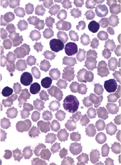
The genetic variability of chronic lymphocytic leukemia (CLL) appears to predict its aggressiveness, according to a study published in Genome Medicine.
Investigators found evidence suggesting that greater variability in gene expression is associated with more aggressive disease.
The team analyzed gene expression in two cohorts of CLL patients—those with IgVH mutations and a good prognosis and those with unmutated CLL who have more aggressive disease.
The researchers examined 70 mutated and 52 unmutated CLL samples, as well as 20 control samples taken from healthy individuals.
Unmutated, aggressive CLL showed increased gene expression variability across individuals, whereas gene expression variability was lower in less aggressive, mutated CLL.
The investigators validated these observations by comparing them against a second sample group consisting of 24 mutated and 36 unmutated CLL samples.
The results suggested that CLL aggressiveness is specifically determined by a set of 500 genes showing increased expression variability across individuals. The genes are involved in processes such as adaptation to the environment, cell death, tumor growth, and drug resistance.
“Our conclusion is that the coefficient of variation for gene expression in CLL efficiently predicts its aggressiveness,” said study author Alfonso Valencia, PhD, of Centro Nacional de Investigaciones Oncologicas (CNIO) in Madrid, Spain.
“More importantly, if further research confirms these findings, a classifier based on the measurement of gene expression variability could be created to predict the disease subtype of CLL patients.”
The researchers said their next step is to discover the mechanisms responsible for the high levels of expression variability for a given gene across individuals.
Understanding the mechanisms underlying this phenomenon is of crucial relevance for oncology research, the investigators said, as it is linked to tumor heterogeneity, a key feature of cancer progression and drug resistance.
The greater the genetic variability in a tumor, the better it will adapt to its environment, and the more probabilities for this tumor to spread, develop resistance to cancer therapies, and metastasize to distant organs. ![]()
Risk of HBV reactivation ‘underappreciated’

Credit: CDC
Reactivation of the hepatitis B virus (HBV) may be more of a risk than we anticipated, investigators have reported in Hepatology.
Their research indicates that HBV reactivation is associated with the use of chemotherapy, high-dose corticosteroids, biologics targeting tumor necrosis factor-alpha (TNF-α), and agents that aren’t really considered immunosuppressive.
HBV reactivation is also fairly common after organ transplant and hematopoietic stem cell transplant (HSCT).
As reactivation of HBV can be fatal, the study authors suggest routine screening of HBV in all patients prior to the start of these treatments.
The researchers noted that, in September 2013, the US Food and Drug Administration (FDA) issued a Drug Safety Communication in an attempt to decrease the risk of HBV reactivation. The communication advised healthcare professionals to screen patients for HBV prior to the administration of ofatumumab or rituximab.
“[T]his may be just the tip of the iceberg,” said Adrian Di Bisceglie, MD, of Saint Louis University School of Medicine in Missouri.
“Our research suggests that the issue of HBV reactivation may be an underappreciated clinical challenge that extends well beyond the use of just two anti-CD20 medications.”
After a systematic literature review, Dr Di Bisceglie and his colleagues identified 504 studies pertaining to reactivation of HBV.
The investigators reviewed 14 studies in which the antiviral agent lamivudine was used to prevent HBV reactivation in HBsAg-positive patients receiving chemotherapy. Among patients who did not receive lamivudine, HBV reactivation occurred in 32%. Thirteen percent of patients experienced liver failure, and 7% died.
The researchers also looked at patients undergoing HSCT. In one study, 61 patients had resolved HBV infection before HSCT. But 12 of these patients (20%) developed reverse seroconversion (reappearance of HBsAg in a person who was HBsAg-negative, anti-HBc-positive prior to HSCT).
The cumulative probability of reverse seroconversion was 9% a year after HSCT, 21.7% at 2 years, and 42.9% at 4 years.
The investigators also noted that high-dose corticosteroids carry a significant risk of HBV reactivation, both as part of combination treatment for malignancies and when used alone to treat benign conditions.
In addition, the researchers found data showing that HBV reactivation has occurred with antitumor agents that are not thought to be particularly immunosuppressive, such as imatinib and thalidomide. The team said this raises questions about the mechanisms by which drugs are causing HBV reactivation.
The investigators also looked at data from 257 patients with active or recovered HBV infection who received treatment with biological therapies targeting TNF-α.
Forty-two percent of the patients had elevations in serum aminotransferase levels, 39% had reappearance of HBV DNA, 16% had signs and symptoms of liver disease, and 5% died of liver failure.
HBV reactivation was more frequent among patients receiving infliximab than etanercept. It was 7-fold higher among patients who were HBsAg-positive (38%) than those who were HBsAg-negative but anti-HBc-positive (5%).
While it remains unclear how HBV reactivation occurs, experts believe a loss of immune control over viral replication may trigger the process.
“Further study and cooperation between various medical disciplines will help broaden understanding of HBV reactivation,” Dr Di Bisceglie concluded. ![]()
Vaccine gets orphan designation for ATLL
The European Medicines Agency (EMA) has granted orphan drug designation for a therapeutic vaccine candidate known as THV02 to treat adult T-cell leukemia/lymphoma (ATLL).
THV02 is an experimental treatment composed of 2 lentiviral vectors to be used in a prime/boost regimen in ATLL patients infected by the HTLV-1 virus.
Both investigational drugs encode the same antigens, derived from 4 proteins of the HTLV-1 virus.
THV02 is intended to induce an immune response against HTLV antigens born by ATLL with the aim of enabling the patients’ immune system to fight leukemic cells.
Preclinical evaluation has suggested that THV02 is safe, and the vaccine has presented an “unprecedented” immunogenicity profile in several models, according to THERAVECTYS, the company developing THV02.
“Preclinical immunogenicity results obtained to date are very promising, and we are really excited by the prospect of bringing a safe and better-tolerated alternative to patients who are desperately in need of a treatment,” said Déborah Revaud, the senior scientist in charge of developing THV02.
The EMA grants orphan designation to drugs in development intended for the treatment, prevention, or diagnosis of life-threatening or chronically debilitating diseases occurring in fewer than 5 in 10,000 people.
The designation allows sponsors to benefit from an accelerated development process, financial incentives, and a 10-year period of market exclusivity once the drug is on the market.
“We are extremely pleased that the European Medicines Agency has granted an orphan drug status to our vaccine candidate against ATLL,” said Emmanuelle Sabbah-Petrover, PhD, head of regulatory affairs at THERAVECTYS.
“We expect to recruit our first patients towards the end of Q3 2015 in Europe and advance further developments in the US and in Japan in 2016.”
Should THV02 demonstrate a convincing safety and efficacy profile during its development against ATLL, THERAVECTYS said it will consider developing the vaccine for HTLV-related infections as treatment and possibly as prophylaxis. ![]()
MicroRNA may be therapeutic target for ALK- ALCL
SAN FRANCISCO—MicroRNA-155 (miR-155) can act as an oncogenic driver in ALK− anaplastic large-cell lymphoma (ALCL) and may therefore be a therapeutic target for the disease, according to a presentation at the 7th Annual T-cell Lymphoma Forum.
Analyzing patient samples and cell lines, researchers discovered that miR-155 is highly expressed in ALK− ALCL but is nearly absent in ALK+ ALCL.
They also found evidence suggesting that miR-155 drives tumor growth in mouse models of ALK− ALCL.
Philipp Staber, MD, PhD, of the Medical University of Vienna in Austria, presented these findings at the meeting.
Dr Staber and his colleagues previously found (Merkel et al, PNAS 2010) that miR-155 was highly expressed in ALK− ALCL. So they decided to take a closer look at the phenomenon.
They assessed miR-155 expression in samples from patients with ALK+ or ALK− ALCL, as well as 6 ALCL cell lines.
miR-155 expression was significantly higher in the ALK− patient samples than in the ALK+ samples (P<0.001). And it was significantly higher (P<0.001) in the ALK− cell lines (DL-40, Mac1, and Mac2a) than in the ALK+ cell lines (K299, SR789, and SUP-M2).
Dr Staber and his colleagues then overexpressed miR-155 in ALK+ ALCL cell lines (K299 and SR789). And they observed a decrease in known target genes of miR-155—C/EBPβ, SOCS1, and SHIP1.
The researchers also observed an inverse correlation between miR-155 host gene promoter methylation and miR-155 expression in an ALCL+ cell line, which suggested that ALK activity has no direct effect on miR-155 levels.
The team treated the K299 cell line with the ALK inhibitor crizotinib at 100 nM, 200 nM, and 400 nM doses and found that miR-155 did not increase at any dose. Dr Staber noted, however, that the researchers were only able to treat cells for a maximum of 24 hours.
The group then discovered that anti-miR-155 mimics could reduce tumor growth in mouse models of ALK- ALCL. Mice were injected with Mac1 or Mac2a cells transfected with anti-miR-155, control RNA, or pre-miR-155 oligos.
In both Mac1 and Mac2 models, tumors were substantially smaller in the anti-mir-155 mice than in the pre-miR-155 mice (P=0.038 and P=0.006, respectively). But tumor growth was not significantly reduced compared to controls.
“Immunohistochemistry in these tumors shows quite a clear picture,” Dr Staber said. “The C/EBPβ target gene is overexpressed when using anti-miR-155, and [expression is decreased] with overexpression of miR-155. And the same is true for SOCS1. STAT3 signaling is increased through overexpression of miR-155.”
The researchers observed the same effect in ALK− ALCL patient samples.
Using ALK+ cell lines (K299 and SR789), the team went on to show that miR-155 suppresses IL-8 expression and induces IL-22 expression, a finding they verified in mice.
“IL-8 is a direct target of C/EBPβ, and C/EBPβ, as shown before, is a target of miR-155, so this makes sense,” Dr Staber said. “On the other hand, IL-22 is a strong inducer of STAT3 signaling, which is strongly induced when we increase miR-155 expression.”
Dr Staber and his colleagues concluded that these findings, when taken together, suggest that miR-155 could be a therapeutic target for ALK− ALCL. ![]()
Combo shows early promise for T-cell lymphomas

SAN FRANCISCO—Preclinical and early phase 1 results suggest the aurora A kinase inhibitor alisertib and the histone deacetylase (HDAC) inhibitor romidepsin have synergistic activity against T-cell lymphomas.
In the preclinical study, the drugs showed synergy in cutaneous T-cell lymphoma (CTCL) cell lines and a benefit over monotherapy in vivo.
In the phase 1 study, romidepsin and alisertib produced clinical benefits in patients with peripheral T-cell lymphoma (PTCL).
Unfortunately, there are currently no good markers for predicting which patients might benefit from this type of combination, potentially because the drugs have multivariate mechanisms of action, said Michelle Fanale, MD, of the University of Texas MD Anderson Cancer Center in Houston.
She presented data on romidepsin and alisertib in combination at the 7th Annual T-cell Lymphoma Forum.
Dr Fanale said she was inspired to test the combination (in a phase 1 trial) after researchers reported promising results with the aurora kinase inhibitors MK-0457 and MK-5108 in combination with the HDAC inhibitor vorinostat (Kretzner et al, Cancer Research 2011).
She noted that aurora kinase inhibitors work mainly through actions at the G2-M transition point, while HDAC inhibitors induce G1-S transition. HDAC inhibitors can also degrade aurora A and B kinases, and the drugs modify kinetochore assembly through hyperacetylation of pericentromeric histones.
“When you actually treat with an HDAC inhibitor by itself, you’re basically getting an increase of this sub-G1 population,” Dr Fanale said. “When you treat with your aurora kinase inhibitor by itself, you’re clearly getting an increase of cells that are arresting at G2/M.”
“When you treat with the combination, you’re actually getting a further increase in the sub-G1, denoting dead cells, and then you’re further getting some increase of cells spreading out now through the G2/M portion as well.”
Preclinical research
Dr Fanale presented preclinical results showing that alisertib is highly synergistic with romidepsin in T-cell, but not B-cell, lymphoma. She was not involved in the research, which was also presented at the recent ASH Annual Meeting (Zullo et al, ASH 2014, abst 4493).
The researchers administered romidepsin at IC10-20 concentrations, with increasing concentrations of alisertib, and incubated cells for 72 hours. A synergy coefficient less than 1 denoted synergy.
The combination demonstrated synergy in the HH (CTCL) cell line when alisertib was given at 100 nM or 1000 nM (0.68 and 0.40, respectively) but not at 50 nM (1.05).
Likewise, the combination demonstrated synergy in the H9 (CTCL) cell line when alisertib was given at 100 nM or 1000 nM (0.66 and 0.46, respectively) but not at 50 nM (1.1).
Romidepsin was shown to cause a mild increase in the percent of cells in G1 compared with alisertib, which significantly increased the percent of cells in G2/M arrest. And live cell imaging showed marked cytokinesis failure following treatment.
“When looking at further markers for apoptosis, when giving the combination, there’s further increase in caspase 3 and PARP cleavage, as well as other pro-apoptotic proteins, including PUMA, and a decrease in the anti-apoptotic protein Bcl-xL,” Dr Fanale noted.
She also pointed out that, in an in vivo xenograft model, alisertib and romidepsin produced significantly better results than those observed with monotherapy or in controls.
Phase 1 trial
The phase 1 trial of romidepsin and alisertib in combination included patients with aggressive B- and T-cell lymphomas (NCT01897012; Fanale et al, ASH 2014, abst 1744).
Twelve patients have been enrolled to date. Ninety-two percent of patients had primary refractory disease, they had a median of 3.5 prior lines of therapy (range, 1 to 7), and none of the patients had received a stem cell transplant.
The patients received treatment as follows:
- Alisertib at 20 mg orally twice daily on days 1-7 and romidepsin at 6 mg/m2 IV on days 1 and 8
- Alisertib at 20 mg orally twice daily on days 1-7 and romidepsin at 8 mg/m2 IV on days 1 and 8
- Alisertib at 20 mg orally twice daily on days 1-7 and romidepsin at 10 mg/m2 IV on days 1 and 8
- Alisertib at 40 mg orally twice daily on days 1-7 and romidepsin at 10 mg/m2 IV on days 1 and 8
- Alisertib at 40 mg orally twice daily on days 1-7 and romidepsin at 12 mg/m2 IV on days 1 and 8
- Alisertib at 40 mg orally twice daily on days 1-7 and romidepsin at 14 mg/m2 IV on days 1 and 8.
The maximum-tolerated dose has not yet been reached. The main side effect was reversible myelosuppression. In the 24 cycles administered, patients experienced grade 3/4 neutropenia (62.5%), anemia (29%), and thrombocytopenia (48%).
Dr Fanale noted that 3 of the 4 patients with T-cell lymphomas had some level of clinical benefit after therapy.
One patient, a heavily pretreated patient with PTCL who was treated at the lowest dose, had a complete response lasting 10 months. The patient had received 7 prior lines of therapy, including romidepsin alone.
Two other patients had stable disease, one with PTCL and one with an overlap diagnosis of B-cell and T-cell lymphoma. The PTCL patient went on to receive a matched, unrelated-donor transplant and is doing well, Dr Fanale said.
“We’ve taken a pause from this clinical trial,” she added. “We plan to reopen it toward T-cell lymphoma patients, potentially exclusively, . . . and also potentially to change a bit of the dosing schema with both romidepsin and alisertib.” ![]()
A better HDAC inhibitor for PTCL?
SAN FRANCISCO—The histone deacetylase (HDAC) inhibitor chidamide is effective and well-tolerated in patients with relapsed or refractory peripheral T-cell lymphoma (PTCL), results of the phase 2 CHIPEL trial suggest.
The study showed that chidamide can elicit higher response rates than those previously observed with the folate analog metabolic inhibitor pralatrexate and the HDAC inhibitor romidepsin.
Chidamide also prolonged survival and posed a lower risk of toxicity compared to pralatrexate and romidepsin.
Yuankai Shi, MD, PhD, of the Chinese Academy of Medical Science & Peking Union Medical College in Beijing, China, presented these results at the 7th Annual T-cell Lymphoma Forum. The trial is sponsored by Chipscreen Biosciences Ltd.
He noted that the CHIPEL study was split into two parts: an exploratory trial and a pivotal trial.
Exploratory trial
The exploratory trial included 19 patients who had a median age of 51 and were a median of 1.41 years from diagnosis. Seventeen patients had PTCL unspecified (PTCL-U), and 2 had other subtypes of PTCL.
The patients received chidamide at 30 mg (n=9) or 50 mg (n=10) twice a week for 2 weeks of a 3-week cycle. The overall response rate was 26% (n=5), and 16% of patients (n=3) had a complete response (CR) or unconfirmed CR (CRu).
Pivotal trial
The pivotal trial included 83 patients who had a median age of 53 and were a median of 1.06 years from diagnosis.
The patients had PTCL-U (29.1%, n=23), nasal NK/T-cell lymphoma (20.3%, n=16), anaplastic large-cell lymphoma (ALCL, 20.3%, n=16), angioimmunoblastic T-cell lymphoma (AITL, 11.4%, n=9), enteropathy-type T-cell lymphoma (2.5%, n=2), CD4-positive PTCL (1.3%, n=1), Lennert-type PTCL (1.3%, n=1), transformed mycosis fungoides (1.3%, n=1), and other subtypes of PTCL (12.7%, n=10).
These patients received chidamide at 30 mg twice a week without a drug-free holiday.
The overall response rate was 29% (n=23) according to investigators and 28% (n=22) according to an independent review committee. Fourteen percent of patients (n=11) had a CR/CRu, according to both sources.
The median progression-free survival was 2.1 months for all patients, 14 months for patients who achieved CR/CRus, 7.7 months in patients with partial responses, and 2.5 months in patients with stable disease.
The median overall survival was 21.4 months in all patients and not reached in patients who had a CR/CRu, partial response, or stable disease.
Overall safety
Of all 102 patients (in both the pivotal and exploratory analyses), 80% had at least one adverse event (AE), 37% had grade 3 or higher AEs, 17% had AEs leading to treatment discontinuation, and 8% had severe AEs.
The most common AEs were thrombocytopenia (50%), leukopenia (37%), neutropenia (19%), fatigue (11%), and fever (11%).
Twenty-four percent of patients had grade 3 or higher thrombocytopenia, 13% had grade 3 or higher leukopenia, and 10% had grade 3 or higher neutropenia.
Chidamide vs other agents
Dr Shi compared response rates with chidamide in the pivotal trial to those previously observed with pralatrexate (O’Connor et al, JCO 2011) and romidepsin (Coiffier et al, J Hematol Oncol 2014).
In patients with PTCL-U, response rates were similar for all 3 drugs (≈30%). In ALCL, the response rate with chidamide was higher (≈50%) than with pralatrexate (≈30%) or romidepsin (≈25%). And in AITL, the response rate with chidamide was higher (≈45%) than with pralatrexate (≈8%) or romidepsin (≈30%).
Chidamide also conferred better overall survival than pralatrexate, romidepsin, and other treatments from previous studies.
The median overall survival was 6.5 months with chemotherapy (Mak et al, JCO 2013), 14.5 months with pralatrexate (O’Connor et al, JCO 2011), 11.3 months with romidepsin (Coiffier et al, J Hematol Oncol 2014), 7.9 months with belinostat (Hyon-Zu Lee, Clinical Review 2009), and 21.4 months with chidamide.
Finally, Dr Shi compared the rates of AEs observed in the chidamide pivotal trial to AEs observed in the pralatrexate and romidepsin trials.
At least one AE occurred in 100% of patients treated with pralatrexate, 96% of patients on romidepsin, and 82% of patients on chidamide. Grade 3 or higher AEs occurred in 74%, 66%, and 39% of patients, respectively. And at least one severe AE occurred in 44%, 46%, and 8% of patients, respectively.
The favorable results observed with chidamide support the Chinese Food and Drug Administration’s December decision to approve the drug (under the brand name Epidaza) for use in patients with PTCL. ![]()
Outcomes still dismal in PTCL, project shows
SAN FRANCISCO—Outcomes remain dismal for the majority of patients with peripheral T-cell lymphoma (PTCL), according to a speaker at the 7th Annual T-cell Lymphoma Forum.
Massimo Federico, MD, of the Università di Modena e Reggio Emilia in Italy, presented an analysis of the first 1000 patients enrolled in the prospective T-Cell Project.
The data showed no improvements in survival for these patients compared to patients included in the retrospective International Peripheral T-Cell Lymphoma
Project.
The International Peripheral T-Cell Lymphoma Project included PTCL patients treated at various institutions between 1990 and 2002.
The T-Cell Project was designed to complement this retrospective analysis, providing prospective international data on PTCL patients.
“The main aim was to verify if a prospective collection of data would allow for more accurate information to better define prognosis of the most frequent subtypes of PTCL—PTCL not otherwise specified (NOS) and angioimmunoblastic T-cell lymphoma (AITL)—and improve our knowledge of clinical and biological characteristics and outcomes of the more uncommon subtypes of PTCL,” Dr Federico said.
He reported that, as of January 12, 2015, 73 institutions were recruiting patients for the project, and 6 institutions were active but not yet recruiting.
Of the 1308 patients registered at that point, 46% were from European countries (Italy, UK, Switzerland, Slovakia, Spain, and France), 20% were from the US, 20% were from South America (Argentina, Brazil, Chile, and Uruguay), and 14% were from the Middle East or Far East (South Korea, Hong Kong, and Israel).
Dr Federico went on to present data from the first 1000 patients registered in the project. The final analysis actually included 943 patients, as some patients withdrew consent, some did not have baseline data available, and some diagnoses could not be confirmed.
So of the 943 patients, 37% had PTCL-NOS, 17% had AITL, 15% had ALK-negative anaplastic large-cell lymphoma (ALCL), 7% had ALK-positive ALCL, 11% had natural killer/T-cell lymphoma (NKTCL), 8% had T-cell receptor γδ T-cell lymphoma, and 5% had other histologies.
The patients’ median age was 56 (range, 18-89), and 61% were male. Twenty-four percent of patients had an ECOG status greater than 1, 48% had B symptoms, and 71% had disease-related discomfort. Sixty-seven percent of patients had stage III-IV disease, 27% had nodal-only disease, 6% had bulky disease, 29% had more than 1 extranodal site, and 19% had bone marrow involvement.
The median follow-up was 41 months (range, 1-91). The 5-year overall survival (OS) was 44%, and the median OS was 39 months.
The 5-year OS was 35% for patients with PTCL-NOS, 42% for those with AITL, 45% for those with ALK-negative ALCL, 80% for those with ALK-positive ALCL, 48% for those with NKTCL (56% for nasal and 33% for extranasal), and 39% for those with T-cell receptor γδ T-cell lymphoma.
In comparison, the International Peripheral T-Cell Lymphoma Project showed a 5-year OS of 32% for patients with PTCL-NOS, 70% for patients with ALK-positive ALCL, and 49% for patients with ALK-negative ALCL (K. Savage et al. Blood 2008). The 5-year OS was 40% for patients with nasal NKTCL and 15% for those with extranasal NKTCL (W. Au et al. Blood 2008).
“[T]he outcome of PTCL continues to be dismal in the majority of cases, [with] no improvement in overall survival compared to older series,” Dr Federico summarized. “Treatment remains challenging, and new therapies are welcome.”
He added that the next steps for the T-Cell Project are to continue registration (with the goal of reaching 2000 assessable cases), extend the network to additional sites (particularly in under-represented areas such as Japan, China, India, and Oceania), and expand the collection of tissue.
“In particular, we intend to create an international tissue catalogue—including paraffin-embedded samples and, if possible, frozen ones—accessible to research groups with a solid reputation in investigating PTCLs at the molecular and translation level.” ![]()
SAN FRANCISCO—Outcomes remain dismal for the majority of patients with peripheral T-cell lymphoma (PTCL), according to a speaker at the 7th Annual T-cell Lymphoma Forum.
Massimo Federico, MD, of the Università di Modena e Reggio Emilia in Italy, presented an analysis of the first 1000 patients enrolled in the prospective T-Cell Project.
The data showed no improvements in survival for these patients compared to patients included in the retrospective International Peripheral T-Cell Lymphoma
Project.
The International Peripheral T-Cell Lymphoma Project included PTCL patients treated at various institutions between 1990 and 2002.
The T-Cell Project was designed to complement this retrospective analysis, providing prospective international data on PTCL patients.
“The main aim was to verify if a prospective collection of data would allow for more accurate information to better define prognosis of the most frequent subtypes of PTCL—PTCL not otherwise specified (NOS) and angioimmunoblastic T-cell lymphoma (AITL)—and improve our knowledge of clinical and biological characteristics and outcomes of the more uncommon subtypes of PTCL,” Dr Federico said.
He reported that, as of January 12, 2015, 73 institutions were recruiting patients for the project, and 6 institutions were active but not yet recruiting.
Of the 1308 patients registered at that point, 46% were from European countries (Italy, UK, Switzerland, Slovakia, Spain, and France), 20% were from the US, 20% were from South America (Argentina, Brazil, Chile, and Uruguay), and 14% were from the Middle East or Far East (South Korea, Hong Kong, and Israel).
Dr Federico went on to present data from the first 1000 patients registered in the project. The final analysis actually included 943 patients, as some patients withdrew consent, some did not have baseline data available, and some diagnoses could not be confirmed.
So of the 943 patients, 37% had PTCL-NOS, 17% had AITL, 15% had ALK-negative anaplastic large-cell lymphoma (ALCL), 7% had ALK-positive ALCL, 11% had natural killer/T-cell lymphoma (NKTCL), 8% had T-cell receptor γδ T-cell lymphoma, and 5% had other histologies.
The patients’ median age was 56 (range, 18-89), and 61% were male. Twenty-four percent of patients had an ECOG status greater than 1, 48% had B symptoms, and 71% had disease-related discomfort. Sixty-seven percent of patients had stage III-IV disease, 27% had nodal-only disease, 6% had bulky disease, 29% had more than 1 extranodal site, and 19% had bone marrow involvement.
The median follow-up was 41 months (range, 1-91). The 5-year overall survival (OS) was 44%, and the median OS was 39 months.
The 5-year OS was 35% for patients with PTCL-NOS, 42% for those with AITL, 45% for those with ALK-negative ALCL, 80% for those with ALK-positive ALCL, 48% for those with NKTCL (56% for nasal and 33% for extranasal), and 39% for those with T-cell receptor γδ T-cell lymphoma.
In comparison, the International Peripheral T-Cell Lymphoma Project showed a 5-year OS of 32% for patients with PTCL-NOS, 70% for patients with ALK-positive ALCL, and 49% for patients with ALK-negative ALCL (K. Savage et al. Blood 2008). The 5-year OS was 40% for patients with nasal NKTCL and 15% for those with extranasal NKTCL (W. Au et al. Blood 2008).
“[T]he outcome of PTCL continues to be dismal in the majority of cases, [with] no improvement in overall survival compared to older series,” Dr Federico summarized. “Treatment remains challenging, and new therapies are welcome.”
He added that the next steps for the T-Cell Project are to continue registration (with the goal of reaching 2000 assessable cases), extend the network to additional sites (particularly in under-represented areas such as Japan, China, India, and Oceania), and expand the collection of tissue.
“In particular, we intend to create an international tissue catalogue—including paraffin-embedded samples and, if possible, frozen ones—accessible to research groups with a solid reputation in investigating PTCLs at the molecular and translation level.”
Distribution of PTCL subtypes varies by race/ethnicity
SAN FRANCISCO—The distribution of peripheral T-cell lymphoma (PTCL) subtypes in the US varies greatly according to race and ethnicity, new research suggests.
The retrospective study showed that the overall incidence of PTCL and its subtypes is lower in American Indians and Alaskan Natives than in other ethnic groups.
And the black population has a significantly higher incidence of PTCL—and the most common subtype, PTCL-not otherwise specified (NOS)—than other populations.
Andrei Shustov, MD, of the University of Washington Medical Center in Seattle, presented these and other findings at the 7th Annual T-cell Lymphoma Forum.
The findings were derived from data collected by the Surveillance, Epidemiology, and End Results (SEER) Cancer Registries, which cover 28% of the US population. The data included patients older than 15 years of age who were treated at 18 centers from 2000 through 2011.
Of all cancer patients registered over the 12-year period, 60% were non-Hispanic whites (n=470,864,199), 17% were Hispanic whites (n=134,464,006), 12% were black (n=92,294,395), 10% were Asian/Pacific Islanders (n=74,973,831), and 1% were American Indian/Alaskan Natives (n=10,802,898).
The overall incidence of PTCL was highest in blacks—2.11 per 100,000 persons per year, compared to 1.63 in non-Hispanic whites, 1.53 in Hispanic whites, 1.46 in Asian/Pacific Islanders, and 0.97 in American Indian/Alaskan Natives.
Although American Indian/Alaskan Natives appear to have the lowest overall rate of PTCLs, some cases may have been misclassified, Dr Shustov noted.
“The data collected for ethnicity in the SEER registry are self-reported, and a lot of American Indian/Alaskan Natives misreport their ethnic background,” he said.
Subtype analyses
PTCL-NOS was the most common subtype among all the racial/ethnic groups. The highest rate of PTCL-NOS (per 100,000 persons per year) was in blacks—at 0.77, compared to 0.47 in non-Hispanic whites, 0.46 in Hispanic whites, 0.45 in Asian/Pacific Islanders, and 0.28 in American Indian/Alaskan Natives.
The proportion of PTCL-NOS cases was 29.5% in non-Hispanic whites, 35.7% in blacks, 29.8% in Asian/Pacific Islanders, 27% in Hispanic whites, and 23.1% in American Indian/Alaskan Natives.
The proportion of angioimmunoblastic T-cell lymphoma cases was 9.9% in non-Hispanic whites, 5.2% in blacks, 15.3% in Asian/Pacific Islanders, 9.9% in Hispanic whites, and 2.6% in American Indian/Alaskan Natives.
The proportion of anaplastic large-cell lymphoma cases was 17.6% in non-Hispanic whites, 17.3% in blacks, 12.4% in Asian/Pacific Islanders, 21.2% in Hispanic whites, and 28.2% in American Indian/Alaskan Natives.
And the proportion of NK/T-cell lymphoma cases was 3.4% in non-Hispanic whites, 2.0% in blacks, 13.9% in Asian/Pacific Islanders, 14.6% in Hispanic whites, and 14.1% in American Indian/Alaskan Natives.
“That data indicates that, given the overall incidence of T-cell lymphoma in Natives is lower than in whites, if you’re a Native American/Alaskan Native [with] T-cell lymphoma, you’re 4 times more likely to have nasal NK-cell lymphoma than non-Hispanic whites,” Dr Shustov said.
He then showed a pairwise comparison of the percentage of PTCL subtypes. All of the racial/ethnic groups were significantly different from one another (P<0.001), except when Hispanic whites were compared to American Indian/Alaskan Natives (P=0.14).
Dr Shustov said this might be explained by the fact that these two groups have similar genetic backgrounds.
David Henry's JCSO podcast, January 2015
In his monthly podcast for The Journal of Community and Supportive Oncology, Dr David Henry looks at Original Reports on the comparison of atropine-diphenoxylate and hyoscyamine in lowering the rates of irinotecan-related cholinergic syndrome; the effects of age and comorbidities in the management of rectal cancer in elderly patients at an institution in Portugal; the impact of a telehealth intervention on quality of life and symptom distress in patients with head and neck cancer; and the beneficial effects of animal-assisted visits on quality of life during multimodal radiation-chemotherapy regimens. He also discusses a Research Report in which the authors attempt, possibly for the first time, to quantify radiation exposure from diagnostic procedures in patients with newly diagnosed breast cancer, as well as two feature articles – a round-up of some of the presentations at the 2014 San Antonio Breast Cancer Symposium and a Journal Club presentation of therapies for lymphoproliferative disorders.
In his monthly podcast for The Journal of Community and Supportive Oncology, Dr David Henry looks at Original Reports on the comparison of atropine-diphenoxylate and hyoscyamine in lowering the rates of irinotecan-related cholinergic syndrome; the effects of age and comorbidities in the management of rectal cancer in elderly patients at an institution in Portugal; the impact of a telehealth intervention on quality of life and symptom distress in patients with head and neck cancer; and the beneficial effects of animal-assisted visits on quality of life during multimodal radiation-chemotherapy regimens. He also discusses a Research Report in which the authors attempt, possibly for the first time, to quantify radiation exposure from diagnostic procedures in patients with newly diagnosed breast cancer, as well as two feature articles – a round-up of some of the presentations at the 2014 San Antonio Breast Cancer Symposium and a Journal Club presentation of therapies for lymphoproliferative disorders.
In his monthly podcast for The Journal of Community and Supportive Oncology, Dr David Henry looks at Original Reports on the comparison of atropine-diphenoxylate and hyoscyamine in lowering the rates of irinotecan-related cholinergic syndrome; the effects of age and comorbidities in the management of rectal cancer in elderly patients at an institution in Portugal; the impact of a telehealth intervention on quality of life and symptom distress in patients with head and neck cancer; and the beneficial effects of animal-assisted visits on quality of life during multimodal radiation-chemotherapy regimens. He also discusses a Research Report in which the authors attempt, possibly for the first time, to quantify radiation exposure from diagnostic procedures in patients with newly diagnosed breast cancer, as well as two feature articles – a round-up of some of the presentations at the 2014 San Antonio Breast Cancer Symposium and a Journal Club presentation of therapies for lymphoproliferative disorders.
FDA approves ibrutinib for WM

Credit: CDC
The US Food and Drug Administration (FDA) has granted approval for ibrutinib (Imbruvica) as the first and only treatment for patients with Waldenstrom’s macroglobulinemia (WM).
The drug is now approved as a single agent for use in all lines of therapy.
This is the fourth indication for ibrutinib, which is also FDA-approved to treat patients with chronic lymphocytic leukemia (CLL) who have received at least one prior therapy, CLL patients with del 17p, and patients with mantle cell lymphoma.
Ibrutinib is being jointly developed and commercialized by Pharmacyclics and Janssen Biotech, Inc.
The latest FDA approval is based on results from a multicenter, phase 2 study in which researchers evaluated the efficacy and tolerability of ibrutinib in 63 patients with previously treated WM.
The response rate, according to an independent review committee, was 62%. Eleven percent of patients had a very good partial response rate, and 51% had a partial response rate.
The median duration of response has not been reached, with a range of 2.8 to 18.8 months.
The most commonly occurring adverse events (>20%) were neutropenia, thrombocytopenia, diarrhea, rash, nausea, muscle spasms, and fatigue.
Six percent of patients discontinued treatment due to adverse events. Events leading to dose reduction occurred in 11% of patients.
For more details, see the full prescribing information.