User login
Method can detect drivers of AML
PHILADELPHIA—Super-enhancer profiling can unearth biomarkers and therapeutic targets for acute myeloid leukemia (AML), according to research presented at the AACR conference Hematologic Malignancies: Translating Discoveries to Novel Therapies.
Researchers used high-throughput ChIP sequencing to identify super-enhancer domains in a cohort of AML patients.
And this revealed both known and previously unknown genes that are important for AML disease biology.
Eric Olson, PhD, and his colleagues from Syros Pharmaceuticals in Watertown, Massachusetts, presented this research during one of the meeting’s poster sessions.
The investigators explained that super-enhancers are a class of densely clustered cis-regulatory elements that are key to initiating and maintaining cell-type-specific gene expression in cancer and other settings. Tumor cells acquire super-enhancers at key oncogenes and at genes that participate in the acquisition of hallmark capabilities in cancer.
So the researchers set out to identify and characterize super-enhancer domains in a cohort of AML patients.
The team collected primary AML samples and performed chromatin fragmentation, chromatin immunoprecipitation, and DNA purification and sequencing.
They then mapped enhancer regions and characterized enhancer profiles. This revealed AML-specific super-enhancers and associated genes.
For example, in one patient, the investigators identified 392 AML-specific super-enhancers, which were associated with 11 genes important for AML disease biology: HOXA7, LMO2, HLX, MYADM, ETV6, AFF1, RUNX1, GFI1, SPI1, MEIS1, and MYB.
In another patient, the team identified 279 AML-specific super-enhancers that were associated with 9 genes: MLLT10, AKT3, FLT3, ETV6, KLF13, RELA, FOSB, BMI1, and RUNX1.
The researchers said these findings suggest that super-enhancer profiling provides a new option for identifying biomarkers and therapeutic targets in AML and other malignancies.
“Syros’s gene control platform can systematically and efficiently identify known and previously unrecognized tumor biomarkers and cancer dependencies directly from patient tissue,” Dr Olson said. “Our data demonstrate unique gene control elements in AML patient subsets that hold promise in the classification and treatment of AML.” ![]()
Murine studies support use of TKIs in ALL subtype

PHILADELPHIA—Experiments in mice reinforce the idea that tyrosine kinase inhibitors (TKIs) can treat patients with Ph-like acute lymphoblastic leukemia (ALL).
Investigators recently identified genomic alterations in Ph-like ALL that suggest these patients might respond to TKIs, and tests in a small number of patients supported this theory.
Now, preclinical results show that kinase fusions in Ph-like ALL activate signaling pathways differently, and this affects sensitivity to TKIs.
Kathryn Roberts, PhD, of St Jude Children’s Research Hospital in Memphis, Tennessee, and her colleagues presented these results at the AACR conference Hematologic Malignancies: Translating Discoveries to Novel Therapies.
“We recently described a subtype of B-cell acute lymphoblastic leukemia with very poor outcome that is characterized by genetic alterations involving tyrosine kinases, termed Ph-like ALL,” Dr Roberts said. “We wanted to examine whether these alterations contribute to the development of Ph-like ALL and determine if they could be targeted with tyrosine kinase inhibitors.”
“We showed, for the first time, that the kinase alterations we tested contribute to the development of Ph-like ALL, and that Ph-like ALL can be treated effectively with tyrosine kinase inhibitors in animal models. These findings provide a strong rationale for treating Ph-like ALL patients with targeted therapies to improve their survival.”
Dr Roberts and her colleagues first introduced kinase alterations—RCSD1-ABL2, SSBP2-CSF1R, or PAX5-JAK2—in IL-7-dependent, Arf-/- mouse pre-B cells expressing IK6.
They found that each fusion conferred cytokine-independent growth in vitro. And mice that received transplants of pre-B cells expressing RCSD1-ABL2 or SSBP2-CSF1R developed ALL with a pre-B immunophenotype.
The investigators then assessed the activation of kinase signaling pathways and TKI sensitivity in Arf-/- pre-B cells and human leukemic cells harvested from xenografted mice expressing ETV6-ABL1, RANBP2-ABL1, PAG1-ABL2, RCSD1-ABL2, SSBP2-CSF1R, IGH-EPOR, ATF7IP-JAK2, and PAX5-JAK2.
In both cell types, signaling pathway activation and TKI sensitivity differed according to the kinase fusion.
Cells expressing ABL1-class kinase fusions (ABL1, ABL2, CSF1R, and PDGFRB) exhibited pSTAT5 activation that was inhibited by imatinib or dasatinib. But in cells expressing ATF7IP-JAK2, PAX5-JAK2, or IGH-EPOR, pSTAT5 activation was only inhibited by ruxolitinib.
Finally, the investigators tested dasatinib in xenograft models of ETV6-ABL1, RCSD1-ABL2, PAG1-ABL2, or SSBP2-CSF1R ALL.
They found that treated mice had significantly lower leukemic burdens and splenic weights than control mice. And STAT5 phosphorylation was attenuated in cells from treated mice.
“Our studies show that different FDA-approved TKIs such as imatinib, dasatinib, ruxolitinib, or crizotinib could potentially be used to treat Ph-like ALL patients, depending on the type of kinase alterations their tumors bear,” Dr Roberts said.
“We were able to gain a better understanding of the genetics underlying Ph-like ALL, and our studies could help identify patients who will not respond optimally to current therapy. By knowing the exact genetic alteration upfront, we may be able to implement different therapeutic strategies to improve the survival rate of future patients with ALL.” ![]()
Drugs demonstrate inconsistent synergy in CLL, MCL

PHILADELPHIA—The Bcl-2 inhibitor ABT-199 and the Bruton tyrosine kinase inhibitor ibrutinib can have a synergistic effect against mantle cell lymphoma (MCL) and chronic lymphocytic leukemia (CLL), preclinical data suggest.
In one set of experiments with MCL and CLL samples, the drugs induced apoptosis at a much higher rate when used together than when used alone.
However, in other experiments with CLL samples, ABT-199 and ibrutinib did not consistently display synergistic cytoxicity.
The researchers said this indicates substantial patient heterogeneity in response to the combination that may be due to variations in the genetic landscape.
Michael J. Weber, PhD, of the University of Virginia School of Medicine in Charlottesville, and his colleagues presented this research at the AACR conference Hematologic Malignancies: Translating Discoveries to Novel Therapies.
“We took an empirical but systematic approach to identify combinations of drugs that might improve the ability of ibrutinib to kill cancer cells,” Dr Weber said. “The combination of ibrutinib and ABT-199 was, by far, the most effective in our assays, and we are in the very earliest stages of planning a clinical trial to test this combination in the clinic.”
In previous studies, Dr Weber and his colleagues found that ibrutinib synergized with both ABT-199 and the proteasome inhibitor carfilzomib to kill MCL cell lines. In this study, the team assessed the effects of exposure to these 2 combinations on samples from patients with MCL or CLL.
The researchers found that apoptosis occurred in 23% of cells exposed to ABT-199 and ibrutinib in combination, compared to 3.8% of cells exposed to ibrutinib and 3% exposed to ABT-199.
The combination of ibrutinib and carfilzomib also increased apoptosis in MCL and CLL cells compared to either agent alone, though to a lesser degree than the ABT-199 combination. Apoptosis occurred in 5.5% of cells exposed to ibrutinib and carfilzomib, 3.8% exposed to ibrutinib, and 1.7% exposed to carfilzomib.
The researchers observed minimal apoptosis in normal T cells, both with the single agents and the combinations.
Further analysis showed that ibrutinib and ABT-199 worked synergistically to cause apoptosis in leukemic cells from 5 of 9 patients with CLL.
According to Dr Weber, the variable response to this combination points to the importance of understanding how these combinations work, so we can match the treatments with the most appropriate patients.
“Ibrutinib and ABT-199 target different pathways involved in promoting cancer cell survival and growth,” Dr Weber said.
“This is very intriguing because, in most instances where cancer cells are resistant to a particular molecularly targeted drug, we find that cancer cells adapt and find new ways to reactivate the pathway being targeted by the drug and that combinations of drugs targeting this pathway in different ways can improve outcomes. Here, we found that targeting a pathway outside the primary pathway was effective.”
This study was funded by the University of Virginia Cancer Center. Dr Weber declared no conflicts of interest. ![]()
Practice gaps and barriers to optimal care of hematologic malignancies in the United States
Objective To identify clinical challenges among hematologists and medical oncologists regarding the provision of care to patients with chronic myeloid leukemia (CML), acute lymphoblastic leukemia (ALL), or B-cell lymphomas.
Methods Hematologists and medical oncologists in active practice in the United States and who have a case load of ≥ 1 patient a year with CML, ALL, or B-cell lymphoma were recruited. The initial qualitative phase consisted of an online case-based survey followed by an interview exploring the contextual and behavioral factors that influence treatment decisions (n = 27). The analysis of qualitative data then informed a quantitative phase, in which 121 participants completed an online survey composed of case vignettes, multiple choice, and semantic differential rating scale questions. The respondents’ answers were compared with recommendations from treatment guidelines and faculty experts.
Results A higher frequency of bone marrow biopsies was reported compared with expert faculty recommendations by 74% of oncologists. Many respondents failed to recognize the clinical relevance of BCR-ABL mutations other than T315I. Respondents reported perceiving difficulties in individualizing treatment and interpreting response to treatment in patients with ALL and B-cell lymphomas. Fewer than 30% of respondents recognized the mechanisms of action of 5 of the 9 promising investigational agents presented.
Limitations Participant self-selection bias is a possibility because participation was voluntary. Practice gaps are not based on clinical data, but hypothetical case situations and self-report.
Conclusions Findings from this study can guide education to address the identified challenges in caring for patients with hematologic malignancies and improving patient care.
Funding This needs assessment was financially supported with an educational research grant from Pfizer Medical Education Group to the Annenberg Center for Health Sciences at Eisenhower.
Click on the PDF icon at the top of this introduction to read the full article.
Exercise boosts anticancer effects of doxorubicin

Credit: Aaron Logan
Exercising during doxorubicin treatment can amplify the drug’s cancer-fighting ability, according to preclinical research.
Previous studies showed that adopting an exercise regimen before receiving doxorubicin could protect against the cardiac side effects associated with the drug.
Now, researchers have reported that exercising during doxorubicin treatment does not protect the heart, but it does help shrink tumors in mice.
Joseph Libonati, PhD, of the University of Pennsylvania in Philadelphia, and his colleagues reported these findings in the American Journal of Physiology—Regulatory, Integrative and Comparative Physiology.
The researchers conducted experiments with 4 groups of mice, all of which received an injection of melanoma cells.
During the next 2 weeks, 2 of the groups received doxorubicin in 2 doses, while the other 2 groups received placebo injections.
A treated group and a placebo group were put on exercise regimens, walking for 45 minutes 5 days a week on mouse-sized treadmills, while the rest of the mice remained sedentary.
After the 2-week trial, the researchers examined the animals’ hearts using echocardiogram and tissue analysis.
As expected, doxorubicin reduced the heart’s function and size and increased fibrosis. Mice that exercised were not protected from this damage.
“We looked, and the exercise didn’t do anything to the heart; it didn’t worsen it, it didn’t help it,” Dr Libonati said. “But the tumor data—I find them actually amazing.”
The mice that received doxorubicin and exercised had significantly smaller tumors after 2 weeks than mice that only received doxorubicin (P<0.05).
Further studies will investigate exactly how exercise enhances the effect of doxorubicin, but the researchers believe it could be, in part, because exercise increases blood flow to the tumor, bringing with it more of the drug in the bloodstream.
“If exercise helps in this way, you could potentially use a smaller dose of the drug and get fewer side effects,” Dr Libonati said.
Gaining a clearer understanding of the many ways that exercise affects various systems of the body could also pave the way for developing drugs that mimic the effects of exercise.
“People don’t take a drug and then sit down all day,” Dr Libonati said. “Something as simple as moving affects how drugs are metabolized. We’re only just beginning to understand the complexities.” ![]()
Idelalisib approved to treat CLL, FL in EU

The European Commission has granted marketing authorization for the PI3K delta inhibitor idelalisib (Zydelig) to treat chronic lymphocytic leukemia (CLL) and follicular lymphoma (FL) in the European Union.
The drug is now approved for use in combination with rituximab for CLL patients who have received at least 1 prior therapy or as first-line treatment in CLL patients who have 17p deletion or TP53 mutation and are not eligible for chemo-immunotherapy.
Idelalisib is also approved as monotherapy for FL patients who were refractory to 2 prior lines of treatment.
These approvals are based on data from 2 clinical trials—Study 116 and Study 101-09.
Study 116: Idelalisib in CLL
This phase 3 trial was stopped early because idelalisib had a significant impact on progression-free survival.
The study included 220 CLL patients who could not receive chemotherapy. Half were randomized to receive idelalisib plus rituximab, and the other half were randomized to rituximab plus placebo.
Patients in the rituximab-idelalisib arm had a much higher overall response rate than patients in the rituximab-placebo arm—81% and 13%, respectively (P<0.001). There were no complete responses.
At 24 weeks, the rate of progression-free survival was 93% in the rituximab-idelalisib arm and 46% in the rituximab-placebo arm (P<0.001). The median progression-free survival was 5.5 months in the rituximab-placebo arm and not reached in the rituximab-idelalisib arm (P<0.001).
At 12 months, the overall survival rate was 92% in the rituximab-idelalisib arm and 80% in the rituximab-placebo arm (P=0.02).
Most adverse events, in either treatment arm, were grade 2 or lower. The most common events in the rituximab-idelalisib arm were pyrexia, fatigue, nausea, chills, and diarrhea. In the rituximab-placebo arm, the most common events were infusion-related reactions, fatigue, cough, nausea, and dyspnea.
There were more serious adverse events in the rituximab-idelalisib arm than in the rituximab-placebo arm—40% and 35%, respectively. The most frequent serious events were pneumonia, pyrexia, and febrile neutropenia (in both treatment arms).
Study 101-09: Idelalisib in FL
This phase 2 trial enrolled 125 patients with indolent non-Hodgkin lymphoma who were refractory to rituximab and chemotherapy containing an alkylating agent. Patients received idelalisib monotherapy.
Of the 72 subjects with FL, 54% achieved a response, and 8% had a complete response. The median duration of response was not reached (range, 0-14.8 months).
Improvements in survival or disease-related symptoms have not been established.
In all patients, the most common grade 3 or higher adverse events were neutropenia (27%), elevations in aminotransferase levels (13%), diarrhea (13%), and pneumonia (7%).
Idelalisib is under development by Gilead Sciences. The drug is already approved in the US for the aforementioned indications, as well as to treat small lymphocytic lymphoma. ![]()
Predicting chemo drugs’ effects on male fertility

Credit: Rhoda Baer
A mathematical formula may help physicians predict which cancer survivors are likely to have normal sperm production following chemotherapy with alkylating agents.
Researchers developed the formula to calculate survivors’ cumulative treatment exposure to alkylating agents as a cyclophosphamide-equivalent dose (CED).
The team used this formula to evaluate a cohort of cancer survivors and found that CEDs of 4 g/m2 or less were associated with normal sperm concentrations.
An account of this research was published in The Lancet Oncology.
“Based on these results, we would recommend pretreatment fertility preservation be offered, whenever clinically possible, to any male whose projected treatment is expected to include a cyclophosphamide-equivalent dose greater than 4 g/m2,” said study author Daniel Green, MD, of St Jude Children’s Research Hospital in Memphis, Tennessee.
Dr Green and his colleagues had studied 214 male survivors of childhood cancer who had a median age of 29. Subjects had received cyclophosphamide and other alkylating agents, but their treatment did not include radiation.
The researchers performed semen analysis in the survivors a median of 21 years from their diagnosis. The analysis showed that 47.6% of survivors had normal sperm concentrations (at least 15 million per milliliter), 27.6% had low sperm counts, and 24.8% produced no sperm.
To determine the impact of alkylating agents on the survivors’ sperm production, the researchers calculated each survivor’s cumulative treatment exposure as a CED.
They did this using the following formula: CED (mg/m2)= 1.0 (cumulative cyclophosphamide dose [mg/m2]) + 0.244 (cumulative ifosfamide dose [mg/m2]) + 0.857 (cumulative procarbazine dose [mg/m2]) + 14.286 (cumulative chlorambucil dose [mg/m2]) + 15.0 (cumulative carmustine dose [mg/m2]) + 16.0 (cumulative lomustine dose [mg/m2]) + 40 (cumulative melphalan dose [mg/m2]) + 50 (cumulative thiotepa dose [mg/m2]) + 100 cumulative chlormethine dose [mg/m2]) + 8.823 (cumulative busulfan dose [mg/m2]).
The researchers could not identify a uniformly safe or toxic dose of these drugs, but they found that survivor sperm concentrations generally decreased as cumulative exposure to alkylating agents increased.
And CEDs of 4 g/m2 or less were associated with normal sperm concentrations. Almost 89% of survivors whose CED was 4 g/m2 or less had a normal sperm count. Their sperm were also more likely than sperm from other survivors to look and behave normally.
Dr Green did note that, although a lower cumulative dose of alkylating agents was associated with normal sperm production, outcomes for individual patients were unpredictable and varied. ![]()
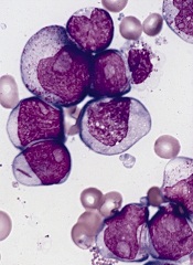
Disrupting equilibrium to fight AML

Credit: Lance Liotta
Understanding the relationship between mutated and wild-type genes could lead to new therapies for acute myeloid leukemia (AML), according to researchers.
The group discovered that RUNX1/ETO, generated by the chromosomal translocation t(8;21), regulates a leukemia-propagating transcriptional network via a dynamic equilibrium with RUNX1.
But depleting RUNX1/ETO prompts the formation of a transcriptional network that promotes myeloid differentiation.
Constanze Bonifer, PhD, of the University of Birmingham in the UK, and her colleagues detailed this work in Cell Reports.
The researchers likened normal hematopoiesis to a production line, with genes acting as regulators to control each step of blood formation. When a mutation occurs in the relevant regulator genes, the finely balanced order of the production line is disrupted, with drastic consequences.
Using digital footprinting and chromatin immunoprecipitation sequencing, the team showed that RUNX1/ETO switches off hundreds of other genes, many of them regulators themselves. As a consequence of the drastically altered production line, normal blood formation cannot happen, and leukemic cells form.
“Understanding how these rogue regulators operate is essential,” Dr Bonifer said. “[T]hese leukemic cells have one mutated gene and one unchanged one that would make the normal regulator. What happens in the leukemic cell is fundamentally a battle for supremacy between the two regulators, and the mutated one wins much of the time.”
“This is compounded by the normal regulator, which tries to compensate for defeat, and, in doing so, changes the output of genes that would be otherwise unaffected by the abnormal regulator. Quite simply, the result is a real mess. The cells are confused and can’t develop into mature blood cells.”
More specifically, the researchers found that the transcriptional program underlying leukemic propagation is regulated by a dynamic equilibrium between RUNX1/ETO and RUNX1 complexes.
Disrupting this equilibrium in t(8;21) cells by depleting RUNX1/ETO led to a global redistribution of transcription factor complexes within pre-existing open chromatin. And this prompted the formation of a transcriptional network that drives myeloid differentiation.
“If targeting [RUNX1/ETO] can reverse the changes it is making to the cellular production line, then it would ultimately point towards new avenues for a more precise treatment of leukemia,” said study author Olaf Heidenreich, PhD, of Newcastle University in the UK.
“Knowing that the production line can be restored to normal function gives us real hope. Of course, that is much easier to do in the lab than it is in the human body. But now we know how this works, we can look to deliver inhibitors to those mutated regulators. Creating one that works is the next step we have to overcome.” ![]()
Credit: Lance Liotta
Understanding the relationship between mutated and wild-type genes could lead to new therapies for acute myeloid leukemia (AML), according to researchers.
The group discovered that RUNX1/ETO, generated by the chromosomal translocation t(8;21), regulates a leukemia-propagating transcriptional network via a dynamic equilibrium with RUNX1.
But depleting RUNX1/ETO prompts the formation of a transcriptional network that promotes myeloid differentiation.
Constanze Bonifer, PhD, of the University of Birmingham in the UK, and her colleagues detailed this work in Cell Reports.
The researchers likened normal hematopoiesis to a production line, with genes acting as regulators to control each step of blood formation. When a mutation occurs in the relevant regulator genes, the finely balanced order of the production line is disrupted, with drastic consequences.
Using digital footprinting and chromatin immunoprecipitation sequencing, the team showed that RUNX1/ETO switches off hundreds of other genes, many of them regulators themselves. As a consequence of the drastically altered production line, normal blood formation cannot happen, and leukemic cells form.
“Understanding how these rogue regulators operate is essential,” Dr Bonifer said. “[T]hese leukemic cells have one mutated gene and one unchanged one that would make the normal regulator. What happens in the leukemic cell is fundamentally a battle for supremacy between the two regulators, and the mutated one wins much of the time.”
“This is compounded by the normal regulator, which tries to compensate for defeat, and, in doing so, changes the output of genes that would be otherwise unaffected by the abnormal regulator. Quite simply, the result is a real mess. The cells are confused and can’t develop into mature blood cells.”
More specifically, the researchers found that the transcriptional program underlying leukemic propagation is regulated by a dynamic equilibrium between RUNX1/ETO and RUNX1 complexes.
Disrupting this equilibrium in t(8;21) cells by depleting RUNX1/ETO led to a global redistribution of transcription factor complexes within pre-existing open chromatin. And this prompted the formation of a transcriptional network that drives myeloid differentiation.
“If targeting [RUNX1/ETO] can reverse the changes it is making to the cellular production line, then it would ultimately point towards new avenues for a more precise treatment of leukemia,” said study author Olaf Heidenreich, PhD, of Newcastle University in the UK.
“Knowing that the production line can be restored to normal function gives us real hope. Of course, that is much easier to do in the lab than it is in the human body. But now we know how this works, we can look to deliver inhibitors to those mutated regulators. Creating one that works is the next step we have to overcome.”
Drug shows activity in models of pediatric ALL
Credit: Aaron Logan
A compound that has demonstrated efficacy in older adults with acute myeloid leukemia (AML) may be a viable option for childhood acute lymphoblastic
leukemia (ALL) as well.
The drug, CPX-351, is a fixed-ratio combination of cytarabine and daunorubicin inside a lipid vesicle.
It previously showed promise in a phase 2 trial of AML patients ages 60 to 75 years. Now, preclinical results suggest CPX-351 can work against aggressive
pediatric ALL too.
The research appears in Pediatric Blood & Cancer. It was supported by the National Cancer Institute.
“Cytarabine and anthracyclines such as daunorubicin are commonly used to treat ALL in pediatric patients, and while these drug are very effective in front-line, multidrug, combination chemotherapy regimens, there remains room for improvement, especially in pediatric patients who relapse within 36 months of diagnosis,” said study author Richard Lock, PhD, of Children’s Cancer Institute in Sydney, Australia.
“We are very encouraged by these preclinical results, indicating that the proprietary CPX-351 formulation of cytarabine and daunorubicin may be an important tool to maximize efficacy outcomes in relapsed ALL, and we believe these data substantiate the need for additional, clinical study in pediatric patients.”
Dr Lock and his colleagues studied CPX-351 in mice inoculated with leukemia cells from 5 children who had died from ALL. Three of the children, 2 with B-precursor ALL and 1 with T-cell ALL, had relapsed disease at the time of biopsy.
The researchers inoculated up to 18 mice for each of the 5 leukemia types and then randomized the mice to treatment or control.
The maximum-tolerated dose of CPX-351 was 5 units/kg (corresponding to 5 mg/kg cytarabine and 2.2 mg/kg daunorubicin). This dose provided clinically relevant plasma drug exposure and correlated to the pharmacokinetic properties observed in patients with AML.
The researchers found that all 5 models of ALL were “highly responsive” to CPX-351. In the 4 models of B-precursor ALL, the median group response was a complete response. In the mice inoculated with T-cell ALL, the median group response was a partial response.
Among treated mice, event-free survival ranged from 32.8 days to 41.9 days. And among controls, event-free survival ranged from 2.4 days to 10.8 days.
Dr Lock and his colleagues believe these results suggest CPX-351 may be a promising treatment for ALL and support its testing in pediatric leukemia patients.
CPX-351 is currently under investigation in a phase 3 trial in older patients with high-risk (secondary) AML.
FDA approves new treatment for PI
Credit: Baxter
The US Food and Drug Administration (FDA) has approved a subcutaneous immune globulin product for use in adults with primary immunodeficiency (PI).
The product, HyQvia, is an immune globulin with a recombinant human hyaluronidase. It requires a single infusion every 3 to 4 weeks and 1 injection site per infusion to deliver a full therapeutic dose of immune globulin.
Current therapies require weekly or bi-weekly treatment with multiple infusion sites per treatment.
Baxter International Inc. expects to launch HyQvia in the US in the coming weeks. The product has been FDA-approved with a black-box warning detailing the risk of thrombosis associated with immune globulin products.
The immune globulin component of HyQvia is a 10% solution prepared from large pools of human plasma consisting of at least 98% IgG. The recombinant human hyaluronidase increases the dispersion and absorption of the immune globulin.
In a phase 3 trial, HyQvia compared well with intravenous human immune globulin 10% (IVIG).
Researchers compared the treatments at different time periods in a cohort of PI patients with a median age of 35 (range, 4-78 years). All 87 patients studied received IVIG, and 83 of the patients received at least 1 dose of HyQvia.
Patients received HyQvia for a median of 366 days and IVIG for a median of 91 days. The median ratio (HyQvia:IVIG) for the IgG dosage administered was 1.088 (range, 0.986–1.382).
Trough IgG concentrations, the incidence of infection, and rates of adverse events were generally similar during the HyQvia treatment period and the IVIG treatment period.
For patients aged 12 years and older, the median IgG Ctrough values with HyQvia were approximately the same as with IVIG. The median trough ratio (HyQvia:IVIG) was 0.985.
For patients younger than 12 (n=11), the median IgG Ctrough values were 10.0 and 9.6 g/L after HyQvia and IVIG, respectively, with a median trough ratio of 1.038.
The overall infection rates were 2.97 per patient-year with HyQvia and 4.51 per patient-year with IVIG.
During the HyQvia treatment period, the rate of acute serious bacterial infection (SBI) was 0.025 per patient-year. The rate of acute SBIs occurring during IVIG treatment was not reported.
In patients age 18 and older (n=59), the rate of acute SBIs was 0.00 per patient-year, and the overall infection rate was 3.20 per patient-year.
For this same patient group, the local adverse reaction rate was 0.286 per infusion.
The rate of systemic adverse events temporally related to an infusion was 0.20 per infusion with HyQvia and 0.33 per infusion with IVIG. There were no serious adverse events reported in these patients with either treatment.
HyQvia was approved in Europe in 2013 for adults with PI syndromes and myeloma or chronic lymphocytic leukemia with severe secondary hypogammaglobulinemia and recurrent infections.
For more details on HyQvia, see the prescribing information.