User login
Obesity affects toxicity of immunotherapy

Immunotherapy that can be effective against tumors in young, thin mice can be lethal to obese ones, according to research published in The Journal of Experimental Medicine.
Investigators conducted experiments in mouse models to determine if there is a subset of cancer patients for whom certain immunotherapies might be especially toxic.
The group found a potential link between body fat and the risk of toxicity from some types of immunotherapy.
“Cancer is primarily considered a disease of the aged, and yet preclinical studies generally use young, lean animal models that may not be reflective of the ‘typical’ cancer patient,” said study author Annie Mirsoian, a PhD candidate at the University of California, Davis in Sacramento.
“Aging is a dynamic process that is characterized by increases in inflammatory factors, as well as a shift in body composition where there is a gradual loss of lean muscle mass and an increase in fat accumulation, which effect how the immune system functions.”
Mirsoian and her colleagues sought to determine if, by adjusting the mouse model to more closely reflect the cancer patient phenotype (advanced age and overweight), they could better understand the discrepancies between animal study outcomes and those in patients in the clinic.
So the team examined aged mice on standard diets and compared those to aged mice that were calorie-restricted throughout their lives.
The investigators found that calorie restriction plays a protective role against toxicity. When aged mice ate their standard diet freely throughout life, they became obese and ultimately experienced lethal adverse reactions after receiving a systemic immunotherapy regimen.
“We know that people who are obese in general are at higher risk for complications from surgery, radiation, and chemotherapy,” said study author Arta Monjazeb, MD, PhD, of the University of California, Davis.
“We know that obese people have higher levels of inflammatory markers in their blood, but there is a lack of data examining the effects of obesity on cancer treatment outcomes.”
In follow-up experiments, the investigators found that young mice that are obese also endure similar toxic consequences, demonstrating that fat is a critical factor in toxic responses to stimulatory anticancer immunotherapy regimens.
“It is important to note, however, that the aged mice on standard diets succumbed to lethality at a quicker rate than young obese mice,” Mirsoian said. “Although our data demonstrate that obesity plays a central role in the development of adverse effects, future studies will focus on examining the aged immune system and cellular characteristics that may have enhanced the sensitivity of these mice to inflammation.”
This study follows an earlier paper, which demonstrated that while young, lean mice tolerate immunotherapy regimens without toxicity, the same regimen for an aged cohort resulted in lethal consequences. The new paper takes a deeper look into how fat deposition throughout aging can be critical in determining treatment tolerance and efficacy.
“Obesity has become an epidemic in our society and is now also affecting younger populations,” Mirsoian said. “Therefore, it’s likely that what the ‘typical’ cancer patient looks like will change. Our findings demonstrate the importance of having preclinical animal models that reflect the clinical scenario.”
“Changing the characteristics of our mouse models allowed for a more accurate determination of possible adverse reactions to therapy and more closely modeled what has been reported in the clinic with stimulatory immunotherapies.”
The investigators said factors like age, fat content, and the types of infections experienced throughout life together shape how the immune system reacts, and they will continue to work on improving their modeling system to reflect these changes.
They project that improvements in mouse modeling may help produce data that can better modulate treatment choices for patients, as well as identify early which patients would benefit from the inclusion of drugs to prevent adverse reactions while maintaining anticancer efficacy. ![]()
Air pollution not to blame for childhood leukemia, study suggests

The increased risk of leukemia reported among children born close to overhead power lines is likely not a result of alterations in air pollution, researchers have reported in the Journal of Radiological Protection.
The group found little evidence to support the “corona-ion hypothesis” which has been cited as a possible explanation for the excess of childhood leukemia cases close to high-voltage overhead power lines in the UK prior to the 1980s.
The hypothesis is based on the fact that high-voltage overhead power lines create charged particles in the surrounding air.
These ionized particles, known as corona ions, can be blown away by the wind and attach to air pollutants, such as those from traffic or smoking.
The corona-ion hypothesis suggests these electrically charged pollutants are more likely to be retained in the airways or lungs, and this could lead to serious health effects, including childhood leukemia.
The researchers previously showed that, on average, there has been no increased risk of leukemia among children born near high-voltage power lines in recent decades. However, the same piece of research confirmed an increased risk prior to the 1980s, which has yet to be explained.
To investigate this theory, John Swanson, of National Grid in London, and his colleagues used data from 7347 children in England and Wales who were born and diagnosed with leukemia between 1968 and 2008, and who lived within 600 m of a high-voltage overhead power line.
The researchers calculated the exposure of each of the subjects to corona ions using a model based on: the voltage of the power line; the distance from the line; how the concentration of corona ions varied with distance from the power lines; and, using data from various meteorological stations, the amount of time and speed that wind blew in each direction around the power lines.
The results did not suggest that exposure to corona ions explained the pattern of increased leukemia rates close to high-voltage overhead power lines previously found in earlier decades.
“We found in earlier studies that, for previous decades, childhood leukemia rates were higher near power lines,” said Kathryn Bunch, of the University of Oxford.
“This new paper seems to show that this wasn’t caused by corona ions, but it leaves us still searching for the true cause, and we are undertaking further investigations of the variation in risk over time.” ![]()
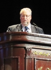
New agents challenge role of transplant in high-risk CLL
Credit: Chad McNeeley
NEW YORK—The role of allogeneic hematopoietic stem cell transplant (HSCT) for patients with high-risk chronic lymphocytic leukemia (CLL) is changing in the age of targeted therapy.
While allogeneic HSCT has been considered standard treatment for these patients, the question arises whether it will maintain its position in the era “of all these wonderful new drugs,” said David Maloney, MD, PhD, of the Fred Hutchinson Cancer Research Center in Seattle, Washington.
Dr Maloney undertook to convince the audience at the Lymphoma & Myeloma 2014 congress that there is still a role for allogeneic transplant in CLL patients.
He noted that early allogeneic transplant trials used myeloablative conditioning regimens, which were “prohibitively toxic.” They have now given way to reduced-intensity regimens.
“But the breakthrough came about when it was realized that the reason that allogeneic transplant could cure patients with CLL had really nothing to do with their conditioning regimen . . . ,” he said. “[I]t was probably the donor T cells providing immunologic activity and graft-vs-host activity that was actually able to provide graft-vs-tumor activity and cure patients.”
Seattle regimen
Dr Maloney described the reduced-intensity regimen used in Seattle—fludarabine and 2 Gy total body irradiation. The single dose of radiation is typically 1/6 of what a myeloablative regimen would be.
“This is truly an outpatient regimen,” he said. “Most patients, 50%, get through this without ever being in the hospital.”
Follow-up at 5 years showed overall survival to be 43%, progression-free survival 36%, complete responses 52%, and relapse 34%.
“This may not look very good,” Dr Maloney said, but these are fludarabine-refractory CLL patients whose expected median survival is around 12 months.
Dr Maloney noted that approximately the same outcomes were achieved whether the graft was from a matched related or unrelated donor, and cytogenetics really didn’t play a huge role in outcome.
The biggest factor affecting outcome was lymph node size. Patients with nodes 5 cm or larger did very poorly. And patients with lymph nodes smaller than 5 cm, irrespective of white cell count or bone marrow infiltration, actually did quite well in comparison to the group with large lymph nodes.
“So the graft-vs-tumor activity seems to be limited in its ability to get rid of bulky lymphadenopathy in this population,” Dr Maloney said.
Prior alemtuzumab therapy was also associated with the worst outcome in terms of relapse and disease progression.
Patients without comorbidities and without bulky lymphadenopathy have a very good outcome, Dr Maloney noted, saying, “You can cure 60% to 70% with an allogeneic transplant.”
He also pointed out that many groups are now doing this type of transplant with related and unrelated donors.
Transplant vs new agents
In addition to offering a potential cure, allogeneic transplant may provide better-functioning hematopoietic and immune systems after transplant than before, especially in those patients who received FCR (fludarabine, mitoxantrone, and rituximab) or other treatments.
Transplant, while potentially curative with a high complete response rate, has early non-relapse mortality around 15% to 20%.
“So this makes it hard to position in this era of pills that you can take,” Dr Maloney said.
He pointed out that while ibrutinib and idelalisib have excellent outcomes and overall survival, “these studies are very, very early . . . but obviously extremely promising.”
A group of European physicians recently published a position paper proposing a treatment algorithm that includes transplant for high-risk CLL patients. The algorithm indicates that relapsed/refractory patients should try the novel agents first.
Then, if patients respond, they can continue with the novel agent or proceed to transplant. Patients with lower-risk disease or those who are a higher transplant risk should probably continue on the novel agent.
Those who are younger with higher-risk disease, such as a 17p deletion, or who are a low transplant risk may be willing to choose transplant earlier.
Patients who do not respond to the novel agents can consider transplant or an alternative salvage regimen.
“[O]bviously, this is extremely controversial,” Dr Maloney said, “and what everyone is going to do is use these new agents to push transplant further down the road. And I think that’s appropriate.”
At the very least, Dr Maloney believes patients deserve a discussion of options early on.
He added that chimeric antigen receptor (CAR) T cells “will likely bump transplant even down another notch” because patients are likely to be willing to take the risk of CAR T cells before they’ll take the risk of chronic graft-vs-host disease with an unrelated donor.” ![]()
EMA grants product orphan status for AML
The European Medicines Agency (EMA) has granted orphan status to Atir, a product consisting of T-cell-depleted donor immune cells, for the treatment of acute myeloid leukemia (AML).
The EMA and the US Food and Drug Administration previously granted Atir orphan status for the prevention of acute graft-vs-host-disease (GVHD) following hematopoietic stem cell transplant (HSCT).
The EMA’s orphan designation provides incentives to support drug development. This includes fee reductions and a 10-year period of market exclusivity in the European Union after product approval.
About Atir
Atir consists of donor immune cells from which the alloreactive T-cells that would otherwise attack the patient’s body have been selectively eliminated.
The product is produced using a molecule known as TH9402 to selectively remove those T cells from the donor graft, while preserving other immune cells. To activate patient-reactive T cells, the graft is mixed (ex vivo) with patient cells.
Then, TH9402 is added. As this phototoxic compound selectively accumulates in activated T cells, the cells can be eliminated by exposing the cell mixture to light of a specific wavelength. The resulting Atir product can be frozen and stored and is infused into the patient in a scheduled procedure.
Trial data
Researchers said Atir proved safe and effective in a phase 1/2 study in which high-risk leukemia patients with very poor prognosis were treated with escalating doses of Atir after a haploidentical HSCT.
The overall survival of 19 patients who received an optimal dose of Atir was 78% at 1 year and 67% at 5 years, rates that compare favorably to outcomes of HSCTs from fully matched donors. The data also suggest that immune cells responsible for the graft-vs-leukemia effect are retained in Atir.
Five-year follow-up data show that none of the 19 patients developed acute grade 3/4 GVHD, compared to an incidence of 30% in matched unrelated HSCTs. In the 9 patients who received an optimal dose of Atir, there was no transplant-related mortality.
Researchers are currently testing Atir in a phase 2 study of patients with AML, acute lymphoblastic leukemia, and myelodysplastic syndrome, to corroborate and extend the safety and efficacy results from the phase 1/2 study. Data from this trial are expected in the second half of 2014.
Atir is under development by Kiadis Pharma. For more information, visit the company’s website. ![]()
Number of cord blood units doesn’t affect survival

Credit: NHS
Single and double cord blood transplants produce similar outcomes, according to a study of young patients with hematologic disorders.
Researchers found that rates of overall and disease-free survival were not significantly different in patients who received a single unit of cord blood and those who received two units.
Other outcome measures, such as neutrophil recovery, relapse, and transplant-related death, were similar between the two groups as well.
However, patients who received a single cord blood unit showed improved platelet recovery, a lower incidence of grade 3-4 acute graft-vs-host disease (GVHD), and a lower rate of extensive chronic GVHD.
John Wagner, Jr, MD, of the University of Minnesota in Minneapolis, and his colleagues reported these results in NEJM. Dr Wagner previously presented results from this study at ASH 2012.
“Based on promising early studies using two cord blood units in adults for whom one unit is often not sufficient, we designed this study in order to determine if the higher number of blood-forming stem cells in two cord blood units might improve survival,” Dr Wagner said. “What we found, however, was that both treatment arms performed very well, with similar rates of white blood cell recovery and survival.”
The researchers enrolled 224 patients, ages 1 to 21 years, with hematologic disorders, including acute and chronic leukemias as well as myelodysplastic syndromes.
Patients were randomized to receive double-unit (n=111) or single-unit (n=113) cord blood transplants after a uniform myeloablative conditioning regimen and immunoprophylaxis for GVHD.
The researchers matched the treatment arms for age, sex, self-reported race, performance status, degree of donor-recipient HLA matching, disease type, and disease status at transplant.
Survival and relapse
The study’s primary endpoint was 1-year survival, which was 65% in the double-unit arm and 73% in the single-unit arm (P=0.17). In a multivariate analysis, the risk of death did not differ significantly between the arms (hazard ratio=1.34, P=0.20).
Similarly, there was no significant difference in 1-year disease-free survival between the double- and single-unit arms—64% and 70%, respectively (P=0.11). In a multivariate analysis, the risk of relapse or death did not differ significantly between arms (hazard ratio=1.48, P=0.08).
It therefore follows that rates of relapse and transplant-related death were similar at 1 year as well. The incidence of relapse was 14% in the double-unit arm and 12% in the single-unit arm (P=0.12). And rates of transplant-related death were 22% and 19%, respectively (P=0.43).
Recovery and GVHD
The incidence of neutrophil recovery was similar between treatment arms—88% in the double-unit arm and 89% in the single-unit arm (P=0.29) at a median of 23 days (range, 11 to 133) and 21 days (range, 11 to 62) after transplant, respectively.
However, the rate of platelet recovery was significantly higher in the single-unit arm—76% vs 65% (P=0.04). Furthermore, the median time to platelet recovery was 58 days (range, 28 to 295) in the single-unit arm and 84 days (range, 22 to 716) in the double-unit arm.
The rate of grade 2-4 acute GVHD was similar between the treatment arms (P=0.78), but patients in the double-unit arm had a higher incidence of grade 3-4 acute GVHD—23% vs 13% (P=0.02).
There was no difference in the incidence of any chronic GVHD at 1 year after transplant—32% in the double-unit arm and 30% in the single-unit arm (P=0.51). But there was a higher incidence of extensive chronic GVHD after double-unit transplant—15% vs 9% (P=0.05).
“This is helpful news for physicians considering the best treatment options for their patients,” said Joanne Kurtzberg, MD, of Duke University Medical Center in Durham, North Carolina.
“We found children who have a cord blood unit with an adequate number of cells do not benefit from receiving two units. This reduces the cost of a cord blood transplant for the majority of pediatric patients needing the procedure. However, for larger children without an adequately dosed single cord blood unit, using two units will provide access to a potentially life-saving transplant.” ![]()
Chlorambucil’s role in untreated CLL debated
NEW YORK—With both the pro and con positions drawing on data from the phase 3 CLL11 trial, two speakers at the Lymphoma & Myeloma2014 congress faced off on whether it’s necessary to use chlorambucil with obinutuzumab in untreated chronic lymphocytic leukemia (CLL).
Myron S. Czuczman, MD, of Roswell Park Cancer Institute in Buffalo, New York, argued in favor of using chlorambucil. And Richard R. Furman, MD, of Weill Cornell Medical College in New York, argued against it.
Obinutuzumab is a glycoengineered, humanized, monoclonal antibody that selectively binds to the extracellular domain of the CD20 antigen on B cells.
It was approved by the US Food and Drug Administration based on initial results from the phase 3 CLL11 study, in which 781 patients were randomized to receive chlorambucil alone or chlorambucil with either obinutuzumab or rituximab.
Pro
Dr Czuczman pointed out that while the obinutuzumab-chlorambucil combination had more toxicity than the rituximab-chlorambucil combination, the overall response rate and complete response rate with obinutuzumab were significantly higher than with rituximab (P<0.0001).
Progression-free survival (PFS), which was the primary endpoint, was significantly higher with either obinutuzumab at 26.7 months, or rituximab, at 16.3 months, than with chlorambucil alone, at 11.1 months.
And in the head-to-head portion of CLL11, PFS with obinutuzumab-chlorambucil was significantly better at 26.7 months than with rituximab-chlorambucil, at 15.2 months (P<0.001).
Dr Czuczman also reviewed data on obinutuzumab combined with drugs other than chlorambucil.
The GALTON trial, a small, phase 1b trial in untreated CLL, compared obinutuzumab plus fludarabine and cyclophosphamide to obinutuzumab plus bendamustine.
Dr Czuczman showed that while there is more toxicity when obinutuzumab is combined with cyclophosphamide or bendamustine than with chlorambucil, “there is not much more activity.”
He said it’s not clear whether obinutuzumab with cyclophosphamide is better than rituximab with cyclophosphamide or if obinutuzumab with bendamustine is better than rituximab with bendamustine in upfront CLL.
“For now,” he said, “chloramubucil should be the only chemo agent combined with obinutuzumab to treat upfront CLL—outside of clinical trial participation.”
Con
Dr Furman also reviewed the CLL11 trial, noting that rituximab did not add very much to chlorambucil, but obinutuzumab did, in terms of overall survival and PFS. He cautioned, however, that additive or synergistic effects cannot be ruled out in the combination studies.
He then reviewed the GAGE trial, which compared 2 doses of single-agent obinutuzumab in untreated CLL. The 2000 mg dose produced a greater overall response rate than the 1000 mg dose, but the difference between the 2 arms was not significant (P=0.08).
PFS was 21 months in the 1000 mg arm and 20 months in the 2000 mg arm (P=0.07). PFS for obinutuzumab plus chlorambucil in the CLL11 trial was 26.7 months.
However, second cancers may be more of an issue with chlorambucil. In CALGB 9011, investigators reported 27 epithelial cancers, 9 with fludarabine, 11 with chlorambucil, and 7 with fludarabine plus chlorambucil.
Dr Furman concluded that while chlorambucil may aid obinutuzumab by reducing bulk, it may be unnecessary if higher doses of the antibody are used. Single-agent obinutuzumab produces a similar PFS as the combination with chlorambucil, and there are greater toxicities with chlorambucil. ![]()
Device monitors methotrexate levels faster

Credit: Juan D. Alfonso
A new device can measure methotrexate levels in a patient’s blood in less than a minute, according to research published in Biosensors and Bioelectronics.
Researchers say this nanoscale device is just as accurate and 10 times less expensive than equipment currently used in hospitals.
It has an optical system that can rapidly gauge the optimal dose of methotrexate a patient needs, thereby reducing the risk of adverse effects.
“While effective, methotrexate is also highly toxic and can damage the healthy cells of patients, hence the importance of closely monitoring the drug’s concentration in the serum of treated individuals to adjust the dosage,” said study author Jean François Masson, PhD, of the University of Montreal in Quebec, Canada.
“The operation of the current [methotrexate monitoring] device is based on a cumbersome, expensive platform that requires experienced personnel because of the many samples that need to be manipulated.”
With this in mind, Dr Masson and his colleagues set out to simplify methotrexate monitoring.
In the course of their research, the team developed and manufactured a miniaturized device that works by surface plasmon resonance. It measures the concentration of serum methotrexate through gold nanoparticles on the surface of a receptacle.
In “competing” with methotrexate to block the enzyme dihydrofolate reductase, the gold nanoparticles change the color of the light detected by the instrument. And the color of the light detected reflects the exact concentration of the drug in the blood sample.
The researchers compared the accuracy of measurements taken with the new device to those taken with equipment used at the Maisonneuve-Rosemont Hospital in Montreal.
“Testing was conclusive,” Dr Masson said. “Not only were the measurements as accurate, but our device took less than 60 seconds to produce results, compared to 30 minutes for current devices.”
Moreover, the comparative tests were performed by lab technicians who were not experienced with surface plasmon resonance and did not encounter major difficulties in operating the new equipment or obtaining the same conclusive results as Dr Masson and his research team.
“In the near future, we can foresee the device in doctors’ offices or even at the bedside, where patients would receive individualized and optimal doses while minimizing the risk of complications,” Dr Masson said.
“While traditional equipment requires an investment of around $100,000, the new mobile device would likely cost 10 times less, around $10,000.” ![]()
Credit: Juan D. Alfonso
A new device can measure methotrexate levels in a patient’s blood in less than a minute, according to research published in Biosensors and Bioelectronics.
Researchers say this nanoscale device is just as accurate and 10 times less expensive than equipment currently used in hospitals.
It has an optical system that can rapidly gauge the optimal dose of methotrexate a patient needs, thereby reducing the risk of adverse effects.
“While effective, methotrexate is also highly toxic and can damage the healthy cells of patients, hence the importance of closely monitoring the drug’s concentration in the serum of treated individuals to adjust the dosage,” said study author Jean François Masson, PhD, of the University of Montreal in Quebec, Canada.
“The operation of the current [methotrexate monitoring] device is based on a cumbersome, expensive platform that requires experienced personnel because of the many samples that need to be manipulated.”
With this in mind, Dr Masson and his colleagues set out to simplify methotrexate monitoring.
In the course of their research, the team developed and manufactured a miniaturized device that works by surface plasmon resonance. It measures the concentration of serum methotrexate through gold nanoparticles on the surface of a receptacle.
In “competing” with methotrexate to block the enzyme dihydrofolate reductase, the gold nanoparticles change the color of the light detected by the instrument. And the color of the light detected reflects the exact concentration of the drug in the blood sample.
The researchers compared the accuracy of measurements taken with the new device to those taken with equipment used at the Maisonneuve-Rosemont Hospital in Montreal.
“Testing was conclusive,” Dr Masson said. “Not only were the measurements as accurate, but our device took less than 60 seconds to produce results, compared to 30 minutes for current devices.”
Moreover, the comparative tests were performed by lab technicians who were not experienced with surface plasmon resonance and did not encounter major difficulties in operating the new equipment or obtaining the same conclusive results as Dr Masson and his research team.
“In the near future, we can foresee the device in doctors’ offices or even at the bedside, where patients would receive individualized and optimal doses while minimizing the risk of complications,” Dr Masson said.
“While traditional equipment requires an investment of around $100,000, the new mobile device would likely cost 10 times less, around $10,000.”
Obese ALL patients more likely to have MRD after induction

Obese youths with acute lymphoblastic leukemia (ALL) are known to have worse outcomes than their lean counterparts.
To gain more insight into this phenomenon, investigators set out to determine if body mass index (BMI) impacted ALL patients’ responses to initial chemotherapy.
The results showed that, following induction chemotherapy, obese patients were more than twice as likely to have minimal residual disease (MRD) than non-obese patients.
“Induction chemotherapy provides a patient’s best chance for remission or a cure,” said principal investigator Steven Mittelman, MD, PhD, of The Saban Research Institute of Children’s Hospital Los Angeles in California.
“Our findings indicate that a patient’s obesity negatively impacts the ability of chemotherapy to kill leukemia cells, reducing the odds of survival.”
The study, which was published in Blood, included 198 patients who were diagnosed with ALL and between the ages of 1 and 21 years.
Each patient’s BMI was converted to a percentile and classified according to the Center for Disease Control and Prevention’s thresholds for overweight (85% to 94%) and obese (greater than 95%). Patients with a BMI less than 85% were considered “lean.”
About one-third of the patients were obese or overweight at the time of diagnosis.
MRD was determined by testing bone marrow specimens at the end of induction therapy, and patients were followed for 2 to 5 years from the time of diagnosis.
The investigators found that lean patients with MRD had similar outcomes to obese patients without evidence of MRD. Obese patients with MRD had the worst outcomes.
Additionally, although nearly a quarter of the patients initially deemed “lean” gained weight and became obese during the first month of treatment, these patients still showed similar outcomes to those who remained lean.
“In addition to increasing a patient’s likelihood of having persistent disease following treatment, obesity appears to add a risk factor that changes the interaction between chemotherapy and residual leukemia cells,” said Hisham Abdel-Azim, MD, also of The Saban Research Institute.
Findings from this study offer new avenues for investigation that include modifying chemotherapy regimens for obese patients and working to change a patient’s weight status beginning at the time of diagnosis.
Armored CAR T cells next on the production line
NEW YORK—Chimeric antigen receptor (CAR) T cells have “remarkable” activity, according to a speaker at the NCCN 9th Annual Congress: Hematologic Malignancies.
“[T]his chimera binds like an antibody, but it acts like a T cell, so it combines the best of both worlds,” said Jae H. Park, MD, of Memorial Sloan Kettering Cancer Center (MSKCC) in New York.
He then traced the evolution of CAR T-cell design, discussed clinical trials using CD19-targed T cells, and described how investigators are working at building a better T cell.
Researchers found that T-cell activation and proliferation require signaling through a costimulatory receptor, such as CD28, 4-1BB, or OX-40. Without costimulation, the T cell becomes unresponsive or undergoes apoptosis.
So based on this observation, Dr Park said, several research groups created second- and third-generation CARs to incorporate the costimulatory signal.
The first generation was typically fused to the CD8 domain. Second-generation CARs include a costimulatory signaling domain, such as CD28, 4-1BB, or OX40. And the third generation contains signaling domains from 2 costimulatory receptors, CD28 with 4-1BB and CD28 with OX40.
The built-in costimulatory signal proved superior to the first-generation CAR T cells.
In NOD/SCID mice inoculated with NALM-6 lymphoma cells, Dr Park said, about 50% more were “cured,” in terms of survival, using a CD80 costimulatory ligand with CD19-targeted T cells compared to those without the ligand.
Clinical trials
Clinical trials using second-generation CD19-targeted T cells in relapsed B-cell acute lymphoblastic leukemia (ALL) at MSKCC produced an overall complete response (CR) rate of 88% in a median of 22.1 days. And 72% of the CRs were minimal residual disease (MRD) negative.
So the CAR T cells produce a “very rapid and deep remission,” Dr Park said.
CAR T-cell therapy, however, comes with adverse events, most notably, cytokine release syndrome (CRS), which results from T-cell activation. CRS causes fevers, hypotension, and neurologic toxicities including mental status changes, obtundation, and seizures.
“CRS is not unique to CAR T-cell therapy,” Dr Park said. “Any therapy that activates T cells can have this type of side effect.”
Dr Park noted that CRS is associated with disease burden at the time of treatment. “The larger the disease burden pre T-cell therapy,” he said, “the more likely [patients are] to develop CRS.”
In the MSKCC trial, no patient with very low disease burden—5% blasts in the bone marrow—developed CRS.
However, there is also a correlation between tumor burden and T-cell expansion, he added. T cells expand much better with a larger disease burden, because there is a greater antigen load.
The investigators found that serum C-reactive protein can serve as a surrogate marker for the severity of CRS. Patients with levels above 20 mg/dL are more likely to experience CRS.
And Dr Park pointed out that CRS symptoms respond pretty rapidly to steroids or interleukin-6 receptor blockade.
CAR T-cell therapy has also been used to treat chronic lymphocytic leukemia, but with much more modest response rates than in ALL. Both University of Pennsylvania and MSKCC trials in CLL have produced overall response rates around 40%.
Building a better T cell
Dr Park described efforts underway to develop the fourth-generation “armored” CAR T cells to overcome the hostile tumor microenvironment, which contains multiple inhibitory factors designed to suppress effector T cells.
Armored T cells can actually secrete some of the inflammatory cytokines to change the tumor microenvironment and overcome the inhibitory effect.
Dr Park described a potential scenario: The armored CAR T cells secrete IL-12, enhance the central memory phenotype, enhance cytotoxicity, enhance persistence, modify the endogenous immune system and T-cell activation, and reactivate tumor-infiltrating lymphocytes.
He said future studies will focus on translation of these armored CAR T cells to the clinical setting in both hematologic and solid tumor malignancies.
CHMP says ponatinib’s benefits outweigh risks

Credit: CDC
The European Medicines Agency’s (EMA) Committee for Medicinal Products for Human Use (CHMP) has adopted its final opinion on ponatinib (Iclusig), saying the drug’s benefits outweigh its risks.
The CHMP recommends that ponatinib continue to be used in accordance with its approved indications.
However, the drug’s product information should be updated with strengthened warnings, particularly about the risk of arterial and venous thrombotic events.
Ponatinib is approved in the European Union (EU) to treat adults with chronic phase, accelerated phase, or blast phase chronic myeloid leukemia (CML) who are resistant to dasatinib or nilotinib, who are intolerant to dasatinib or nilotinib and for whom subsequent treatment with imatinib is not clinically appropriate, or who have the T315I mutation.
The drug is also approved to treat adults with Philadelphia-chromosome positive acute lymphoblastic leukemia who are resistant to dasatinib, who cannot tolerate dasatinib and subsequent treatment with imatinib is not clinically appropriate, or who have the T315I mutation.
Roughly a year ago, follow-up data revealed that ponatinib-treated patients had a higher incidence of arterial and venous thrombotic events than was observed when the drug first gained approval. So one ponatinib trial was discontinued, and the rest were placed on partial clinical hold.
Then, ponatinib was pulled from the US market. The drug ultimately returned to the marketplace with new recommendations designed to decrease the risk of thrombotic events. The EMA also revised its recommendations for ponatinib but kept the drug on the market.
PRAC review and recommendations
Because of these risks, the EMA’s Pharmacovigilance Risk Assessment Committee (PRAC) conducted an 11-month review of the available data on ponatinib and consulted with a scientific advisory group.
The PRAC assessed the available data on the nature, frequency, and severity of arterial and venous thrombotic events. And the committee concluded that the benefits of ponatinib outweigh its risks.
The PRAC said the risk of thrombotic events is likely dose-related, but there are insufficient data to formally recommend using lower doses of ponatinib. And there is a risk that lower doses might not be as effective in all patients and in long-term treatment.
The PRAC therefore concluded that the recommended starting dose of ponatinib should remain 45 mg once a day.
However, the committee also recommended updates to the product information to provide healthcare professionals with the latest evidence, in case they want to consider reducing the dose in patients with chronic phase CML who are responding well to treatment and who might be at particular risk of thrombotic events.
In addition, PRAC recommended that healthcare professionals stop ponatinib if there has been no response after 3 months of treatment and monitor patients for high blood pressure or signs of heart problems.
The CHMP concurred with these recommendations and is forwarding them to the European Commission. The commission is expected to issue a final, legally binding decision on ponatinib in December 2014, which will be valid throughout the EU.
A new study on the safety and benefits of ponatinib is in the works to help clarify if lower doses of the drug carry a lower risk of thrombotic events while still having a beneficial effect in patients with chronic phase CML.