User login
AML study shows transcription factors are ‘druggable’

In developing a compound that exhibits activity against a type of acute myeloid leukemia (AML), investigators have shown that transcription factors are, in fact, “druggable.”
The compound, AI-10-49, selectively binds to the mutated transcription factor CBFβ-SMMHC, and this prevents CBFβ-SMMHC from binding to another transcription factor, RUNX1.
In this way, AI-10-49 restores RUNX1 transcriptional activity, which leads to the death of inv(16) AML cells in vitro and in vivo.
“This drug that we’ve developed is . . . targeting a class of proteins that hasn’t been targeted for drug development very much in the past,” said study author John H. Bushweller, PhD, of the University of Virginia Health System in Charlottesville.
“It’s really a new paradigm, a new approach to try to treat these diseases. This class of proteins is very important for determining how much of many other proteins are made, so it’s a unique way of changing the way the cell behaves.”
Dr Bushweller and his colleagues described their work in Science.
The investigators noted that CBFβ-SMMHC is expressed in AML with the chromosome inversion inv(16)(p13q22). And CBFβ-SMMHC outcompetes wild-type CBFβ for binding to RUNX1, thereby deregulating RUNX1 activity in hematopoiesis and inducing AML.
“When you target a mutated protein in a cancer, you would ideally like to inhibit that mutated form of the protein but not affect the normal form of the protein that’s still there,” Dr Bushweller said. “In the case of [AI-10-49], we’ve achieved that.”
He and his colleagues tested AI-10-49 in 11 leukemia cells lines and found that only ME-1 cells were highly sensitive to the treatment. AI-10-49 effectively dissociated RUNX1 from CBFβ-SMMHC in ME-1 cells, with 90% dissociation after 6 hours of treatment.
But the drug had a modest effect on CBFβ-RUNX1 association. It also had negligible activity in normal human bone marrow cells.
The investigators then tested AI-10-49 in a mouse model of inv(16) AML. Control mice (vehicle-treated) succumbed to leukemia in a median of 33.5 days, compared to a median of 61 days for mice that received AI-10-49.
Dr Bushweller and his colleagues did not evaluate toxicity after long-term exposure to AI-10-49, but they found no evidence of toxicity after 7 days of AI-10-49 treatment.
Next, the investigators tested AI-10-49 in 4 primary inv(16) AML cell samples. They saw a reduction in leukemia cell viability when AI-10-49 was administered at 5 μM and 10 μM concentrations. Bivalent AI-10-49 was more potent than monovalent AI-10-47.
The team said these results suggest that direct inhibition of CBFβ-SMMHC may be an effective therapeutic approach for inv(16) AML, and the findings provide support for therapies targeting transcription factors. ![]()
Israel approves ponatinib for CML, Ph+ ALL

Photo courtesy of the US FDA
The Israeli Ministry of Health has granted regulatory approval for the kinase inhibitor ponatinib (Iclusig) to treat certain adults with chronic myeloid leukemia (CML) or Philadelphia chromosome-positive acute lymphoblastic leukemia (Ph+ ALL).
The drug can now be used to treat adults with any phase of CML who have the T315I mutation or are resistant to/cannot tolerate dasatinib or nilotinib and for whom subsequent treatment with imatinib is not clinically appropriate.
Ponatinib is also approved to treat patients with Ph+ ALL who have the T315I mutation or are resistant to/cannot tolerate dasatinib and for whom subsequent treatment with imatinib is not clinically appropriate.
Ariad Pharmaceuticals, Inc., the company developing ponatinib, said the drug should be available in Israel in the second quarter of 2015.
Trial results
The Ministry of Health’s decision to approve ponatinib was based on results from the phase 2 PACE trial, which included patients with CML or Ph+ ALL who were resistant to or intolerant of prior tyrosine kinase inhibitor therapy, or who had the T315I mutation.
The median follow-up times were 15.3 months in chronic-phase CML patients, 15.8 months in accelerated-phase CML patients, and 6.2 months in patients with blast-phase CML or Ph+ ALL.
In chronic-phase CML, the primary endpoint was major cytogenetic response, and it occurred in 56% of patients. Among chronic-phase patients with the T315I mutation, 70% achieved a major cytogenetic response. Among patients who had failed treatment with dasatinib or nilotinib, 51% achieved a major cytogenetic response.
In accelerated-phase CML, the primary endpoint was major hematologic response. This occurred in 57% of all patients in this group, 50% of patients with the T315I mutation, and 58% of patients who had failed treatment with dasatinib or nilotinib.
The primary endpoint was major hematologic response in blast-phase CML/Ph+ ALL as well. Thirty-four percent of all patients in this group met this endpoint, as did 33% of patients with the T315I mutation and 35% of patients who had failed treatment with dasatinib or nilotinib.
Common non-hematologic adverse events included rash (38%), abdominal pain (38%), headache (35%), dry skin (35%), constipation (34%), fatigue (27%), pyrexia (27%), nausea (26%), arthralgia (25%), hypertension (21%), increased lipase (19%), and increased amylase (7%).
Hematologic events of any grade included thrombocytopenia (42%), neutropenia (24%), and anemia (20%). Serious adverse events of arterial thromboembolism, including arterial stenosis, occurred in patients with cardiovascular risk factors.
Safety issues
Extended follow-up data from the PACE trial, collected in 2013, suggested ponatinib can increase the risk of thrombotic events. When these data came to light, officials in the European Union and the US, where ponatinib had already been approved, began to investigate the drug.
Ponatinib was pulled from the US market for a little over 2 months, and trials of the drug were placed on partial hold while the Food and Drug Administration evaluated the drug’s safety. Ponatinib went back on the market in January 2014, with new safety measures in place.
The drug was not pulled from the market in the European Union, but the European Medicine’s Agency released recommendations for safer use of ponatinib. The Committee for Medicinal Products for Human Use reviewed data on ponatinib and decided the drug’s benefits outweigh its risks. ![]()
Long-term endocrine effects common after reduced-intensity chemotherapy in children
SAN DIEGO – Remain vigilant for endocrine side effects after reduced-intensity conditioning for hematopoietic stem cell transplants in children.
Traditionally, hematopoietic stem cell transplants (HSCT) for leukemia and other malignancies are preceded by myeloablation with high-dose chemotherapy and radiation. Over the past decade, however, that approach has been supplanted by reduced-intensity conditioning (RIC), a gentler method for less aggressive diseases that don’t require a cancer to be wiped out before HSCT. RIC usually involves low-dose chemotherapy without radiation.

The hope is to reduce side effects with the gentler approach, but that’s not always how it works, according to a retrospective study of 120 children followed for a mean of 3.2 years at the Cincinnati Children’s Hospital Medical Center, which was presented at a poster session at the meeting of the Endocrine Society.
The children had RIC with campath, fludarabine, and melphalan – but no radiation – prior to one HSCT for hemophagocytic lymphohistiocytosis/X-linked lymphoproliferative syndrome (HLH/XLP), primary immune deficiency, or other generally nonmalignant conditions.
During follow-up, almost a quarter had thyroid problems and nearly three-fourths were vitamin-D deficient. Children less than 2 years old when transplanted started to catch up to their peers on growth charts, but children over 2 years old fell further behind. Hypogonadism might be a problem, too; the investigators plan to follow the children to see how they fare during puberty.
“We were surprised” by the results, said senior investigator Dr. Susan Rose, professor of endocrinology at the University of Cincinnati.
“We really thought that, with [RIC], not as many of them would have problems after their transplants, and they would grow better. [Many] were coming off steroids” after the procedure, “which usually leads children to have catch-up growth. Instead, we are seeing their growth is slower than normal. The occurrence of endocrinopathy after [RIC] is probably lower than with high-dose chemotherapy combined with radiation, but we still have to monitor [these children] and not be cavalier” about follow-up, she said.
“We can’t expect there to be no or fewer endocrine complications just because there was a less intensive [conditioning] regimen. We must, as endocrinologists, be vigilant about monitoring and screening” these patients, said endocrinologist and lead author Dr. Jonathan Howell, assistant professor of pediatrics at the university.
The children were an average of 6 years old when transplanted. They were shorter than average both before (height-for-age Z-score [HAZ] = –1.33) and after (HAZ = –1.35).
Children with HLH/XLP who were under 2 years old at transplant improved from an HAZ of –3.36 to –1.35 afterwards, but older children fell from an HAZ of –0.61 to –0.99.
Older children with HLH/XLP lost weight after transplant (body mass index Z-score [BMI-Z], 1.29 vs. 0.61), as did those transplanted for metabolic or genetic disorders (BMI-Z, 0.56 vs. –0.77). There was a trend towards increased BMI-Z in toddlers.
Seventy-seven children had their thyroids checked after HSCT: 11 had primary hypothyroidism, five had central hypothyroidism, and two were hyperthyroid. Also, 48 of the 68 children assessed for 25-OH vitamin D had levels below 30 ng/mL.
Fourteen children broke bones after their transplants and six were diagnosed with avascular necrosis. The team is looking into whether it had something to do with endocrine dysfunction.
The investigators had no disclosures and no external funding for their work.
SAN DIEGO – Remain vigilant for endocrine side effects after reduced-intensity conditioning for hematopoietic stem cell transplants in children.
Traditionally, hematopoietic stem cell transplants (HSCT) for leukemia and other malignancies are preceded by myeloablation with high-dose chemotherapy and radiation. Over the past decade, however, that approach has been supplanted by reduced-intensity conditioning (RIC), a gentler method for less aggressive diseases that don’t require a cancer to be wiped out before HSCT. RIC usually involves low-dose chemotherapy without radiation.
The hope is to reduce side effects with the gentler approach, but that’s not always how it works, according to a retrospective study of 120 children followed for a mean of 3.2 years at the Cincinnati Children’s Hospital Medical Center, which was presented at a poster session at the meeting of the Endocrine Society.
The children had RIC with campath, fludarabine, and melphalan – but no radiation – prior to one HSCT for hemophagocytic lymphohistiocytosis/X-linked lymphoproliferative syndrome (HLH/XLP), primary immune deficiency, or other generally nonmalignant conditions.
During follow-up, almost a quarter had thyroid problems and nearly three-fourths were vitamin-D deficient. Children less than 2 years old when transplanted started to catch up to their peers on growth charts, but children over 2 years old fell further behind. Hypogonadism might be a problem, too; the investigators plan to follow the children to see how they fare during puberty.
“We were surprised” by the results, said senior investigator Dr. Susan Rose, professor of endocrinology at the University of Cincinnati.
“We really thought that, with [RIC], not as many of them would have problems after their transplants, and they would grow better. [Many] were coming off steroids” after the procedure, “which usually leads children to have catch-up growth. Instead, we are seeing their growth is slower than normal. The occurrence of endocrinopathy after [RIC] is probably lower than with high-dose chemotherapy combined with radiation, but we still have to monitor [these children] and not be cavalier” about follow-up, she said.
“We can’t expect there to be no or fewer endocrine complications just because there was a less intensive [conditioning] regimen. We must, as endocrinologists, be vigilant about monitoring and screening” these patients, said endocrinologist and lead author Dr. Jonathan Howell, assistant professor of pediatrics at the university.
The children were an average of 6 years old when transplanted. They were shorter than average both before (height-for-age Z-score [HAZ] = –1.33) and after (HAZ = –1.35).
Children with HLH/XLP who were under 2 years old at transplant improved from an HAZ of –3.36 to –1.35 afterwards, but older children fell from an HAZ of –0.61 to –0.99.
Older children with HLH/XLP lost weight after transplant (body mass index Z-score [BMI-Z], 1.29 vs. 0.61), as did those transplanted for metabolic or genetic disorders (BMI-Z, 0.56 vs. –0.77). There was a trend towards increased BMI-Z in toddlers.
Seventy-seven children had their thyroids checked after HSCT: 11 had primary hypothyroidism, five had central hypothyroidism, and two were hyperthyroid. Also, 48 of the 68 children assessed for 25-OH vitamin D had levels below 30 ng/mL.
Fourteen children broke bones after their transplants and six were diagnosed with avascular necrosis. The team is looking into whether it had something to do with endocrine dysfunction.
The investigators had no disclosures and no external funding for their work.
AT ENDO 2015
Key clinical point: Remain vigilant for endocrine side effects after reduced-intensity conditioning for hematopoietic stem cell transplants in children.
Major finding: Of the 77 children assessed for thyroid function after HSCT with reduced-intensity conditioning, 11 had evidence of primary hypothyroidism, five had central hypothyroidism, and two had evidence of primary hyperthyroidism.
Data source: Review of 120 pediatric hematopoietic stem cell transplants at the Cincinnati Children’s Hospital Medical Center.
Disclosures: The investigators had no disclosures and no external funding for their work.
Germline mutations linked to ALL

Photo by Rhoda Baer
New research suggests that heritable mutations in the gene ETV6 can predispose people to acute lymphoblastic leukemia (ALL).
Somatic mutations in ETV6 have previously been implicated in the development of ALL and other hematologic malignancies.
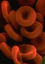
The new study, published in Nature Genetics, has shown that germline mutations in ETV6 are associated with thrombocytopenia, red blood cell (RBC) macrocytosis, and predisposition to ALL.
The research began with a family (family 1) that had autosomal dominant thrombocytopenia (67,000-132,000 platelets/μL), high RBC mean corpuscular volume (MCV, 92.5-101.5 fl), and 2 cases of B-cell-precursor ALL.
“All of them had big red blood cells, low platelet counts, and propensity to bleed,” said study author Christopher Porter, MD, of the University of Colorado Denver.
This familial link to abnormal blood dynamics and predisposition to ALL implied a common genetic denominator. To find it, the researchers performed whole-exome sequencing and found a heterozygous single-nucleotide change in ETV6, c.641C>T, encoding a p.Pro214Leu substitution in the central domain.
The researchers then screened 23 other families with similar phenotypes as the first and identified 2 families with ETV6 mutations.
One family (family 2) had members with platelet counts ranging from 44,000 to 115,000 platelets/μL, RBC MCVs ranging from 88 to 97 fl, and a member with ALL. All affected family members had a c.641C>T mutation identical to the one identified in family 1.
Another family (family 3) had members with platelet counts ranging from 99,000 to 101,000 platelets/μL and RBC MCVs ranging from 93 to 98 fl but no malignancies. Members of this family had a c.1252A>G transition producing a p.Arg418Gly substitution in the DNA-binding domain, with alternative splicing and exon skipping.
The researchers said these mutations partially disrupt ETV6 transcriptional repression in vitro and cause aberrant cytoplasmic localization of both mutant and endogenous ETV6, suggesting a dominant-negative effect. The mutations also impair megakaryocyte development and proplatelet formation in culture.
Dr Porter and his colleagues noted that another team of researchers recently discovered germline missense mutations in ETV6 in 3 unrelated families. Dr Porter’s group hopes future work will show the prevalence of germline mutations in ETV6.
“It’s not common in a general population, but we think it might be much more common in people who develop ALL,” Dr Porter said. “[Our] paper highlights this gene in the development of leukemia. By studying this mutation, we should be able to gather a better understanding of how leukemia develops.” ![]()
Photo by Rhoda Baer
New research suggests that heritable mutations in the gene ETV6 can predispose people to acute lymphoblastic leukemia (ALL).
Somatic mutations in ETV6 have previously been implicated in the development of ALL and other hematologic malignancies.
The new study, published in Nature Genetics, has shown that germline mutations in ETV6 are associated with thrombocytopenia, red blood cell (RBC) macrocytosis, and predisposition to ALL.
The research began with a family (family 1) that had autosomal dominant thrombocytopenia (67,000-132,000 platelets/μL), high RBC mean corpuscular volume (MCV, 92.5-101.5 fl), and 2 cases of B-cell-precursor ALL.
“All of them had big red blood cells, low platelet counts, and propensity to bleed,” said study author Christopher Porter, MD, of the University of Colorado Denver.
This familial link to abnormal blood dynamics and predisposition to ALL implied a common genetic denominator. To find it, the researchers performed whole-exome sequencing and found a heterozygous single-nucleotide change in ETV6, c.641C>T, encoding a p.Pro214Leu substitution in the central domain.
The researchers then screened 23 other families with similar phenotypes as the first and identified 2 families with ETV6 mutations.
One family (family 2) had members with platelet counts ranging from 44,000 to 115,000 platelets/μL, RBC MCVs ranging from 88 to 97 fl, and a member with ALL. All affected family members had a c.641C>T mutation identical to the one identified in family 1.
Another family (family 3) had members with platelet counts ranging from 99,000 to 101,000 platelets/μL and RBC MCVs ranging from 93 to 98 fl but no malignancies. Members of this family had a c.1252A>G transition producing a p.Arg418Gly substitution in the DNA-binding domain, with alternative splicing and exon skipping.
The researchers said these mutations partially disrupt ETV6 transcriptional repression in vitro and cause aberrant cytoplasmic localization of both mutant and endogenous ETV6, suggesting a dominant-negative effect. The mutations also impair megakaryocyte development and proplatelet formation in culture.
Dr Porter and his colleagues noted that another team of researchers recently discovered germline missense mutations in ETV6 in 3 unrelated families. Dr Porter’s group hopes future work will show the prevalence of germline mutations in ETV6.
“It’s not common in a general population, but we think it might be much more common in people who develop ALL,” Dr Porter said. “[Our] paper highlights this gene in the development of leukemia. By studying this mutation, we should be able to gather a better understanding of how leukemia develops.” ![]()
Understanding the role of del(7q) in MDS

Image by James Thomson
A new study has improved researchers’ understanding of a genetic defect associated with myelodysplastic syndromes (MDS), and the team hopes this will ultimately help us correct the defect in patients.
The researchers used induced pluripotent stem cells (iPSCs) to study del(7q), a chromosomal abnormality found in patients with MDS.
This provided the team with new insight into how the deletion contributes to MDS development.
Steven D. Nimer, MD, of the Sylvester Comprehensive Cancer Center at the University of Miami in Florida, and his colleagues described this work in Nature Biotechnology.
To determine how del(7q) contributes to MDS development, the researchers isolated hematopoietic cells from a patient with del(7q) and reprogrammed them into iPSCs. These iPSCs recapitulated MDS-associated phenotypes, including impaired hematopoietic differentiation.
The researchers also showed that, by engineering heterozygous chromosome 7q loss, they could recapitulate in normal iPSCs the characteristics they observed in the del(7q) iPSCs.
So the team was not surprised to find that disease phenotypes were rescued when del(7q) iPSC lines acquired a duplicate copy of chromosome 7q material.
The researchers also found that hemizygosity of chromosome 7q reduced the expression of many genes in the chromosome7q-deleted region. But gene expression was restored upon chromosome 7q dosage correction.
“[W]e were able to pinpoint a region on chromosome 7 that is critical and were able to identify candidate genes residing there that may cause this disease,” said study author Eirini Papapetrou, MD, PhD, of the Icahn School of Medicine at Mount Sinai in New York, New York.
Focusing on hits residing in this region, 7q32.3–7q36.1, the researchers found 10 genes that were enriched (by >1.5-fold) recurrently.
Further investigation confirmed that 4 of these genes—HIPK2, ATP6V0E2, LUC7L2, and EZH2—could partially rescue the emergence of CD45+ hematopoietic progenitors when overexpressed in del(7q) iPSCs.
The researchers conducted additional experiments focusing on EZH2 alone and found that haploinsufficiency of EZH2 decreases cells’ hematopoietic potential.
But it seems the hematopoietic defects caused by chromosome 7q hemizygosity are mediated through the haploinsufficiency of EZH2 in combination with 1 or more additional genes, which might include LUC7L2, HIPK2, ATP6V0E2, and/or other genes. ![]()
Enzyme keeps HSCs functional to prevent anemia
Preclinical research suggests an enzyme found in hematopoietic stem cells (HSCs) is key to maintaining periods of inactivity, thereby decreasing the odds that HSCs will divide too often and acquire mutations or cell damage.
Experiments showed that animals lacking this enzyme, inositol trisphosphate 3-kinase B (Itpkb), experience dangerous HSC activation and ultimately succumb to lethal anemia.
“These HSCs remain active too long and then disappear,” said Karsten Sauer, PhD, of The Scripps Research Institute in La Jolla, California.
“As a consequence, the mice lose their red blood cells and die.”
With this new understanding of Itpkb, Dr Sauer and his colleagues believe they are closer to improving therapies for diseases such as bone marrow failure syndrome, anemia, leukemia, and lymphoma.
The team described their research in Blood.
The group set out to investigate the mechanisms that activate and deactivate HSCs. They focused on Itpkb because it is produced in HSCs, and the enzyme is known to dampen activating signaling in other cells.
“We hypothesized that Itpkb might do the same in HSCs to keep them at rest,” Dr Sauer said. “Moreover, Itpkb is an enzyme whose function can be controlled by small molecules. This might facilitate drug development if our hypothesis were true.”
The researchers started with a strain of mice that lacked the gene to produce Itpkb. As expected, these mice developed hyperactive HSCs. Eventually, the mutant HSCs exhausted themselves and stopped producing progenitor cells, so the mice developed severe anemia and died.
Dr Sauer and his colleagues linked the abnormal behavior of the mutant HSCs to a chain of events at the molecular level.
Itpkb’s job is to attach phosphates to molecules called inositols, which then send messages to other parts of the cell. The researchers found that Itpkb can turn one inositol, IP3, into another inositol known as IP4.
This is significant because IP4 controls cell proliferation, cellular metabolism, and aspects of the immune system. The study showed that IP4 also protects HSCs by dampening PI3K/Akt/mTOR signaling.
To confirm this finding, the researchers treated the animals with the mTOR inhibitor rapamycin. The drug halted the abnormal signaling process and prevented the excessive division of HSCs lacking Itpkb. This supported the notion that Itpkb maintains HSCs’ quiescence by dampening PI3K/Akt/mTOR signaling.
Dr Sauer said future research in his lab will focus on studying whether Itpkb has a similar function in human HSCs.
“A major question is whether we can translate our findings into innovative therapies,” he said. “If we can show that Itpkb also keeps human HSCs healthy, this could open avenues to target Itpkb to improve HSC function in bone marrow failure syndromes and immunodeficiencies or to increase the success rates of HSC transplantation therapies for leukemias and lymphomas.” ![]()
Pesticides may cause NHL, other cancers

Photo by John Messina
The International Agency for Research on Cancer (IARC), the specialized cancer agency of the World Health Organization, has found evidence suggesting that 5 organophosphate pesticides may be carcinogenic.
The IARC classified the herbicide glyphosate and the insecticides malathion and diazinon as “probably carcinogenic” to humans and the insecticides tetrachlorvinphos and parathion as “possibly carcinogenic” to humans.
A summary of these findings has been published in The Lancet Oncology.
Glyphosate
For the herbicide glyphosate, the IARC found limited evidence of carcinogenicity in humans. Case-control studies of occupational exposure to glyphosate in the US, Canada, and Sweden showed increased risks for non-Hodgkin lymphoma (NHL).
However, the Agricultural Health Study (AHS) showed no significantly increased risk of NHL in subjects exposed to glyphosate.
A study of community residents showed increases in blood markers of chromosomal damage after glyphosate formulations were sprayed nearby. And glyphosate was shown to cause DNA and chromosomal damage in human cells, although bacterial mutagenesis tests were negative.
In studies of male mice, glyphosate increased the incidence of renal tubule carcinoma and hemangiosarcoma. Glyphosate also increased the incidence of pancreatic islet-cell adenoma in male rats, and a glyphosate formulation promoted skin tumors in mice.
The IARC said glyphosate has the highest global production volume of all herbicides. It is used in agriculture, forestry, urban, and home applications.
Glyphosate has been detected in the air during spraying, in water, and in food. The general population is exposed to the chemical primarily by living near sprayed areas, home use, and diet. But the IARC said the level of exposure observed is generally low.
Malathion
The IARC classified malathion as “probably carcinogenic” for humans based on limited evidence linking the insecticide to NHL and prostate cancer. Occupational use of malathion was associated with an increased risk of prostate cancer in a Canadian case-control study and in the AHS.
Studies of occupational exposures in the US, Canada, and Sweden revealed positive associations between malathion and NHL. However, results of the AHS did not show an association between the insecticide and NHL.
Studies showed that malathion induced DNA and chromosomal damage in humans and animals, although bacterial mutagenesis tests were negative. Results also suggested malathion disrupts hormone pathways.
Experiments in mice showed malathion increased the incidence of hepatocellular adenoma or carcinoma (combined). In rats, the insecticide increased the incidence of thyroid carcinoma in males, hepatocellular adenoma or carcinoma (combined) in females, and mammary gland adenocarcinoma after subcutaneous injection in females.
The IARC said malathion is used in “substantial volumes throughout the world” to control insects in agricultural and residential areas.
Workers may be exposed to malathion during the use and production of the product. The general population may be exposed if they live near sprayed areas, use the product at home, or consume food exposed to the chemical.
Diazinon
The IARC classified diazinon as “probably carcinogenic” for humans based on limited evidence linking the insecticide to NHL, leukemia, and lung cancer.
Two multicenter, case-control studies of agricultural exposures suggested a positive association between diazinon and NHL. The AHS showed positive associations with specific subtypes of NHL but no overall increased risk of NHL. The AHS also suggested an increased risk of leukemia and lung cancer in subjects exposed to diazinon.
Evidence suggested that diazinon induced DNA or chromosomal damage in human and mammalian cells in vitro. In vivo, diazinon increased the incidence of hepatocellular carcinoma in mice and leukemia or lymphoma (combined) in rats, but only in males receiving the low dose in each study.
Diazinon has been used to control insects in agricultural and residential areas. The IARC said production volumes have been relatively low and decreased further after 2006 due to restrictions in the US and the European Union (EU). There was limited information on the use of this pesticide in other countries.
Tetrachlorvinphos
The insecticide tetrachlorvinphos was classified as “possibly carcinogenic” to humans based on convincing evidence that the agent causes cancer in lab animals. The IARC said the evidence in humans was inadequate.
However, tetrachlorvinphos was shown to induce hepatocellular tumors (benign or malignant) in mice, renal tubule tumors (benign or malignant) in male mice, and spleen hemangioma in male rats.
Tetrachlorvinphos is banned in the EU. In the US, the insecticide is still used on livestock and pets (in flea collars). The IARC said there was no information available on tetrachlorvinphos use in other countries.
Parathion
The insecticide parathion was classified as “possibly carcinogenic” to humans based on convincing evidence that the agent causes cancer in lab animals.
Researchers have observed associations between the insecticide and cancers in several tissues in occupational studies. But the IARC said the evidence that parathion is carcinogenic in humans remains sparse.
Experiments in mice showed that parathion increased the incidence of bronchioloalveolar adenoma and/or carcinoma in males and lymphoma in females. In rats, parathion induced adrenal cortical adenoma or carcinoma (combined), malignant pancreatic tumors, and thyroid follicular cell adenoma in males, and mammary gland adenocarcinoma (after subcutaneous injection in females).
Parathion use has been severely restricted since the 1980s, and all authorized uses of this chemical were cancelled in the EU and the US by 2003. ![]()
Study supports sequential MRD monitoring in certain patients

Photo by Chad McNeeley
Based on results of a prospective study, investigators are advocating sequential minimal residual disease (MRD) monitoring in pediatric patients with acute lymphoblastic leukemia (ALL) who have detectable MRD after remission induction therapy.
The researchers said their findings show that MRD levels during remission induction treatment have important therapeutic indications, even in the context of MRD-guided therapy, as patients with higher MRD levels have worse outcomes.
Ching-Hon Pui, MD, of St. Jude Children’s Research Hospital in Memphis, Tennessee, and his colleagues described the findings in The Lancet Oncology.
The team analyzed 498 pediatric patients newly diagnosed with ALL, 492 of whom (99%) attained a complete remission following induction therapy and 491 of whom were monitored for MRD.
The researchers first estimated patients’ risk of relapse according to baseline clinical and laboratory features, provisionally classifying them as having a low, standard, or high risk of relapse.
But the investigators also took MRD levels into consideration. They measured MRD on days 19 and 46 of remission induction, on week 7 of maintenance treatment, and on weeks 17, 48, and 120 (the end of treatment).
The team found that 10-year event-free survival (EFS) was significantly worse for patients with 1% or greater MRD levels on day 19, regardless of their initial risk assessment. Ten-year EFS was 64.1% in these patients, compared to 90.7% in patients with lower or no detectable MRD (P<0.001).
Thirteen percent of patients who were deemed low-risk initially and 28% of patients deemed standard-risk initially had 1% or higher MRD levels on day 19. And these levels were associated with worse 10-year EFS.
In the provisional low-risk group, EFS was 69.2% in the high-MRD patients and 95.5% in the low-MRD patients (P<0.001). And in the provisional standard-risk group, 10-year EFS was 65.1% and 82.9%, respectively (P=0.01).
MRD levels at day 46 also appeared to have a bearing on EFS. For patients in the provisional low-risk group who had 1% or higher MRD on day 19 but became MRD-negative on day 46, 10-year EFS was 88.9%, compared to 59.2% for other provisionally low-risk patients who had detectable MRD on day 46 (P=0.02).
MRD levels on days 19 and 46 led to the reclassification of 50 patients from low-risk to a higher risk group that warranted more intensive therapy. The researchers credited the change with boosting survival.
“This analysis shows that MRD-directed therapy clearly contributed to the unprecedented high rates of long-term survival that patients in this study achieved,” Dr Pui said. “MRD proved to be a powerful way to identify high-risk patients who needed more intensive therapy and helped us avoid over-treatment of low-risk patients by reducing their exposure to chemotherapy.”
Still, MRD assessments at days 19 and 46 were not perfect predictors of patient outcomes. Of the patients who were MRD negative after remission induction, MRD re-emerged in 6 patients—4 of the 382 patients studied on week 7, 1 of the 448 studied at week 17, and 1 of the 437 studied at week 48. All but 1 of these patients died despite additional treatment.
On the other hand, relapse occurred in 2 of the 11 patients who had decreasing MRD levels between the end of induction and week 7 of maintenance therapy and were treated with chemotherapy alone.
Taking these results together, the investigators concluded that measuring MRD at days 19 and 46 was sufficient to guide the treatment of most pediatric ALL patients. However, MRD measurements should continue to guide treatment for patients with detectable MRD on day 46. ![]()
Photo by Chad McNeeley
Based on results of a prospective study, investigators are advocating sequential minimal residual disease (MRD) monitoring in pediatric patients with acute lymphoblastic leukemia (ALL) who have detectable MRD after remission induction therapy.
The researchers said their findings show that MRD levels during remission induction treatment have important therapeutic indications, even in the context of MRD-guided therapy, as patients with higher MRD levels have worse outcomes.
Ching-Hon Pui, MD, of St. Jude Children’s Research Hospital in Memphis, Tennessee, and his colleagues described the findings in The Lancet Oncology.
The team analyzed 498 pediatric patients newly diagnosed with ALL, 492 of whom (99%) attained a complete remission following induction therapy and 491 of whom were monitored for MRD.
The researchers first estimated patients’ risk of relapse according to baseline clinical and laboratory features, provisionally classifying them as having a low, standard, or high risk of relapse.
But the investigators also took MRD levels into consideration. They measured MRD on days 19 and 46 of remission induction, on week 7 of maintenance treatment, and on weeks 17, 48, and 120 (the end of treatment).
The team found that 10-year event-free survival (EFS) was significantly worse for patients with 1% or greater MRD levels on day 19, regardless of their initial risk assessment. Ten-year EFS was 64.1% in these patients, compared to 90.7% in patients with lower or no detectable MRD (P<0.001).
Thirteen percent of patients who were deemed low-risk initially and 28% of patients deemed standard-risk initially had 1% or higher MRD levels on day 19. And these levels were associated with worse 10-year EFS.
In the provisional low-risk group, EFS was 69.2% in the high-MRD patients and 95.5% in the low-MRD patients (P<0.001). And in the provisional standard-risk group, 10-year EFS was 65.1% and 82.9%, respectively (P=0.01).
MRD levels at day 46 also appeared to have a bearing on EFS. For patients in the provisional low-risk group who had 1% or higher MRD on day 19 but became MRD-negative on day 46, 10-year EFS was 88.9%, compared to 59.2% for other provisionally low-risk patients who had detectable MRD on day 46 (P=0.02).
MRD levels on days 19 and 46 led to the reclassification of 50 patients from low-risk to a higher risk group that warranted more intensive therapy. The researchers credited the change with boosting survival.
“This analysis shows that MRD-directed therapy clearly contributed to the unprecedented high rates of long-term survival that patients in this study achieved,” Dr Pui said. “MRD proved to be a powerful way to identify high-risk patients who needed more intensive therapy and helped us avoid over-treatment of low-risk patients by reducing their exposure to chemotherapy.”
Still, MRD assessments at days 19 and 46 were not perfect predictors of patient outcomes. Of the patients who were MRD negative after remission induction, MRD re-emerged in 6 patients—4 of the 382 patients studied on week 7, 1 of the 448 studied at week 17, and 1 of the 437 studied at week 48. All but 1 of these patients died despite additional treatment.
On the other hand, relapse occurred in 2 of the 11 patients who had decreasing MRD levels between the end of induction and week 7 of maintenance therapy and were treated with chemotherapy alone.
Taking these results together, the investigators concluded that measuring MRD at days 19 and 46 was sufficient to guide the treatment of most pediatric ALL patients. However, MRD measurements should continue to guide treatment for patients with detectable MRD on day 46.
Group traces clonal evolution of B-ALL

In tracing the clonal evolution of B-cell acute lymphoblastic leukemia (B-ALL) from diagnosis to relapse, researchers discovered that clonal diversity is comparable in both states.
They also identified mutations associated with B-ALL relapse and found that clonal survival is not dependent upon mutation burden.
In most of the cases the researchers analyzed, a single, minor clone survived therapy, acquired additional mutations, and drove disease relapse.
Jinghui Zhang, PhD, of St. Jude Children’s Research Hospital in Memphis, Tennessee, and her colleagues recounted these findings in Nature Communications.
The researchers performed deep, whole-exome sequencing on cell samples from 20 young patients (ages 2 to 19) with relapsed B-ALL. The samples were collected at diagnosis, remission, and relapse.
“[W]e wanted to find out the underlying mechanism leading to cancer relapse,” Dr Zhang said. “When the cancer recurs, is it a completely different cancer, or is it an extension, or change, arising from pre-existing cancer?”
The researchers were able to detect the mutations in both the “rising” and “falling” clones—those that survive therapy and those that don’t—at the different disease stages and pinpoint the mutations that drove the leukemia.
Seven genes were highly likely to be mutated in relapsed disease—NT5C2, CREBBP, WHSC1, TP53, USH2A, NRAS, and IKZF1.
The researchers also characterized how diverse those mutations were at diagnosis and relapse. They found that B-ALL cells were mutating just as wildly and diversely in one phase of disease as in the other.
“This finding was interesting, because most people think that the clone that has the most mutations is more likely to survive therapy and evolve, but that doesn’t seem to be the case,” Dr Zhang said.
In most cases, relapse was driven by a minor subclone that had survived therapy and was present at an extremely low level. The researchers said this finding suggests a need to change the way we assess patients after treatment to determine the likelihood of relapse.
“When we are analyzing for the level of minimum residual disease in monitoring remission in patients, we should not only pay attention to the mutations in the predominant clone,” Dr Zhang said. “We should also be tracking what kinds of mutations exist in the minor subclones.”
Group reprograms B-ALL cells into macrophage-like cells

pseudopodia to engulf particles
Investigators have reported methods for reprogramming leukemia cells into non-leukemic, macrophage-like cells.
The team reprogrammed cells derived from patients with BCR-ABL1+ precursor B-cell acute lymphoblastic leukemia (B-ALL) by exposing the cells to myeloid differentiation-promoting cytokines or by transient expression of certain transcription factors.
The group described this work in Proceedings of the National Academy of Sciences.
The research began when the investigators were trying to keep patient-derived leukemia cells alive in culture.
“We were throwing everything at them to help them survive,” said Ravindra Majeti, MD, PhD, of Stanford University School of Medicine in California.
Then, James Scott McClellan, MD, PhD, also of Stanford University School of Medicine, mentioned that some of the cells were changing shape and size, morphing into what looked like macrophages.
Dr Majeti concurred with that observation, but the reasons for the changed cells were a mystery. That is, until he remembered an old research paper, which showed that early B-cell mouse progenitor cells could be forced to become macrophages when exposed to certain transcription factors.
So he and his colleagues set out to confirm that they could transform leukemic cells into macrophage-like cells.
The team isolated CD19+CD34+ blasts from 12 adults with BCR-ABL1+ B-ALL and cultured the blasts in the presence of myeloid-differentiation-promoting cytokines. This resulted in CD14high/CD19low cells that expressed the surface markers and had the functional properties typical of normal macrophages.
The investigators also cultured B-ALL cells with the myeloid transcription factor C/EBPα or the myeloid/lymphoid transcription factor PU.1. Both factors were able to reprogram blasts into macrophage-like cells.
Experiments in mice revealed that reprogramming the blasts into macrophage-like cells eliminated their leukemogenicity.
And the investigators’ final experiments suggested that myeloid reprogramming occurs, to some degree, in humans. In samples from patients with BCR-ABL1+ B-ALL, the team found primary CD14+ monocytes/macrophages that had the BCR-ABL1+ translocation and clonally recombined VDJ regions.
Dr Majeti and his colleagues said they have reason to hope that, when the leukemic cells become macrophage-like cells, they are not only neutralized but may actually assist in fighting the leukemia.
“Because the macrophage cells came from the cancer cells, they will already carry with them the chemical signals that will identify the cancer cells, making an immune attack against the cancer more likely,” Dr Majeti said.