User login
Genetic analysis identifies prognostic markers in CLL
A genetic analysis of patients with chronic lymphocytic leukemia treated with frontline, rituximab-based regimens found that deletion 11q22 and unmutated IgVH status may predict worse prognosis.
Michaela Spunarova, MD, of Masaryk University, Brno, Czech Republic, and colleagues conducted a genetic analysis of 177 patients with chronic lymphocytic leukemia (CLL). The results of the analysis were published in Leukemia Research.
The study focused on patients with CLL with an intact TP53 gene, looking at recurrently muted genes in CLL, genomic aberrations by fluorescence in situ hybridization, and IgVH status, according to the researchers.
The team analyzed the effects of these mutations on progression-free survival (PFS) following frontline treatment with bendamustine and rituximab (BR) or fludarabine, cyclophosphamide, and rituximab (FCR) therapeutic regimens.
Dr. Spunarova and colleagues used next-generation sequencing to analyze DNA from the patient samples. Data on 11q22, 13q14, trisomy 12, and IgVH mutation status were also considered in the analyses of PFS.
After analysis, the researchers validated that unmutated IgVH status is an indicator of poor prognosis in CLL patients with wild-type TP53 treated with frontline FCR.
When looking at both BR and FCR regimens, a single 11q22 deletion, lacking an ATM mutation on the other allele, resulted in the shortest PFS, at a median of just 16 months.
“Based on our data, special attention should be given to CLL patients harboring a sole 11q22 deletion, with no ATM mutation on the other allele, who manifest particularly short PFS,” they noted.
The researchers acknowledged a key limitation of the study was the small sample size. As a result, the results should be interpreted in a careful manner.
The study was funded by the Ministry of Health of the Czech Republic. The authors reported having no conflicts of interest.
SOURCE: Spunarova M et al. Leuk Res. 2019 Jun;81:75-81.
FROM LEUKEMIA RESEARCH
FDA: Faulty hematology analyzers face class I recall
The Food and Drug Administration is alerting laboratories and providers to a class I recall on Beckman Coulter hematology analyzers because of the potential for inaccurate platelet count results.

A class I recall indicates reasonable probability of serious adverse health consequences or death associated with use, according to the FDA.
The recall is related to the devices’ platelet analyzing function; among other uses, these devices help assess patients fitness for surgery, so a faulty reading on platelet counts could result in increased risk for life-threatening bleeding during a procedure in patients who have unidentified severe thrombocytopenia, according to a statement from the agency.
“Because this may cause serious injury, or even death, to a patient, we are urging health care professionals to be aware of the potential for inaccurate diagnostic results with these analyzers and to take appropriate actions including the use of alternative diagnostic testing or confirming analyzer results with manual scanning or estimate of platelets,” Tim Stenzel, MD, PhD, director of the Office of In Vitro Diagnostics and Radiological Health in the FDA’s Center for Devices and Radiological Health, said in the statement.
The recall applies to the UniCel DxH 800 Coulter Cellular Analysis System, UniCel DxH 600 Coulter Cellular Analysis System, and UniCel DxH 900 Coulter Cellular Analysis System. The faulty devices were first identified in 2018, and the manufacturer released an urgent medical device correction letter at that time. The company has more recently released a software patch for the devices, but the FDA has not yet assessed whether it resolves the problem. The agency has released detailed actions and recommendations related to these devices.
At this time, the FDA is unaware of any serious adverse events that have been directly linked to these devices, but the agency recommends that any events be reported through its MedWatch reporting system.
The Food and Drug Administration is alerting laboratories and providers to a class I recall on Beckman Coulter hematology analyzers because of the potential for inaccurate platelet count results.
A class I recall indicates reasonable probability of serious adverse health consequences or death associated with use, according to the FDA.
The recall is related to the devices’ platelet analyzing function; among other uses, these devices help assess patients fitness for surgery, so a faulty reading on platelet counts could result in increased risk for life-threatening bleeding during a procedure in patients who have unidentified severe thrombocytopenia, according to a statement from the agency.
“Because this may cause serious injury, or even death, to a patient, we are urging health care professionals to be aware of the potential for inaccurate diagnostic results with these analyzers and to take appropriate actions including the use of alternative diagnostic testing or confirming analyzer results with manual scanning or estimate of platelets,” Tim Stenzel, MD, PhD, director of the Office of In Vitro Diagnostics and Radiological Health in the FDA’s Center for Devices and Radiological Health, said in the statement.
The recall applies to the UniCel DxH 800 Coulter Cellular Analysis System, UniCel DxH 600 Coulter Cellular Analysis System, and UniCel DxH 900 Coulter Cellular Analysis System. The faulty devices were first identified in 2018, and the manufacturer released an urgent medical device correction letter at that time. The company has more recently released a software patch for the devices, but the FDA has not yet assessed whether it resolves the problem. The agency has released detailed actions and recommendations related to these devices.
At this time, the FDA is unaware of any serious adverse events that have been directly linked to these devices, but the agency recommends that any events be reported through its MedWatch reporting system.
Call for 2019 AVAHO Abstracts
The AVAHO Program Planning Committee is pleased to announce the call for abstracts for our meeting in Minneapolis, Minnesota, September 20-22, 2019. Abstracts should be submitted online here: https://www.avaho.org/call-for-abstracts.
Abstracts should not exceed 350 words, excluding title and titles should not exceed 300 characters. Illustrations, tables or bullet points are not permitted.
The following categories of abstracts are suitable for submission:
- Research (eg, clinical trials, laboratory studies, translational studies, descriptive studies, qualitative studies)
- Evidence-Based Projects
- Quality Improvement Projects and initiatives
- Clinical Practice (eg, best clinical practice exemplar, case study, disease management, palliative care (non-research), survivorship (nonresearch), symptom management (non-research)
- Program Initiatives (eg, workforce, infrastructure, workflow)
- Projects in Progress
The first author is considered the primary author, is responsible for the content and integrity of the project, and will serve as the contact person by the AVAHO administrator and Planning Committee. Authors may submit more than one abstract; however, they may be first author on only one abstract. At least one author must be a member of AVAHO. One author must present the abstract at the meeting. Accepted abstracts will be published in a special edition of Federal Practitioner.
The electronic poster session was viewed very favorably by attendees last year and will be used again this year. This requires more logistic hurdles and thus, the June 1 submission deadline is firm.
Each poster will be assigned to one of two time slots for presentation. One author must be present to give a brief presentation of the poster. Authors are encouraged to record their poster presentation to provide attendees another option for viewing your poster. This recording does NOT replace the obligation to be present IN PERSON during the assigned poster session. Details regarding this recording will be forthcoming.
All abstracts must be submitted by June 1, 2019. This is a firm deadline. No extensions.
Family history plays a large role in bleeding disorder diagnosis in women
Disease severity and family history appear to play a significant role in the age of diagnosis for women with congenital bleeding disorders, according to recent survey findings.
A European multinational survey has identified delays in diagnosis and other challenges faced by girls and women with congenital bleeding disorders.
“The aim of this survey, carried out by the European Haemophilia Consortium (EHC), was to provide the patient voice of their lived experiences with congenital bleeding disorders,” wrote Declan Noone of the EHC in Brussels, and colleagues. The findings were published in Haemophilia.
The researchers conducted a survey of 709 girls and women with various congenital bleeding disorders from 32 countries, primarily located in Western Europe. Most respondents were adults, with just 3.8% under age 18 years.
The questionnaire was administered to eligible patients at various hemophilia treatment centers. More than half of respondents were hemophilia carriers and nearly 28% had von Willebrand disease.
The survey explored the effects of bleeding disorders on several activities of daily life, including symptoms, physical activity, and reproductive ability.
After analysis, the researchers found that overall the median age at diagnosis of a bleeding disorder was 16 years (range, 2-28 years) among respondents. Having a family history of a bleeding disorder resulted in a significantly younger median age at diagnosis (6 years; range, 0-26 years) versus those without a family history (17 years; range 5-28 years; P less than .01).
Disease severity also appears to play a role. Women with type 3 von Willebrand disease had a median age of diagnosis of 1 year old, compared with 19.3 years old for type 2 disease (P less than .01).
Respondents reported a substantial disease burden on activities of daily life, especially for women with platelet function disorders and other factor deficiency.
Women without a known family history of a bleeding disorders reported a significantly greater impact on their physical life, social life, and romantic life (P less than .01 for all domains), compared with women with a family history of bleeding disorders.
There were no statistically significant differences across types of bleeding disorders on questions related to reproductive life. However, the researchers reported that “surprisingly,” 25% of women reported that having a bleeding disorder “has had a severe impact on their decision or has prevented them from having children.
“The bleeding symptom of biggest impact on daily life is [heavy menstrual bleeding], reported by 55% of women,” the researchers wrote.
The researchers acknowledged that a key limitation of the survey was the composition of the sample: predominantly of patients from Western Europe. As a result, the findings may not be generalizable to all patient populations.
No funding sources were reported. The authors reported having no conflicts of interest.
SOURCE: Noone D et al. Haemophilia. 2019 Apr 29. doi: 10.1111/hae.13722.
FROM HAEMOPHILIA
Lenalidomide may reduce risk of progression from SMM to MM
Lenalidomide can reduce the risk of progression from smoldering multiple myeloma (SMM) to multiple myeloma (MM), according to a phase 2/3 trial.
At 3 years, the rate of progression-free survival (PFS) was 91% in SMM patients randomized to lenalidomide and 66% in those randomized to observation.
However, more than half of patients randomized to lenalidomide discontinued treatment because of toxicity.
These results are scheduled to be presented at the annual meeting of the American Society of Clinical Oncology.
Sagar Lonial, MD, of Winship Cancer Institute, Emory University, Atlanta, discussed the results in a press briefing in advance of the meeting.
A prior trial suggested that lenalidomide plus dexamethasone can improve time to MM development and overall survival in patients with high-risk SMM (Mateos MV et al. NEJM 2013). However, inferior imaging was used in this trial, and the addition of dexamethasone hindered researchers’ ability to isolate the effects of lenalidomide, Dr. Lonial said.
With their trial (NCT01169337), Dr. Lonial and colleagues tested lenalidomide alone and screened patients using magnetic resonance imaging.
The trial enrolled patients with intermediate or high-risk SMM in two phases. In phase 2, all 44 patients received lenalidomide at 25 mg daily on days 1-21 of a 28-day cycle. They also received aspirin at 325 mg on days 1-28.
In the phase 3 portion of the trial, 182 patients were randomized to observation or lenalidomide and aspirin at the aforementioned dose and schedule. Patients were stratified according to time since SMM diagnosis – 1 year or less vs. more than 1 year.
Safety
Dr. Lonial said, in general, lenalidomide was “very well tolerated.” However, 80% of patients in phase 2 and 51% in phase 3 discontinued lenalidomide due to toxicity.
The rates of treatment-related adverse events (AEs) in the phase 2 portion were 34.1% for grade 3 AEs, 11.4% for grade 4, and 4.5% for grade 5. In the phase 3 portion, 35.2% of patients had grade 3 treatment-related AEs, and 5.7% had grade 4 treatment-related AEs.
Common AEs in phase 3 were grade 4 neutrophil count decrease (4.5%) and grade 3 infections (20.5%), hypertension (9.1%), fatigue (6.8%), skin AEs (5.7%), dyspnea (5.7%), and hypokalemia (3.4%).
Efficacy
“It is worth noting that about 50% of patients had an objective response to lenalidomide in both the phase 2 and the phase 3 trial,” Dr. Lonial said. “I think it’s also important to realize that, in the phase 2 portion of this study, of the 44 patients enrolled, 78% of them did not progress to myeloma with a median follow-up of over 5 years.”
In phase 2, PFS was 98% at 1 year, 87% at 3 years, and 78% at 5 years.
In phase 3, PFS was 98% in the lenalidomide arm and 89% in the observation arm at 1 year. At 2 years, PFS was 93% in the lenalidomide arm and 76% in the observation arm. At 3 years, PFS was 91% in the lenalidomide arm and 66% in the observation arm.
“What’s really quite interesting is that each [risk] group appeared to benefit almost equally from the early intervention of lenalidomide as a single agent,” Dr. Lonial said. “[W]hile the high-risk group may be the target now, this may be a fertile area for investigation in the intermediate-risk group as well.”
Dr. Lonial has relationships with AbbVie, Amgen, Bristol-Myers Squibb, Celgene, GlaxoSmithKline, Janssen Oncology, Juno Therapeutics, Merck, Novartis, and Takeda. The trial was funded by the National Institutes of Health.
SOURCE: Lonial S et al. ASCO 2019. Abstract 8001.
REPORTING FROM ASCO 2019
FDA approves venetoclax/obinutuzumab combo for CLL
The Food and Drug Administration has approved the combination of venetoclax (Venclexta) plus obinutuzumab (Gazyva) for patients with previously untreated chronic lymphocytic leukemia (CLL) or small lymphocytic lymphoma.
The approval provides a chemotherapy-free, fixed duration treatment. The FDA based the approval on the results of the phase 3 CLL14 trial, which will be presented at the 2019 annual meeting of the American Society of Clinical Oncology.
Researchers randomized 432 patients to either a 12-month duration of venetoclax with a 6-month duration of obinutuzumab or to a 6-month duration of obinutuzumab plus chlorambucil and another 6-month duration of chlorambucil.
The newly approved combination reduced the risk of disease progression or death (progression-free survival as assessed by an independent review committee) by 67%, compared with obinutuzumab/chlorambucil (hazard ratio, 0.33; P less than .0001).
Venetoclax/obinutuzumab also had a higher rate of minimal residual disease negativity in bone marrow and peripheral blood, compared to the other combination, according to Genentech.
The most common adverse reactions of any grade reported for venetoclax/obinutuzumab were neutropenia, diarrhea, fatigue, nausea, anemia, and upper respiratory tract infection.
QOL concerns prompt second-line therapy in children with ITP
NEW ORLEANS – In a survey of pediatric hematologists, quality of life was the most frequently cited reason for starting second-line therapy in children with immune thrombocytopenia.
Quality of life (QOL) was an indication for second-line treatment in nearly three-quarters of patients studied, and it ranked among the top three indications – along with bleeding frequency and bleeding severity – for treatment in more than half of patients.
Kristin A. Shimano, MD, of the department of pediatrics at the University of California, San Francisco, presented these results at the annual meeting of the American Society of Pediatric Hematology/Oncology.
Dr. Shimano and colleagues surveyed hematologists treating children in the ICON1 study (Am J Hematol. 2019 Apr 3. doi: 10.1002/ajh.25479).
The study enrolled 120 children receiving second-line immune thrombocytopenia (ITP) treatment at 21 centers. The median age at enrollment was 11.7 years (range, 1.2-17.8 years). About half of patients (53%) had chronic ITP, 31% had persistent ITP, and 16% had newly diagnosed ITP. The median number of prior treatments was three (range, zero to eight).
At study entry, the hematologists were asked to provide reasons that patients required second-line treatment. The list of 12 possible reasons included patient or parent QOL; bleeding severity; bleeding frequency; severity of thrombocytopenia; chronicity of ITP; high baseline activity level; involvement in sports; patient age; distance from medical center; and parent, patient, or physician anxiety. The hematologists were asked to choose all reasons that applied and to rank the top three reasons.
QOL was chosen as a reason to treat in 73% of patients (n = 88). QOL was among the top three reasons in 57% of patients (n = 68) and was the most important reason in 27% of patients (n = 32).
The severity and frequency of bleeding were ranked among the top three indications as well. Bleeding severity was a top indication in 29% of cases (n = 35), and bleeding frequency was a top indication in 40% of cases (n = 48).
Reasons for starting second-line treatment varied depending on patients’ phase of disease.
Bleeding severity was significantly more likely to be an indication for treatment among patients who had newly diagnosed or persistent ITP (69%), rather than chronic ITP (31%; P = .0025). Bleeding frequency was also significantly more likely to be an indication among patients with newly diagnosed or persistent ITP (63% vs. 37%; P = .0054).
Conversely, QOL was significantly more likely to be an indication for patients with chronic ITP (65%) rather than newly diagnosed or persistent ITP (35%, P = .0056). Sports participation was a more likely indication among patients with chronic ITP as well (75% vs. 26%, P = .017).
Indications for treatment also varied according to baseline platelet counts. For example, QOL was an indication for treatment in 42% of patients with baseline platelet counts less than 10 x 109/L and 78% of patients with platelet counts of 20 x 109/L or greater. So the higher the baseline platelet count, the more likely QOL was an indication for treatment (P = .006).
On the other hand, the importance hematologists placed on QOL did not appear to correlate with actual health-related QOL as assessed by the Kids ITP Tool. There was no difference reported in baseline health-related QOL, according to the tool, in children for whom QOL was ranked versus unranked by hematologists.
This finding suggests physicians may not be adequately assessing the impact of ITP on QOL, Dr. Shimano said.
“Better clinical measures of the impact of ITP on patient quality of life are needed to assess both need for treatment and treatment response,” she said. “Understanding the effects of individual second-line treatments on quality of life is critical for this patient population in order to best tailor therapy for each patient.”
Dr. Shimano reported involvement in an investigator-initiated trial for eltrombopag in children with ITP. The study, which has not yet opened, is funded by Novartis.
SOURCE: Shimano KA et al. ASPHO 2019, Abstract 2012.
REPORTING FROM THE 2019 ASPHO CONFERENCE
Key clinical point: Quality of life was the most frequently cited reason for starting second-line therapy in children with immune thrombocytopenia.
Major finding: Quality of life was chosen as a reason to treat in 73% of patients, it was among the top three reasons in 57% of patients, and it was the most important reason in 27%.
Study details: A survey of hematologists treating 120 children in an observational study.
Disclosures: The speaker reported involvement in an investigator-initiated trial for eltrombopag in children with ITP. The study, which has not yet opened, is funded by Novartis.
Source: Shimano KA et al. ASPHO 2019, Abstract 2012.
Master trial seeks to aid drug development for pediatric AML
NEW ORLEANS – Researchers are organizing a master trial in an attempt to improve the treatment of pediatric acute myeloid leukemia (AML).
The Pediatric Acute Leukemia (PedAL) trial is an effort to collect data on all pediatric AML patients. The plan is to use these data to match patients to clinical trials, better understand pediatric AML, and bring new treatments to this population.
E. Anders Kolb, MD, of Nemours Center for Cancer and Blood Disorders in Wilmington, Del., described the initiative at the annual meeting of the American Society of Pediatric Hematology/Oncology.
Dr. Kolb noted that several drugs have been approved to treat adult AML in the last 2 years, but most of them are not approved for use in children.
“What we see in childhood AML is a lot different than what we see in adult AML, and this challenges the paradigm that we have traditionally followed where we use the adult as the 'preclinical model' for pediatric AML,” he said. “I think we are learning more and more that children have a unique disease, unique targets, and need unique therapies.”
The PedAL initiative is an attempt to address these unique needs. PedAL is part of the Leukemia & Lymphoma Society’s Children’s Initiative, and it involves researchers from academic centers and the Children’s Oncology Group.
The PedAL initiative includes preclinical, biomarker, and informatics research, as well as the master clinical trial. The main goal of the master trial is to collect genomic, proteomic, metabolomic, flow cytometry, and clinical data from all children with AML and use these data to match patients to clinical trials.
The PedAL trial will leverage Project:EveryChild, an effort by the Children’s Oncology Group to study every child with cancer. Each child enrolled in this program has an identification number that follows the child through all clinical interventions.
The goal is that Project:EveryChild will capture all pediatric AML patients at the time of diagnosis, although patients can join the project at any time. Then, sequencing, clinical, and other data will be collected from these patients and stored in a data commons.
If patients relapse after standard or other therapies, the GEARBOX algorithm (genomic eligibility algorithm at relapse for better outcomes) can be used to match the patient’s information to clinical trial eligibility criteria and provide a list of appropriate trials.
Dr. Kolb said this process should reduce logistical barriers and get relapsed patients to trials more quickly. Additionally, the data collected through PedAL should help researchers design better trials for pediatric patients with relapsed AML.
“Ultimately, we’ll create the largest data set that will give us a better understanding of all the risks and benefits associated with postrelapse AML,” Dr. Kolb said. “No matter what happens to the patient, no matter where that patient enrolls, we’re going to have the capacity to collect data and present that data to the community for analysis for improved understanding of outcomes.”
Dr. Kolb and his colleagues are already working with researchers in Europe and Japan to make this a global effort and create an international data commons. In addition, the researchers are planning to collaborate with the pharmaceutical industry to unite efforts in pediatric AML drug development.
“We can’t just test drugs in kids because they worked in adults,” Dr. Kolb said. “We really need to maintain the integrity of the science and ask relevant questions in children but do so with the intent to make sure these drugs are licensed for use in kids.”
Dr. Kolb reported having no conflicts of interest. The PedAL trial is sponsored by the Leukemia & Lymphoma Society.
REPORTING FROM 2019 ASPHO CONFERENCE
Next-generation sequencing test detects pathogens with high sensitivity
NEW ORLEANS – A next-generation sequencing (NGS) test for pathogen detection demonstrated higher sensitivity than conventional testing methods in a cohort of diverse pediatric patients, according to researchers.
The NGS test, which detects sequences of circulating cell-free DNA in plasma, detected pathogens with 92% sensitivity, compared with 64% sensitivity for all conventional testing methods combined (P less than .01).
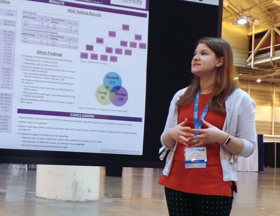
“While I think we can all recognize that specificity is important, I think sensitivity is more important to be able to get at sources of infection,” said Jenna Rossoff, MD, of Ann & Robert H. Lurie Children’s Hospital of Chicago.
Dr. Rossoff and her colleagues conducted this study and presented the results in a poster at the annual meeting of the American Society of Pediatric Hematology/Oncology.
Lurie Children’s Hospital began using a commercially available NGS pathogen test, the Karius test, in 2016. Dr. Rossoff and her colleagues set out to evaluate how the test affected patient care by conducting a retrospective analysis of tests performed from December 2016 through August 2018.
The researchers studied 100 NGS tests performed for 79 pediatric patients. The patients had a median age of 11 years (range, 0.5-24 years).
Most patients (n = 60) were immunocompromised, largely due to a hematologic malignancy (n = 16), primary immune deficiency (n = 13), hematopoietic cell transplant (n = 10), or solid organ transplant (n = 7).
The remaining 19 patients were immunocompetent, and 9 of them had no underlying diagnosis. The most common diagnosis for this group was neurologic disorder (n = 6).
Results
Of the 100 NGS tests evaluated, 70 were positive for any organism, and 56 of these were deemed clinically relevant.
“What I think is quite remarkable is that, of those clinically relevant organisms, tests on 14, which is 25% of those, were able to identify clinically relevant or pathogenic organisms when no other conventional testing modality was able to identify them,” Dr. Rossoff said. “And these were often in patients who underwent invasive procedures to try to get at the source of their infectious disease.”
In fact, the study included 42 patients who underwent 54 invasive diagnostic procedures, and 32 of those procedures could have been avoided based on positive NGS results, according to Dr. Rossoff and her colleagues.
Dr. Rossoff noted that the most common sites of infection were the bloodstream and respiratory tract, but the NGS test was able to identify pathogens in the bone, skin, cerebrospinal fluid, and urinary tract. She also pointed out that NGS results were available “in a fairly timely manner,” as 86% of test results were available within 48 hours of sample receipt.
Dr. Rossoff and her colleagues did not receive any funding for this study, but they were previously involved in a study funded by Karius, the company that commercialized the NGS test.
SOURCE: Rossoff J et al. ASPHO 2019. Abstract 439.
NEW ORLEANS – A next-generation sequencing (NGS) test for pathogen detection demonstrated higher sensitivity than conventional testing methods in a cohort of diverse pediatric patients, according to researchers.
The NGS test, which detects sequences of circulating cell-free DNA in plasma, detected pathogens with 92% sensitivity, compared with 64% sensitivity for all conventional testing methods combined (P less than .01).
“While I think we can all recognize that specificity is important, I think sensitivity is more important to be able to get at sources of infection,” said Jenna Rossoff, MD, of Ann & Robert H. Lurie Children’s Hospital of Chicago.
Dr. Rossoff and her colleagues conducted this study and presented the results in a poster at the annual meeting of the American Society of Pediatric Hematology/Oncology.
Lurie Children’s Hospital began using a commercially available NGS pathogen test, the Karius test, in 2016. Dr. Rossoff and her colleagues set out to evaluate how the test affected patient care by conducting a retrospective analysis of tests performed from December 2016 through August 2018.
The researchers studied 100 NGS tests performed for 79 pediatric patients. The patients had a median age of 11 years (range, 0.5-24 years).
Most patients (n = 60) were immunocompromised, largely due to a hematologic malignancy (n = 16), primary immune deficiency (n = 13), hematopoietic cell transplant (n = 10), or solid organ transplant (n = 7).
The remaining 19 patients were immunocompetent, and 9 of them had no underlying diagnosis. The most common diagnosis for this group was neurologic disorder (n = 6).
Results
Of the 100 NGS tests evaluated, 70 were positive for any organism, and 56 of these were deemed clinically relevant.
“What I think is quite remarkable is that, of those clinically relevant organisms, tests on 14, which is 25% of those, were able to identify clinically relevant or pathogenic organisms when no other conventional testing modality was able to identify them,” Dr. Rossoff said. “And these were often in patients who underwent invasive procedures to try to get at the source of their infectious disease.”
In fact, the study included 42 patients who underwent 54 invasive diagnostic procedures, and 32 of those procedures could have been avoided based on positive NGS results, according to Dr. Rossoff and her colleagues.
Dr. Rossoff noted that the most common sites of infection were the bloodstream and respiratory tract, but the NGS test was able to identify pathogens in the bone, skin, cerebrospinal fluid, and urinary tract. She also pointed out that NGS results were available “in a fairly timely manner,” as 86% of test results were available within 48 hours of sample receipt.
Dr. Rossoff and her colleagues did not receive any funding for this study, but they were previously involved in a study funded by Karius, the company that commercialized the NGS test.
SOURCE: Rossoff J et al. ASPHO 2019. Abstract 439.
REPORTING FROM 2019 ASPHO CONFERENCE
Widespread hyperpigmented plaques

The differential diagnosis included psoriasis, drug eruption, and a cutaneous T-cell lymphoma.
A drug eruption could have been due to an over-the-counter medication or supplement, so the lack of improvement from stopping the antihypertensive medication did not rule out this diagnosis. Psoriasis does not always show erythema in persons of color, but these plaques were not typical of psoriasis. (There also were some flat patches that were even less typical of psoriasis.)
The FP performed a 4-mm punch biopsy on one of the hyperpigmented plaques on the abdomen. A 4-mm punch biopsy is generally an ideal method for determining the cause of an unknown skin rash, and it is usually better to choose a lesion on the upper body rather than below the waist if the rash is widespread. (See the Watch & Learn video on “Punch biopsy.”)
The FP also prescribed a 1-pound tub of 0.1% triamcinolone ointment for symptomatic relief as this could help any of the possible diagnoses being considered. The pathology report came back as mycosis fungoides, the most common type of cutaneous T-cell lymphoma.
The patient was sent to Hematology/Oncology for further evaluation and treatment. Mycosis fungoides can have both patches and plaques and frequently involves the trunk more than the extremities (which was the situation in this case). It is important to consider uncommon diagnoses like this in the differential when the initial diagnosis does not appear to be responding to treatment or there is something atypical about the presentation of an expected diagnosis.
Photos and text for Photo Rounds Friday courtesy of Richard P. Usatine, MD. This case was adapted from: Chacon G, Nayar A, Usatine R, Smith M. Cutaneous T-cell lymphoma. In: Usatine R, Smith M, Mayeaux EJ, et al. Color Atlas and Synopsis of Family Medicine. 3rd ed. New York, NY: McGraw-Hill; 2019:1124-1131.
To learn more about the newest 3rd edition of the Color Atlas and Synopsis of Family Medicine, see: https://www.amazon.com/Color-Atlas-Synopsis-Family-Medicine/dp/1259862046/
You can get the Color Atlas of Family Medicine app by clicking on this link: usatinemedia.com
