User login
Bringing you the latest news, research and reviews, exclusive interviews, podcasts, quizzes, and more.
Powered by CHEST Physician, Clinician Reviews, MDedge Family Medicine, Internal Medicine News, and The Journal of Clinical Outcomes Management.
Asthma Blunted Worst H1N1 Outcomes
Asthmatics hospitalized during the H1N1 influenza pandemic had less-severe outcomes than did nonasthmatics, U.K. researchers have found.
People with asthma saw a lower risk of dying or requiring intensive care than did nonasthmatics, including people without any comorbidities. They were just as sick as nonasthmatics at admission. And the difference in outcomes could not be wholly ascribed to asthmatics’ use of inhaled steroids: Nonasthmatics taking inhaled steroids for other conditions did not see any protective effect.
A multivariate analysis showed asthma itself to be an independent factor for less-severe outcomes in patients hospitalized with H1N1.
This finding, from a prospective cohort of 1,520 people admitted to 75 different U.K. hospitals during the pandemic, was "surprising," said Dr. Malcolm Semple, the University of Liverpool, England, who presented his findings to the European Respiratory Society annual congress in Amsterdam.
However, it did align with recent results from a global analysis of 70,000 H1N1 patients, in which asthma was also seen to be associated with less severe outcomes (PLoS Med 2011;8:e1001053).
"Respiratory viruses cause exacerbations of asthma, and so it would be tempting to assume that these people were admitted with exacerbations of asthma – that they were less sick than the rest," Dr. Semple said in an interview. "But if anything, they were more sick, with more dyspnea and the same amount of radiological changes of pneumonia" as the other patients admitted to hospital.
"Do steroids protect everyone? No. But do steroids protect asthmatics? Emphatically, yes."
For their research, Dr. Semple and his colleagues used data from a prospective cohort in a study funded by the U.K. Department of Health during the H1N1 pandemic of 2009 and 2010. The cohort was used to provide real-time information on the pandemic and its clinical features to the Department of Health, and data collected were standardized to include information on age, comorbidities, inhaled steroid use, time from symptom onset to admission, and medications administered in hospital.
"It was something innovative that we had never tried before," Dr. Semple said of the cohort, adding that its large size "allowed us to catch this signal – it would be very hard to replicate this study in a non–pandemic situation."
The researchers found that the asthmatics, who comprised a quarter of the cohort (n = 385), were half as likely as nonasthmatics to die or require intensive care (11.2% vs. 19.8%; unadjusted odds ratio, 0.51) despite similar rates of pneumonia at admission.
Three variables – inhaled steroid use, admission within the first 4 days of symptoms, and systemic steroid use – were all seen as contributing to less severe outcomes for asthmatics. However, even after adjusting for these, simply having asthma was still associated with a 45% reduced likelihood of death or intensive care (adjusted OR, 0.55).
Asthmatics taking inhaled steroids were significantly less likely to die or require intensive care (7.4%) than were those not taking inhaled steroids (15.4%).
But inhaled steroids protected only the asthmatics in the study. About a fifth of the patients taking inhaled steroids were not taking them for asthma – and these patients saw no benefit, meaning that the findings do not support the use of inhaled corticosteroids in nonasthmatics.
"Do steroids protect everyone? No. But do steroids protect asthmatics? Emphatically, yes," Dr. Semple said. "The implication for practice is that, if you have a diagnosis of asthma, you should adhere to the published guidance if you’re a physician; and if you’re a patient, you should do as you’re told and take your steroids regularly."
Dr. Semple and his colleagues’ study was funded by the U.K. Department of Health. The researchers disclosed no conflicts of interest.
Asthmatics hospitalized during the H1N1 influenza pandemic had less-severe outcomes than did nonasthmatics, U.K. researchers have found.
People with asthma saw a lower risk of dying or requiring intensive care than did nonasthmatics, including people without any comorbidities. They were just as sick as nonasthmatics at admission. And the difference in outcomes could not be wholly ascribed to asthmatics’ use of inhaled steroids: Nonasthmatics taking inhaled steroids for other conditions did not see any protective effect.
A multivariate analysis showed asthma itself to be an independent factor for less-severe outcomes in patients hospitalized with H1N1.
This finding, from a prospective cohort of 1,520 people admitted to 75 different U.K. hospitals during the pandemic, was "surprising," said Dr. Malcolm Semple, the University of Liverpool, England, who presented his findings to the European Respiratory Society annual congress in Amsterdam.
However, it did align with recent results from a global analysis of 70,000 H1N1 patients, in which asthma was also seen to be associated with less severe outcomes (PLoS Med 2011;8:e1001053).
"Respiratory viruses cause exacerbations of asthma, and so it would be tempting to assume that these people were admitted with exacerbations of asthma – that they were less sick than the rest," Dr. Semple said in an interview. "But if anything, they were more sick, with more dyspnea and the same amount of radiological changes of pneumonia" as the other patients admitted to hospital.
"Do steroids protect everyone? No. But do steroids protect asthmatics? Emphatically, yes."
For their research, Dr. Semple and his colleagues used data from a prospective cohort in a study funded by the U.K. Department of Health during the H1N1 pandemic of 2009 and 2010. The cohort was used to provide real-time information on the pandemic and its clinical features to the Department of Health, and data collected were standardized to include information on age, comorbidities, inhaled steroid use, time from symptom onset to admission, and medications administered in hospital.
"It was something innovative that we had never tried before," Dr. Semple said of the cohort, adding that its large size "allowed us to catch this signal – it would be very hard to replicate this study in a non–pandemic situation."
The researchers found that the asthmatics, who comprised a quarter of the cohort (n = 385), were half as likely as nonasthmatics to die or require intensive care (11.2% vs. 19.8%; unadjusted odds ratio, 0.51) despite similar rates of pneumonia at admission.
Three variables – inhaled steroid use, admission within the first 4 days of symptoms, and systemic steroid use – were all seen as contributing to less severe outcomes for asthmatics. However, even after adjusting for these, simply having asthma was still associated with a 45% reduced likelihood of death or intensive care (adjusted OR, 0.55).
Asthmatics taking inhaled steroids were significantly less likely to die or require intensive care (7.4%) than were those not taking inhaled steroids (15.4%).
But inhaled steroids protected only the asthmatics in the study. About a fifth of the patients taking inhaled steroids were not taking them for asthma – and these patients saw no benefit, meaning that the findings do not support the use of inhaled corticosteroids in nonasthmatics.
"Do steroids protect everyone? No. But do steroids protect asthmatics? Emphatically, yes," Dr. Semple said. "The implication for practice is that, if you have a diagnosis of asthma, you should adhere to the published guidance if you’re a physician; and if you’re a patient, you should do as you’re told and take your steroids regularly."
Dr. Semple and his colleagues’ study was funded by the U.K. Department of Health. The researchers disclosed no conflicts of interest.
Asthmatics hospitalized during the H1N1 influenza pandemic had less-severe outcomes than did nonasthmatics, U.K. researchers have found.
People with asthma saw a lower risk of dying or requiring intensive care than did nonasthmatics, including people without any comorbidities. They were just as sick as nonasthmatics at admission. And the difference in outcomes could not be wholly ascribed to asthmatics’ use of inhaled steroids: Nonasthmatics taking inhaled steroids for other conditions did not see any protective effect.
A multivariate analysis showed asthma itself to be an independent factor for less-severe outcomes in patients hospitalized with H1N1.
This finding, from a prospective cohort of 1,520 people admitted to 75 different U.K. hospitals during the pandemic, was "surprising," said Dr. Malcolm Semple, the University of Liverpool, England, who presented his findings to the European Respiratory Society annual congress in Amsterdam.
However, it did align with recent results from a global analysis of 70,000 H1N1 patients, in which asthma was also seen to be associated with less severe outcomes (PLoS Med 2011;8:e1001053).
"Respiratory viruses cause exacerbations of asthma, and so it would be tempting to assume that these people were admitted with exacerbations of asthma – that they were less sick than the rest," Dr. Semple said in an interview. "But if anything, they were more sick, with more dyspnea and the same amount of radiological changes of pneumonia" as the other patients admitted to hospital.
"Do steroids protect everyone? No. But do steroids protect asthmatics? Emphatically, yes."
For their research, Dr. Semple and his colleagues used data from a prospective cohort in a study funded by the U.K. Department of Health during the H1N1 pandemic of 2009 and 2010. The cohort was used to provide real-time information on the pandemic and its clinical features to the Department of Health, and data collected were standardized to include information on age, comorbidities, inhaled steroid use, time from symptom onset to admission, and medications administered in hospital.
"It was something innovative that we had never tried before," Dr. Semple said of the cohort, adding that its large size "allowed us to catch this signal – it would be very hard to replicate this study in a non–pandemic situation."
The researchers found that the asthmatics, who comprised a quarter of the cohort (n = 385), were half as likely as nonasthmatics to die or require intensive care (11.2% vs. 19.8%; unadjusted odds ratio, 0.51) despite similar rates of pneumonia at admission.
Three variables – inhaled steroid use, admission within the first 4 days of symptoms, and systemic steroid use – were all seen as contributing to less severe outcomes for asthmatics. However, even after adjusting for these, simply having asthma was still associated with a 45% reduced likelihood of death or intensive care (adjusted OR, 0.55).
Asthmatics taking inhaled steroids were significantly less likely to die or require intensive care (7.4%) than were those not taking inhaled steroids (15.4%).
But inhaled steroids protected only the asthmatics in the study. About a fifth of the patients taking inhaled steroids were not taking them for asthma – and these patients saw no benefit, meaning that the findings do not support the use of inhaled corticosteroids in nonasthmatics.
"Do steroids protect everyone? No. But do steroids protect asthmatics? Emphatically, yes," Dr. Semple said. "The implication for practice is that, if you have a diagnosis of asthma, you should adhere to the published guidance if you’re a physician; and if you’re a patient, you should do as you’re told and take your steroids regularly."
Dr. Semple and his colleagues’ study was funded by the U.K. Department of Health. The researchers disclosed no conflicts of interest.
'On-Off' Sleep Switches Shed Light on Brain Disorders
SAN DIEGO – A series of "on-off" switches regulates sleep, clarifying many of the mechanisms underlying narcolepsy, cataplexy, and REM sleep behavior disorder, according to Dr. Clifford B. Saper.
The states of sleep, wakefulness, rapid eye movement and non-REM sleep can best be understood as "flip-flop" mechanisms of brain circuitry, akin to light switches, said Dr. Saper, professor of neurology and neuroscience at Harvard Medical School and head of the department of neurology at Beth Israel Deaconess Medical Center in Boston.

"Each side inhibits the other" in an ascending arousal pathway to the cortex, facilitating rapid transitions from one state to the other.
Normally, human beings spend 99% of the 24-hour day fully awake or fully asleep, and just 1% of the time transitioning. This is due to an on-off switch that regulates arousal and sleep, Dr. Saper said at the annual meeting of the American Neurological Association.
"One of the problems with a flip-flop switch is that it has a tendency, sometimes, to fall into the wrong position too easily. One can imagine driving down a boring road and flipping into the wrong state and suddenly being asleep behind the wheel of a car," he said.
To prevent such an occurrence, the brain stabilizes wakefulness by the use of orexins, or hypocretins, which are neuropeptides produced by excitatory neurons in the lateral region of the hypothalamus.
Narcolepsy, in which patients do fall asleep essentially at the "flip of a switch," is the result of a single neurotransmitter deficit in sleep’s "master switch," the ventrolateral preoptic nucleus, Dr. Saper explained.
A similar "flip-flop" switch regulates the normally rapid transition between REM and non-REM (slow-wave) sleep, he said.
The development of REM sleep behavior disorder (in which patients make jerky motor maneuvers as they act out dreams during sleep) and cataplexy – atonic lapses in muscle control from a waking state – are opposites on a spectrum, both indicative of triggering of the on-off mechanism at an inappropriate point in the cycle.
Of great interest to Dr. Saper is an evolving apparent link between the development of REM sleep behavior disorder in young adulthood and later development of Parkinson’s disease, a phenomenon that occurs in about half of REM behavior disorder patients within 12 years.
He noted that Dr. Ronald B. Postuma and his associates at Montreal General Hospital have identified early markers of Parkinson’s disease in idiopathic REM sleep behavior disorder patients, including difficulties with visual and olfactory discrimination tasks and subthreshold but low scores on the Unified Parkinson's Disease Rating Scale.
The connection has led some researchers to suspect that synucleinopathies such as Parkinson’s disease and dementia with Lewy bodies may begin at the brainstem level of the locus coeruleus or the subcoeruleus complex and slowly progress in an ascending pathway to the basal ganglia over years or decades, offering the possibility of introducing neuroprotective therapy to stop that progression.
Dr. Saper reported having no relevant financial disclosures.
parkinsons sleep
SAN DIEGO – A series of "on-off" switches regulates sleep, clarifying many of the mechanisms underlying narcolepsy, cataplexy, and REM sleep behavior disorder, according to Dr. Clifford B. Saper.
The states of sleep, wakefulness, rapid eye movement and non-REM sleep can best be understood as "flip-flop" mechanisms of brain circuitry, akin to light switches, said Dr. Saper, professor of neurology and neuroscience at Harvard Medical School and head of the department of neurology at Beth Israel Deaconess Medical Center in Boston.
"Each side inhibits the other" in an ascending arousal pathway to the cortex, facilitating rapid transitions from one state to the other.
Normally, human beings spend 99% of the 24-hour day fully awake or fully asleep, and just 1% of the time transitioning. This is due to an on-off switch that regulates arousal and sleep, Dr. Saper said at the annual meeting of the American Neurological Association.
"One of the problems with a flip-flop switch is that it has a tendency, sometimes, to fall into the wrong position too easily. One can imagine driving down a boring road and flipping into the wrong state and suddenly being asleep behind the wheel of a car," he said.
To prevent such an occurrence, the brain stabilizes wakefulness by the use of orexins, or hypocretins, which are neuropeptides produced by excitatory neurons in the lateral region of the hypothalamus.
Narcolepsy, in which patients do fall asleep essentially at the "flip of a switch," is the result of a single neurotransmitter deficit in sleep’s "master switch," the ventrolateral preoptic nucleus, Dr. Saper explained.
A similar "flip-flop" switch regulates the normally rapid transition between REM and non-REM (slow-wave) sleep, he said.
The development of REM sleep behavior disorder (in which patients make jerky motor maneuvers as they act out dreams during sleep) and cataplexy – atonic lapses in muscle control from a waking state – are opposites on a spectrum, both indicative of triggering of the on-off mechanism at an inappropriate point in the cycle.
Of great interest to Dr. Saper is an evolving apparent link between the development of REM sleep behavior disorder in young adulthood and later development of Parkinson’s disease, a phenomenon that occurs in about half of REM behavior disorder patients within 12 years.
He noted that Dr. Ronald B. Postuma and his associates at Montreal General Hospital have identified early markers of Parkinson’s disease in idiopathic REM sleep behavior disorder patients, including difficulties with visual and olfactory discrimination tasks and subthreshold but low scores on the Unified Parkinson's Disease Rating Scale.
The connection has led some researchers to suspect that synucleinopathies such as Parkinson’s disease and dementia with Lewy bodies may begin at the brainstem level of the locus coeruleus or the subcoeruleus complex and slowly progress in an ascending pathway to the basal ganglia over years or decades, offering the possibility of introducing neuroprotective therapy to stop that progression.
Dr. Saper reported having no relevant financial disclosures.
SAN DIEGO – A series of "on-off" switches regulates sleep, clarifying many of the mechanisms underlying narcolepsy, cataplexy, and REM sleep behavior disorder, according to Dr. Clifford B. Saper.
The states of sleep, wakefulness, rapid eye movement and non-REM sleep can best be understood as "flip-flop" mechanisms of brain circuitry, akin to light switches, said Dr. Saper, professor of neurology and neuroscience at Harvard Medical School and head of the department of neurology at Beth Israel Deaconess Medical Center in Boston.
"Each side inhibits the other" in an ascending arousal pathway to the cortex, facilitating rapid transitions from one state to the other.
Normally, human beings spend 99% of the 24-hour day fully awake or fully asleep, and just 1% of the time transitioning. This is due to an on-off switch that regulates arousal and sleep, Dr. Saper said at the annual meeting of the American Neurological Association.
"One of the problems with a flip-flop switch is that it has a tendency, sometimes, to fall into the wrong position too easily. One can imagine driving down a boring road and flipping into the wrong state and suddenly being asleep behind the wheel of a car," he said.
To prevent such an occurrence, the brain stabilizes wakefulness by the use of orexins, or hypocretins, which are neuropeptides produced by excitatory neurons in the lateral region of the hypothalamus.
Narcolepsy, in which patients do fall asleep essentially at the "flip of a switch," is the result of a single neurotransmitter deficit in sleep’s "master switch," the ventrolateral preoptic nucleus, Dr. Saper explained.
A similar "flip-flop" switch regulates the normally rapid transition between REM and non-REM (slow-wave) sleep, he said.
The development of REM sleep behavior disorder (in which patients make jerky motor maneuvers as they act out dreams during sleep) and cataplexy – atonic lapses in muscle control from a waking state – are opposites on a spectrum, both indicative of triggering of the on-off mechanism at an inappropriate point in the cycle.
Of great interest to Dr. Saper is an evolving apparent link between the development of REM sleep behavior disorder in young adulthood and later development of Parkinson’s disease, a phenomenon that occurs in about half of REM behavior disorder patients within 12 years.
He noted that Dr. Ronald B. Postuma and his associates at Montreal General Hospital have identified early markers of Parkinson’s disease in idiopathic REM sleep behavior disorder patients, including difficulties with visual and olfactory discrimination tasks and subthreshold but low scores on the Unified Parkinson's Disease Rating Scale.
The connection has led some researchers to suspect that synucleinopathies such as Parkinson’s disease and dementia with Lewy bodies may begin at the brainstem level of the locus coeruleus or the subcoeruleus complex and slowly progress in an ascending pathway to the basal ganglia over years or decades, offering the possibility of introducing neuroprotective therapy to stop that progression.
Dr. Saper reported having no relevant financial disclosures.
parkinsons sleep
parkinsons sleep
EXPERT ANALYSIS FROM THE ANNUAL MEETING OF THE AMERICAN NEUROLOGICAL ASSOCIATION
FDA: Increased PAH Risk Seen With Dasatinib
Treatment with the leukemia drug dasatinib has been associated with an increased risk for pulmonary arterial hypertension, which can occur at any time after starting treatment, the Food and Drug Administration announced on Oct. 11.
None of the cases was fatal, and PAH "may be reversible" if treatment is discontinued, according to the statement, posted on the agency’s MedWatch site.
Dasatinib, a kinase inhibitor marketed as Sprycel by Bristol-Myers Squibb, is approved for treating certain adults with Philadelphia chromosome-positive chronic myeloid leukemia (CML) or acute lymphoblastic leukemia (ALL). It is an oral therapy, administered once daily.
Since dasatinib was approved in 2006, the BMS global pharmacovigilance database has identified cases of PAH in treated patients, the statement said. In 12 of these cases, right heart catheterization confirmed the diagnosis, and dasatinib was considered "the most likely cause," the FDA said. These patients had developed symptoms at various time intervals after starting treatment, including more than 12 months afterward, and they often were taking other medications or had comorbidities, so "there may be a combination of factors contributing to the development of PAH" in patients taking dasatinib, the FDA said.
Because dyspnea, fatigue, hypoxia, fluid retention, and other PAH symptoms overlap with those of other conditions, "a diagnosis of Sprycel-associated PAH should be considered" if other causes have been ruled out in symptomatic patients, the FDA advises. Health care professionals should also evaluate patients for signs and symptoms of underlying cardiopulmonary disease before starting treatment and during treatment. The drug should be permanently discontinued if a diagnosis of PAH is confirmed.
Improvements in hemodynamic and clinical parameters were observed following discontinuation in some patients, the FDA statement said.
This information has been added to the drug’s prescribing information. Serious adverse events associated with dasatinib should be reported to the FDA’s MedWatch program at 800-332-1088.
Treatment with the leukemia drug dasatinib has been associated with an increased risk for pulmonary arterial hypertension, which can occur at any time after starting treatment, the Food and Drug Administration announced on Oct. 11.
None of the cases was fatal, and PAH "may be reversible" if treatment is discontinued, according to the statement, posted on the agency’s MedWatch site.
Dasatinib, a kinase inhibitor marketed as Sprycel by Bristol-Myers Squibb, is approved for treating certain adults with Philadelphia chromosome-positive chronic myeloid leukemia (CML) or acute lymphoblastic leukemia (ALL). It is an oral therapy, administered once daily.
Since dasatinib was approved in 2006, the BMS global pharmacovigilance database has identified cases of PAH in treated patients, the statement said. In 12 of these cases, right heart catheterization confirmed the diagnosis, and dasatinib was considered "the most likely cause," the FDA said. These patients had developed symptoms at various time intervals after starting treatment, including more than 12 months afterward, and they often were taking other medications or had comorbidities, so "there may be a combination of factors contributing to the development of PAH" in patients taking dasatinib, the FDA said.
Because dyspnea, fatigue, hypoxia, fluid retention, and other PAH symptoms overlap with those of other conditions, "a diagnosis of Sprycel-associated PAH should be considered" if other causes have been ruled out in symptomatic patients, the FDA advises. Health care professionals should also evaluate patients for signs and symptoms of underlying cardiopulmonary disease before starting treatment and during treatment. The drug should be permanently discontinued if a diagnosis of PAH is confirmed.
Improvements in hemodynamic and clinical parameters were observed following discontinuation in some patients, the FDA statement said.
This information has been added to the drug’s prescribing information. Serious adverse events associated with dasatinib should be reported to the FDA’s MedWatch program at 800-332-1088.
Treatment with the leukemia drug dasatinib has been associated with an increased risk for pulmonary arterial hypertension, which can occur at any time after starting treatment, the Food and Drug Administration announced on Oct. 11.
None of the cases was fatal, and PAH "may be reversible" if treatment is discontinued, according to the statement, posted on the agency’s MedWatch site.
Dasatinib, a kinase inhibitor marketed as Sprycel by Bristol-Myers Squibb, is approved for treating certain adults with Philadelphia chromosome-positive chronic myeloid leukemia (CML) or acute lymphoblastic leukemia (ALL). It is an oral therapy, administered once daily.
Since dasatinib was approved in 2006, the BMS global pharmacovigilance database has identified cases of PAH in treated patients, the statement said. In 12 of these cases, right heart catheterization confirmed the diagnosis, and dasatinib was considered "the most likely cause," the FDA said. These patients had developed symptoms at various time intervals after starting treatment, including more than 12 months afterward, and they often were taking other medications or had comorbidities, so "there may be a combination of factors contributing to the development of PAH" in patients taking dasatinib, the FDA said.
Because dyspnea, fatigue, hypoxia, fluid retention, and other PAH symptoms overlap with those of other conditions, "a diagnosis of Sprycel-associated PAH should be considered" if other causes have been ruled out in symptomatic patients, the FDA advises. Health care professionals should also evaluate patients for signs and symptoms of underlying cardiopulmonary disease before starting treatment and during treatment. The drug should be permanently discontinued if a diagnosis of PAH is confirmed.
Improvements in hemodynamic and clinical parameters were observed following discontinuation in some patients, the FDA statement said.
This information has been added to the drug’s prescribing information. Serious adverse events associated with dasatinib should be reported to the FDA’s MedWatch program at 800-332-1088.
FDA Approves CFC-Free Inhaler for COPD
An inhalation spray containing ipratropium bromide and albuterol sulfate had been approved for patients with chronic obstructive pulmonary disease, according to a statement issued Oct. 7 by the Food and Drug Administration.
The product, marketed as Combivent Respimat inhalation spray, does not contain chlorofluorocarbons (CFCs) and "is a suitable alternative for patients who are currently using Combivent (ipratropium bromide and albuterol sulfate) inhalation aerosol," according to the statement, issued by the Division of Drug Information (DDI) in the FDA’s Center for Drugs, Evaluation and Research (CDER). Combivent inhalation aerosol, which contains CFCs, will not be available after Dec. 31, 2013. Like other inhalers that contain CFCs that deplete the ozone layer, the inhaler is being phased out because of the Montreal Protocol on Substances that Deplete the Ozone Layer, which makes it illegal to sell or make substances that decrease the ozone layer.
Ipratropium is an anticholinergic bronchodilator, and albuterol is a selective beta 2 adrenergic bronchodilator. Combivent inhalers are indicated for people with COPD, on a regular bronchodilator, who continue to have evidence of bronchospasm and who require a second bronchodilator.
An inhalation spray containing ipratropium bromide and albuterol sulfate had been approved for patients with chronic obstructive pulmonary disease, according to a statement issued Oct. 7 by the Food and Drug Administration.
The product, marketed as Combivent Respimat inhalation spray, does not contain chlorofluorocarbons (CFCs) and "is a suitable alternative for patients who are currently using Combivent (ipratropium bromide and albuterol sulfate) inhalation aerosol," according to the statement, issued by the Division of Drug Information (DDI) in the FDA’s Center for Drugs, Evaluation and Research (CDER). Combivent inhalation aerosol, which contains CFCs, will not be available after Dec. 31, 2013. Like other inhalers that contain CFCs that deplete the ozone layer, the inhaler is being phased out because of the Montreal Protocol on Substances that Deplete the Ozone Layer, which makes it illegal to sell or make substances that decrease the ozone layer.
Ipratropium is an anticholinergic bronchodilator, and albuterol is a selective beta 2 adrenergic bronchodilator. Combivent inhalers are indicated for people with COPD, on a regular bronchodilator, who continue to have evidence of bronchospasm and who require a second bronchodilator.
An inhalation spray containing ipratropium bromide and albuterol sulfate had been approved for patients with chronic obstructive pulmonary disease, according to a statement issued Oct. 7 by the Food and Drug Administration.
The product, marketed as Combivent Respimat inhalation spray, does not contain chlorofluorocarbons (CFCs) and "is a suitable alternative for patients who are currently using Combivent (ipratropium bromide and albuterol sulfate) inhalation aerosol," according to the statement, issued by the Division of Drug Information (DDI) in the FDA’s Center for Drugs, Evaluation and Research (CDER). Combivent inhalation aerosol, which contains CFCs, will not be available after Dec. 31, 2013. Like other inhalers that contain CFCs that deplete the ozone layer, the inhaler is being phased out because of the Montreal Protocol on Substances that Deplete the Ozone Layer, which makes it illegal to sell or make substances that decrease the ozone layer.
Ipratropium is an anticholinergic bronchodilator, and albuterol is a selective beta 2 adrenergic bronchodilator. Combivent inhalers are indicated for people with COPD, on a regular bronchodilator, who continue to have evidence of bronchospasm and who require a second bronchodilator.
Single-Dose Azithromycin is Safe Option for Treating Pneumonia
CHICAGO – Compressing the usual 3-day course of intravenous azithromycin into a single high dose appears to be a safe, efficacious alternative for treating community-acquired pneumonia in adult outpatients.
In an open-label, randomized trial, investigators in Austria compared the same total dose of azithromycin either split over 3 days or given as a single infusion among 72 outpatients with community-acquired pneumonia (CAP).
Trial results, reported at the annual Interscience conference on Antimicrobial Agents and Chemotherapy, showed that the two regimens had similar efficacy and adverse effect profiles. In both groups, nearly 90% of patients were cured.
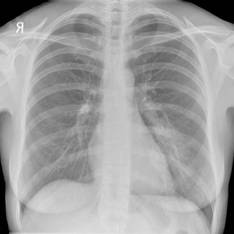
The single-dose strategy ensures high levels of the drug independent of patient compliance, according to presenting author Dr. Rainer Gattringer of the Elisabethinen Hospital and Analyse BioLab, both in Linz, Austria. Additionally, it may reduce treatment costs.
"I think maybe physicians are a little bit afraid because it’s not written in the description of the drug that you can use it at 1.5 g as a single dose," he proposed in an interview. "They say, I have no experience with it, and they don’t know about side effects and so on."
But the drug appeared safe and well tolerated with the protocol used in the trial: administration in a liter of infusion solution given over an extended period of 4 hours. "That [protocol] is I think the main thing," Dr. Gattringer said. "So don’t be afraid, try it."
The patients studied were 45 years old, on average, and 65% were men, according to data reported in a poster session. They were fairly healthy, Dr. Gattringer noted; the CRB-65 (confusion, respiratory rate, blood pressure, and age 65 years or older) score was 0, 1, and 2 in 33.3%, 51.4%, and 15.3%, respectively. Overall, 18% had previously received oral antibiotic therapy.
The most common pathogens identified were Streptococcus pneumoniae (22.2% of patients), Mycoplasma pneumoniae (6.9%), Legionella pneumophila (6.9%), Coxiella burnetii (1.4%), and Cytomegalovirus (1.4%). No pathogen was identified in 59.7% of cases.
The patients received either a single IV dose of 1.5 g azithromycin (diluted in 1 L of lactated Ringer solution and given as a 4-hour infusion) or the conventional dose schedule of 500 mg azithromycin IV once daily for 3 days.
Azithromycin is especially useful in patients who have an allergy to beta-lactam antibiotics, and the intravenous formulation is attractive given that oral macrolides, including azithromycin, have low bioavailability, Dr. Gattringer noted at the conference, which was sponsored by the American Society for Microbiology.
The rate of clinical cure – defined as resolution of fever within 72 hours; disappearance of other clinical and laboratory signs of inflammation, including achievement of C-reactive protein level of less than 1 mg/dL; and no need for additional antimicrobial therapy at 10 days – was statistically indistinguishable between the single-dose group and the conventional-regimen group (87.2% vs. 87.9%).
With the single-dose strategy, "you can send the patient home and you can achieve the right level of the drug in the patient," he concluded.
Dr. Gattringer reported having no conflicts of interest related to the trial. The trial was supported by a grant from Pfizer Research, manufacturer of Zithromax (azithromycin).
CHICAGO – Compressing the usual 3-day course of intravenous azithromycin into a single high dose appears to be a safe, efficacious alternative for treating community-acquired pneumonia in adult outpatients.
In an open-label, randomized trial, investigators in Austria compared the same total dose of azithromycin either split over 3 days or given as a single infusion among 72 outpatients with community-acquired pneumonia (CAP).
Trial results, reported at the annual Interscience conference on Antimicrobial Agents and Chemotherapy, showed that the two regimens had similar efficacy and adverse effect profiles. In both groups, nearly 90% of patients were cured.
The single-dose strategy ensures high levels of the drug independent of patient compliance, according to presenting author Dr. Rainer Gattringer of the Elisabethinen Hospital and Analyse BioLab, both in Linz, Austria. Additionally, it may reduce treatment costs.
"I think maybe physicians are a little bit afraid because it’s not written in the description of the drug that you can use it at 1.5 g as a single dose," he proposed in an interview. "They say, I have no experience with it, and they don’t know about side effects and so on."
But the drug appeared safe and well tolerated with the protocol used in the trial: administration in a liter of infusion solution given over an extended period of 4 hours. "That [protocol] is I think the main thing," Dr. Gattringer said. "So don’t be afraid, try it."
The patients studied were 45 years old, on average, and 65% were men, according to data reported in a poster session. They were fairly healthy, Dr. Gattringer noted; the CRB-65 (confusion, respiratory rate, blood pressure, and age 65 years or older) score was 0, 1, and 2 in 33.3%, 51.4%, and 15.3%, respectively. Overall, 18% had previously received oral antibiotic therapy.
The most common pathogens identified were Streptococcus pneumoniae (22.2% of patients), Mycoplasma pneumoniae (6.9%), Legionella pneumophila (6.9%), Coxiella burnetii (1.4%), and Cytomegalovirus (1.4%). No pathogen was identified in 59.7% of cases.
The patients received either a single IV dose of 1.5 g azithromycin (diluted in 1 L of lactated Ringer solution and given as a 4-hour infusion) or the conventional dose schedule of 500 mg azithromycin IV once daily for 3 days.
Azithromycin is especially useful in patients who have an allergy to beta-lactam antibiotics, and the intravenous formulation is attractive given that oral macrolides, including azithromycin, have low bioavailability, Dr. Gattringer noted at the conference, which was sponsored by the American Society for Microbiology.
The rate of clinical cure – defined as resolution of fever within 72 hours; disappearance of other clinical and laboratory signs of inflammation, including achievement of C-reactive protein level of less than 1 mg/dL; and no need for additional antimicrobial therapy at 10 days – was statistically indistinguishable between the single-dose group and the conventional-regimen group (87.2% vs. 87.9%).
With the single-dose strategy, "you can send the patient home and you can achieve the right level of the drug in the patient," he concluded.
Dr. Gattringer reported having no conflicts of interest related to the trial. The trial was supported by a grant from Pfizer Research, manufacturer of Zithromax (azithromycin).
CHICAGO – Compressing the usual 3-day course of intravenous azithromycin into a single high dose appears to be a safe, efficacious alternative for treating community-acquired pneumonia in adult outpatients.
In an open-label, randomized trial, investigators in Austria compared the same total dose of azithromycin either split over 3 days or given as a single infusion among 72 outpatients with community-acquired pneumonia (CAP).
Trial results, reported at the annual Interscience conference on Antimicrobial Agents and Chemotherapy, showed that the two regimens had similar efficacy and adverse effect profiles. In both groups, nearly 90% of patients were cured.
The single-dose strategy ensures high levels of the drug independent of patient compliance, according to presenting author Dr. Rainer Gattringer of the Elisabethinen Hospital and Analyse BioLab, both in Linz, Austria. Additionally, it may reduce treatment costs.
"I think maybe physicians are a little bit afraid because it’s not written in the description of the drug that you can use it at 1.5 g as a single dose," he proposed in an interview. "They say, I have no experience with it, and they don’t know about side effects and so on."
But the drug appeared safe and well tolerated with the protocol used in the trial: administration in a liter of infusion solution given over an extended period of 4 hours. "That [protocol] is I think the main thing," Dr. Gattringer said. "So don’t be afraid, try it."
The patients studied were 45 years old, on average, and 65% were men, according to data reported in a poster session. They were fairly healthy, Dr. Gattringer noted; the CRB-65 (confusion, respiratory rate, blood pressure, and age 65 years or older) score was 0, 1, and 2 in 33.3%, 51.4%, and 15.3%, respectively. Overall, 18% had previously received oral antibiotic therapy.
The most common pathogens identified were Streptococcus pneumoniae (22.2% of patients), Mycoplasma pneumoniae (6.9%), Legionella pneumophila (6.9%), Coxiella burnetii (1.4%), and Cytomegalovirus (1.4%). No pathogen was identified in 59.7% of cases.
The patients received either a single IV dose of 1.5 g azithromycin (diluted in 1 L of lactated Ringer solution and given as a 4-hour infusion) or the conventional dose schedule of 500 mg azithromycin IV once daily for 3 days.
Azithromycin is especially useful in patients who have an allergy to beta-lactam antibiotics, and the intravenous formulation is attractive given that oral macrolides, including azithromycin, have low bioavailability, Dr. Gattringer noted at the conference, which was sponsored by the American Society for Microbiology.
The rate of clinical cure – defined as resolution of fever within 72 hours; disappearance of other clinical and laboratory signs of inflammation, including achievement of C-reactive protein level of less than 1 mg/dL; and no need for additional antimicrobial therapy at 10 days – was statistically indistinguishable between the single-dose group and the conventional-regimen group (87.2% vs. 87.9%).
With the single-dose strategy, "you can send the patient home and you can achieve the right level of the drug in the patient," he concluded.
Dr. Gattringer reported having no conflicts of interest related to the trial. The trial was supported by a grant from Pfizer Research, manufacturer of Zithromax (azithromycin).
FROM THE ANNUAL INTERSCIENCE CONFERENCE ON ANTIMICROBIAL AGENTS AND CHEMOTHERAPY
Major Finding: The cure rate did not differ between patients given the conventional 3-day regimen of IV azithromycin (87.9%) and patients given a single high IV dose of azithromycin (87.2%).
Data Source: An open-label randomized trial among 72 adult outpatients with community-acquired pneumonia.
Disclosures: Dr. Gattringer reported having no relevant conflicts of interest. The trial was supported by a grant from Pfizer Research.
ECMO Reduced Hospital Mortality in Flu Patients
Hospital mortality rates were 55% lower in 2009 influenza A(H1N1) patients with severe acute respiratory distress syndrome who received extracorporeal membrane oxygenation compared with non-ECMO controls, based on data from a cohort study of 80 patients.
Data from a recent randomized trial showed that patients with acute respiratory distress syndrome (ARDS) who were transferred to an ECMO center were more likely to survive without severe disability compared with non-ECMO patients, but the role of the treatment remains controversial because of the increased costs associated with its use, said Dr. Moronke Noah of Glenfield Hospital in Leicester, England, and colleagues.
In this study, the researchers used data from the Swine Flu Triage study (SwiFT) to compare mortality rates in patients with ARDS resulting from the 2009 H1N1 flu who received ECMO with non-ECMO controls. SwiFT was a prospective study of patients with suspected or confirmed 2009 H1N1 flu who were referred for critical care.
The findings were simultaneously presented at the European Society of Intensive Care Medicine and published online Oct. 5 in JAMA (Epub doi:10.1001/jama.2011.1471).
The researchers reviewed data from 80 patients who were referred for ECMO. They used three different matching techniques: individual matching, propensity scoring, and GenMatch matching. A total of 59 patients were matched with non-ECMO controls using individual matching, 75 were matched with non-ECMO controls using propensity scoring, and 75 were matched with non-ECMO controls using GenMatch matching. GenMatch "combines propensity score matching with multivariate matching," the authors explained.
The hospital mortality rates were significantly lower among ECMO patients than among non-ECMO patients in each of the three matching techniques. Hospital mortality rates were 24% in ECMO patients and 53% in non-ECMO patients when individual matching was used; 24% and 47%, respectively, when propensity scoring was used; and 24% and 51%, respectively, when GenMatch was used.
The findings were limited by the possible role of unobserved confounding variables, and by the lack of data on the exact treatment protocols of non-ECMO patients, the researchers noted.
However, "the unique value of this study lies in the homogeneity of the patients with H1N1-related ARDS and the matching methods used," they said. The consistency of the results across all three matching methods strengthens the role of ECMO in reducing hospital mortality in these patients, they said.
The SwiFT study was supported by the U.K. National Institute for Health Research. Dr. Noah had no financial conflicts to disclose, but many of the study coauthors have received reimbursement or grant support from multiple pharmaceutical companies and institutions.
Hospital mortality rates were 55% lower in 2009 influenza A(H1N1) patients with severe acute respiratory distress syndrome who received extracorporeal membrane oxygenation compared with non-ECMO controls, based on data from a cohort study of 80 patients.
Data from a recent randomized trial showed that patients with acute respiratory distress syndrome (ARDS) who were transferred to an ECMO center were more likely to survive without severe disability compared with non-ECMO patients, but the role of the treatment remains controversial because of the increased costs associated with its use, said Dr. Moronke Noah of Glenfield Hospital in Leicester, England, and colleagues.
In this study, the researchers used data from the Swine Flu Triage study (SwiFT) to compare mortality rates in patients with ARDS resulting from the 2009 H1N1 flu who received ECMO with non-ECMO controls. SwiFT was a prospective study of patients with suspected or confirmed 2009 H1N1 flu who were referred for critical care.
The findings were simultaneously presented at the European Society of Intensive Care Medicine and published online Oct. 5 in JAMA (Epub doi:10.1001/jama.2011.1471).
The researchers reviewed data from 80 patients who were referred for ECMO. They used three different matching techniques: individual matching, propensity scoring, and GenMatch matching. A total of 59 patients were matched with non-ECMO controls using individual matching, 75 were matched with non-ECMO controls using propensity scoring, and 75 were matched with non-ECMO controls using GenMatch matching. GenMatch "combines propensity score matching with multivariate matching," the authors explained.
The hospital mortality rates were significantly lower among ECMO patients than among non-ECMO patients in each of the three matching techniques. Hospital mortality rates were 24% in ECMO patients and 53% in non-ECMO patients when individual matching was used; 24% and 47%, respectively, when propensity scoring was used; and 24% and 51%, respectively, when GenMatch was used.
The findings were limited by the possible role of unobserved confounding variables, and by the lack of data on the exact treatment protocols of non-ECMO patients, the researchers noted.
However, "the unique value of this study lies in the homogeneity of the patients with H1N1-related ARDS and the matching methods used," they said. The consistency of the results across all three matching methods strengthens the role of ECMO in reducing hospital mortality in these patients, they said.
The SwiFT study was supported by the U.K. National Institute for Health Research. Dr. Noah had no financial conflicts to disclose, but many of the study coauthors have received reimbursement or grant support from multiple pharmaceutical companies and institutions.
Hospital mortality rates were 55% lower in 2009 influenza A(H1N1) patients with severe acute respiratory distress syndrome who received extracorporeal membrane oxygenation compared with non-ECMO controls, based on data from a cohort study of 80 patients.
Data from a recent randomized trial showed that patients with acute respiratory distress syndrome (ARDS) who were transferred to an ECMO center were more likely to survive without severe disability compared with non-ECMO patients, but the role of the treatment remains controversial because of the increased costs associated with its use, said Dr. Moronke Noah of Glenfield Hospital in Leicester, England, and colleagues.
In this study, the researchers used data from the Swine Flu Triage study (SwiFT) to compare mortality rates in patients with ARDS resulting from the 2009 H1N1 flu who received ECMO with non-ECMO controls. SwiFT was a prospective study of patients with suspected or confirmed 2009 H1N1 flu who were referred for critical care.
The findings were simultaneously presented at the European Society of Intensive Care Medicine and published online Oct. 5 in JAMA (Epub doi:10.1001/jama.2011.1471).
The researchers reviewed data from 80 patients who were referred for ECMO. They used three different matching techniques: individual matching, propensity scoring, and GenMatch matching. A total of 59 patients were matched with non-ECMO controls using individual matching, 75 were matched with non-ECMO controls using propensity scoring, and 75 were matched with non-ECMO controls using GenMatch matching. GenMatch "combines propensity score matching with multivariate matching," the authors explained.
The hospital mortality rates were significantly lower among ECMO patients than among non-ECMO patients in each of the three matching techniques. Hospital mortality rates were 24% in ECMO patients and 53% in non-ECMO patients when individual matching was used; 24% and 47%, respectively, when propensity scoring was used; and 24% and 51%, respectively, when GenMatch was used.
The findings were limited by the possible role of unobserved confounding variables, and by the lack of data on the exact treatment protocols of non-ECMO patients, the researchers noted.
However, "the unique value of this study lies in the homogeneity of the patients with H1N1-related ARDS and the matching methods used," they said. The consistency of the results across all three matching methods strengthens the role of ECMO in reducing hospital mortality in these patients, they said.
The SwiFT study was supported by the U.K. National Institute for Health Research. Dr. Noah had no financial conflicts to disclose, but many of the study coauthors have received reimbursement or grant support from multiple pharmaceutical companies and institutions.
FROM JAMA
Major Finding: Extracorporeal membrane oxygenation reduced hospital mortality by 55% in patients with severe acute respiratory distress resulting from the 2009 H1N1 flu, compared with controls who did not receive ECMO.
Data Source: A cohort study of 80 adults with 2009 H1N1 influenza and severe acute respiratory distress.
Disclosures: The SwiFT study was supported by the National Institute for Health Research. Dr. Noah had no financial conflicts to disclose, but many of the study coauthors have received reimbursement or grant support from multiple pharmaceutical companies and institutions.
Asthma Care Measures Don't Reflect Outcomes in Children
Hospital compliance with the Children’s Asthma Care set of process measures did not correlate with asthma patients’ clinical outcomes in a study of more than 37,000 asthma patients who were admitted to 30 U.S. children’s hospitals, according to a study reported in the Oct. 5 issue of JAMA.
Because compliance with these process measures was not associated with improved outcomes, it "cannot serve as a means to evaluate and compare the quality of care provided for patients admitted with asthma exacerbations," said Dr. Rustin B. Morse of Phoenix Children’s Hospital and the University of Arizona, Phoenix, and his associates.
The Joint Commission considers the Children’s Asthma Care (CAC) measure set to be an "accountability measure," appropriate for use in determining accreditation, in public reporting of hospital performance, and in pay-for-performance efforts. But the findings of this study instead suggest that the CAC measure set does not meet the Joint Commission criteria for accountability measures and should be "reconsidered," Dr. Morse and his colleagues said.
They assessed time trends in compliance with the CAC measure set using data on a random sample of 37,267 pediatric inpatients with 45,499 admissions for asthma exacerbations during a 33-month period at 30 freestanding children’s hospitals across the country.
The CAC measure set includes three measures: whether patients received asthma relievers on admission (CAC-1), whether they received systemic corticosteroids on admission (CAC-2), and whether they were discharged with a complete home management plan of care (CAC-3). Compliance is measured quarterly by a review of the medical records of a random sample of patients.
Compliance with CAC-1 and CAC-2 was quite high, exceeding 95% in all but 1 of the 11 quarters assessed, and was consistent across hospitals. Because there were so few cases of poor compliance, no analysis could be performed to examine whether better compliance correlated with improved clinical outcomes.
In contrast, compliance with CAC-3 was not as high and varied substantially among hospitals. Mean CAC-3 compliance was only 41% during the first three quarters of the study and improved to 73% in the final three quarters.
This allowed an analysis of the relationship between compliance with CAC-3 and clinical outcomes. But no significant association was found between CAC-3 compliance and improved outcomes at 7 days, 30 days, or 90 days after discharge, the investigators said (JAMA 2011;306:1454-60).
There also was no association between compliance and clinical outcomes when hospitals in the highest-performing quartile were compared with those in the lowest-performing quartile.
One of Dr. Morse’s associates reported ties to the Robert Wood Johnson Foundation, the National Institute of Allergy and Infectious Diseases, the Child Health Corporation of America, and the Pediatric Research in Inpatient Settings Network. Two reported grants from the Agency for Healthcare ResearcDr. Homer reported no financial conflicts of interest.
The findings of Dr. Morse and coauthors demonstrate that the "use of a written discharge management plan no longer meets the criteria for a high-quality measure," Dr. Charles J. Homer said.
The authors showed that the Joint Commission’s CAC-3 measure (a written plan for managing asthma given to the patient at discharge) "should be retired" as a measure of hospital performance.
They also showed that compliance with CAC measures 1 and 2 is nearly universal within a subset of freestanding children’s hospitals. However, more than two-thirds of hospitalizations occur at other types of facilities, and the performance of these measures is yet to be documented in other settings.
Dr. Homer is with the National Initiative for Children’s Healthcare Quality and the department of pediatrics at Harvard Medical School and Children’s Hospital Boston. He reported no financial conflicts of interest. These remarks were adapted from his editorial accompanying Dr. Morse’s report (JAMA 2011;306:1487-8).
The findings of Dr. Morse and coauthors demonstrate that the "use of a written discharge management plan no longer meets the criteria for a high-quality measure," Dr. Charles J. Homer said.
The authors showed that the Joint Commission’s CAC-3 measure (a written plan for managing asthma given to the patient at discharge) "should be retired" as a measure of hospital performance.
They also showed that compliance with CAC measures 1 and 2 is nearly universal within a subset of freestanding children’s hospitals. However, more than two-thirds of hospitalizations occur at other types of facilities, and the performance of these measures is yet to be documented in other settings.
Dr. Homer is with the National Initiative for Children’s Healthcare Quality and the department of pediatrics at Harvard Medical School and Children’s Hospital Boston. He reported no financial conflicts of interest. These remarks were adapted from his editorial accompanying Dr. Morse’s report (JAMA 2011;306:1487-8).
The findings of Dr. Morse and coauthors demonstrate that the "use of a written discharge management plan no longer meets the criteria for a high-quality measure," Dr. Charles J. Homer said.
The authors showed that the Joint Commission’s CAC-3 measure (a written plan for managing asthma given to the patient at discharge) "should be retired" as a measure of hospital performance.
They also showed that compliance with CAC measures 1 and 2 is nearly universal within a subset of freestanding children’s hospitals. However, more than two-thirds of hospitalizations occur at other types of facilities, and the performance of these measures is yet to be documented in other settings.
Dr. Homer is with the National Initiative for Children’s Healthcare Quality and the department of pediatrics at Harvard Medical School and Children’s Hospital Boston. He reported no financial conflicts of interest. These remarks were adapted from his editorial accompanying Dr. Morse’s report (JAMA 2011;306:1487-8).
Hospital compliance with the Children’s Asthma Care set of process measures did not correlate with asthma patients’ clinical outcomes in a study of more than 37,000 asthma patients who were admitted to 30 U.S. children’s hospitals, according to a study reported in the Oct. 5 issue of JAMA.
Because compliance with these process measures was not associated with improved outcomes, it "cannot serve as a means to evaluate and compare the quality of care provided for patients admitted with asthma exacerbations," said Dr. Rustin B. Morse of Phoenix Children’s Hospital and the University of Arizona, Phoenix, and his associates.
The Joint Commission considers the Children’s Asthma Care (CAC) measure set to be an "accountability measure," appropriate for use in determining accreditation, in public reporting of hospital performance, and in pay-for-performance efforts. But the findings of this study instead suggest that the CAC measure set does not meet the Joint Commission criteria for accountability measures and should be "reconsidered," Dr. Morse and his colleagues said.
They assessed time trends in compliance with the CAC measure set using data on a random sample of 37,267 pediatric inpatients with 45,499 admissions for asthma exacerbations during a 33-month period at 30 freestanding children’s hospitals across the country.
The CAC measure set includes three measures: whether patients received asthma relievers on admission (CAC-1), whether they received systemic corticosteroids on admission (CAC-2), and whether they were discharged with a complete home management plan of care (CAC-3). Compliance is measured quarterly by a review of the medical records of a random sample of patients.
Compliance with CAC-1 and CAC-2 was quite high, exceeding 95% in all but 1 of the 11 quarters assessed, and was consistent across hospitals. Because there were so few cases of poor compliance, no analysis could be performed to examine whether better compliance correlated with improved clinical outcomes.
In contrast, compliance with CAC-3 was not as high and varied substantially among hospitals. Mean CAC-3 compliance was only 41% during the first three quarters of the study and improved to 73% in the final three quarters.
This allowed an analysis of the relationship between compliance with CAC-3 and clinical outcomes. But no significant association was found between CAC-3 compliance and improved outcomes at 7 days, 30 days, or 90 days after discharge, the investigators said (JAMA 2011;306:1454-60).
There also was no association between compliance and clinical outcomes when hospitals in the highest-performing quartile were compared with those in the lowest-performing quartile.
One of Dr. Morse’s associates reported ties to the Robert Wood Johnson Foundation, the National Institute of Allergy and Infectious Diseases, the Child Health Corporation of America, and the Pediatric Research in Inpatient Settings Network. Two reported grants from the Agency for Healthcare ResearcDr. Homer reported no financial conflicts of interest.
Hospital compliance with the Children’s Asthma Care set of process measures did not correlate with asthma patients’ clinical outcomes in a study of more than 37,000 asthma patients who were admitted to 30 U.S. children’s hospitals, according to a study reported in the Oct. 5 issue of JAMA.
Because compliance with these process measures was not associated with improved outcomes, it "cannot serve as a means to evaluate and compare the quality of care provided for patients admitted with asthma exacerbations," said Dr. Rustin B. Morse of Phoenix Children’s Hospital and the University of Arizona, Phoenix, and his associates.
The Joint Commission considers the Children’s Asthma Care (CAC) measure set to be an "accountability measure," appropriate for use in determining accreditation, in public reporting of hospital performance, and in pay-for-performance efforts. But the findings of this study instead suggest that the CAC measure set does not meet the Joint Commission criteria for accountability measures and should be "reconsidered," Dr. Morse and his colleagues said.
They assessed time trends in compliance with the CAC measure set using data on a random sample of 37,267 pediatric inpatients with 45,499 admissions for asthma exacerbations during a 33-month period at 30 freestanding children’s hospitals across the country.
The CAC measure set includes three measures: whether patients received asthma relievers on admission (CAC-1), whether they received systemic corticosteroids on admission (CAC-2), and whether they were discharged with a complete home management plan of care (CAC-3). Compliance is measured quarterly by a review of the medical records of a random sample of patients.
Compliance with CAC-1 and CAC-2 was quite high, exceeding 95% in all but 1 of the 11 quarters assessed, and was consistent across hospitals. Because there were so few cases of poor compliance, no analysis could be performed to examine whether better compliance correlated with improved clinical outcomes.
In contrast, compliance with CAC-3 was not as high and varied substantially among hospitals. Mean CAC-3 compliance was only 41% during the first three quarters of the study and improved to 73% in the final three quarters.
This allowed an analysis of the relationship between compliance with CAC-3 and clinical outcomes. But no significant association was found between CAC-3 compliance and improved outcomes at 7 days, 30 days, or 90 days after discharge, the investigators said (JAMA 2011;306:1454-60).
There also was no association between compliance and clinical outcomes when hospitals in the highest-performing quartile were compared with those in the lowest-performing quartile.
One of Dr. Morse’s associates reported ties to the Robert Wood Johnson Foundation, the National Institute of Allergy and Infectious Diseases, the Child Health Corporation of America, and the Pediatric Research in Inpatient Settings Network. Two reported grants from the Agency for Healthcare ResearcDr. Homer reported no financial conflicts of interest.
FROM JAMA
Major Finding: Compliance with two of the three CAC process measures was so high that no analysis could be performed to assess whether it correlated with patient outcomes, and compliance with the third measure did not correlate with patient outcomes.
Data Source: A cross-sectional study assessing 30 U.S. children’s hospitals’ compliance with the CAC measures set in a sample of 37,267 pediatric asthma patients seen during a 33-month period.
Disclosures: One of Dr. Morse’s associates reported ties to the Robert Wood Johnson Foundation, the National Institute of Allergy and Infectious Diseases, the Child Health Corporation of America, and the Pediatric Research in Inpatient Settings Network; two reported grants from the Agency for Healthcare Research and Quality.
Moxifloxacin Proves Noninferior in COPD Exacerbation Tx
CHICAGO – Moxifloxacin works as well as the combination of amoxicillin and clavulanic acid in the treatment of acute exacerbations of complicated chronic obstructive pulmonary disease, new data show. But the former may have the edge in cases in which a bacterial pathogen is identified.
In a randomized, double-blind, noninferiority trial among 1,352 patients who had complicated COPD and an exacerbation, about a fifth of patients had a clinical failure of their antibiotic therapy, no matter which regimen they received, according to results reported at the annual Interscience Conference on Antimicrobial Agents and Chemotherapy.
However, secondary analyses showed that in the subset of patients in whom a bacterial pathogen was identified in sputum before antibiotic therapy was started, clinical failure was significantly less likely for those who received moxifloxacin, which is a member of the fluoroquinolone class of antibiotics.
"I think, clinically, what this translates to is, yes, you can use either antibiotic in this group," lead investigator Dr. Sanjay Sethi said in an interview at the conference, which was sponsored by the American Society for Microbiology. But in patients in whom a bacterial pathogen is identified or strongly suspected, "Moxifloxacin does better in that subgroup."
The difference seems to be driven by better bacterial eradication with moxifloxacin, he added. "So when you have a well-defined pathogen, you eradicate the bacterium [and] you get a clinical difference in outcomes."
Both antibiotics were well tolerated, according to Dr. Sethi. Moxifloxacin may have a slight advantage in terms of convenience, as it was given once daily for 5 days, compared with twice daily for 7 days for the amoxicillin–clavulanic acid. Comparative costs will depend on the local market and availability of generic formulations.
Dr. Sethi uses the quinolones as well as amoxicillin plus clavulanic acid. When he is clinically sure that a patient has a predominant pathogen, or when a biomarker such as a C-reactive protein or procalcitonin level indicates "that this could very likely be bacterial, then I would tend to go for the ‘moxi,’ " based on such data, he added.
Some guidelines, such as the Global Initiative for Chronic Obstructive Lung Disease (GOLD) guidelines, list the two antibiotic regimens as equal alternatives for patients with complicated COPD, such as those having underlying severe airflow obstruction, recurrent exacerbations, or comorbid cardiac disease, according to Dr. Sethi, chief of the division of pulmonary, critical care, and sleep medicine at the State University of New York at Buffalo.
"What this study tells me is, I think that we did that right," that is, listing the two regimens as alternatives, he commented. "These are secondary analyses, but there could be a subgroup with a predominant pathogen [in whom] there may be differences between the two."
In a novel finding, the study also showed that early bacterial eradication at the end of antibiotic therapy increased the likelihood of clinical cure at 8 weeks later, no matter which antibiotic the patients received. This association "has not been very well described in COPD exacerbations," Dr. Sethi noted.
Patients with COPD were eligible for the trial, called MAESTRAL (Moxifloxacin vs. Amoxicillin–Clavulanic Acid in Treatment of Acute Exacerbation of Chronic Bronchitis), if they were aged 60 years or older, had an Anthonisen type 1 exacerbation (with purulent sputum, increased sputum volume, and increased dyspnea), had an forced expiratory volume in 1 second (FEV1) of less than or equal to 60% of predicted, and had experienced more than two exacerbations in the last year that required systemic antibiotics and/or systemic corticosteroids.
The subjects were randomized in equal numbers to receive moxifloxacin (400 mg orally once daily for 5 days), or amoxicillin plus clavulanic acid (875 and 125 mg orally twice daily for 7 days). Sputum samples were collected before start of therapy and serially thereafter for Gram staining and culture.
The trial’s primary end point was the rate of clinical failure (defined as the need for additional or alternative treatment for the exacerbation with systemic antibiotics, systemic corticosteroids, and/or hospitalization) within 8 weeks of the end of antibiotic therapy. The investigators defined noninferiority as a difference in the rate of this end point of no more than 6% between groups.
The patients studied were 70 years old on average, and 80% were men. Slightly more than a third used systemic corticosteroids. Their mean FEV1 was about 980 mL, and their mean number of exacerbations in the previous year had been 2.5.
Study results, reported in a poster session, showed that the rate of clinical failure was noninferior with moxifloxacin, compared with amoxicillin–clavulanic acid, in both the intent-to-treat population (20.4% vs. 21.6%) and the per-protocol population (20.6% vs. 22.0%).
Slightly fewer than half of patients had at least one potentially pathogenic bacterium isolated from sputum before starting antibiotic therapy, most often Haemophilus influenzae, Pseudomonas aeruginosa, Streptococcus pneumoniae, Moraxella catarrhalis, and Staphylococcus aureus.
In this subset of patients, the rate of clinical failure was lower with moxifloxacin than with amoxicillin–clavulanic acid in both the intent-to-treat population (19.0% vs. 25.4%; P = .02) and the per-protocol population (19.2% vs. 26.1%; P = .03). Further analysis showed higher rates of bacterial eradication with moxifloxacin, mainly driven by higher rates of eradication of H. influenzae (89.2% vs. 66.7%).
In the study population overall, patients who had bacteriologic eradication at the end of antibiotic therapy, compared with bacteriologic persistence or superinfection, were more likely to be cured 8 weeks later (79.7% vs. 54.7%; P less than .001). The finding was the same in the two treatment groups individually.
The rate of drug-related adverse events was 7.8% with moxifloxacin and 6.1% with amoxicillin–clavulanic acid. The most common events were headache, diarrhea, and fever.
Dr. Sethi reported that he is an investigator for and scientific advisor to Bayer HealthCare Pharmaceuticals, which manufactured the moxifloxacin, and has received speaker honoraria and consulting fees from the company. The trial was supported by Bayer HealthCare Pharmaceuticals.
CHICAGO – Moxifloxacin works as well as the combination of amoxicillin and clavulanic acid in the treatment of acute exacerbations of complicated chronic obstructive pulmonary disease, new data show. But the former may have the edge in cases in which a bacterial pathogen is identified.
In a randomized, double-blind, noninferiority trial among 1,352 patients who had complicated COPD and an exacerbation, about a fifth of patients had a clinical failure of their antibiotic therapy, no matter which regimen they received, according to results reported at the annual Interscience Conference on Antimicrobial Agents and Chemotherapy.
However, secondary analyses showed that in the subset of patients in whom a bacterial pathogen was identified in sputum before antibiotic therapy was started, clinical failure was significantly less likely for those who received moxifloxacin, which is a member of the fluoroquinolone class of antibiotics.
"I think, clinically, what this translates to is, yes, you can use either antibiotic in this group," lead investigator Dr. Sanjay Sethi said in an interview at the conference, which was sponsored by the American Society for Microbiology. But in patients in whom a bacterial pathogen is identified or strongly suspected, "Moxifloxacin does better in that subgroup."
The difference seems to be driven by better bacterial eradication with moxifloxacin, he added. "So when you have a well-defined pathogen, you eradicate the bacterium [and] you get a clinical difference in outcomes."
Both antibiotics were well tolerated, according to Dr. Sethi. Moxifloxacin may have a slight advantage in terms of convenience, as it was given once daily for 5 days, compared with twice daily for 7 days for the amoxicillin–clavulanic acid. Comparative costs will depend on the local market and availability of generic formulations.
Dr. Sethi uses the quinolones as well as amoxicillin plus clavulanic acid. When he is clinically sure that a patient has a predominant pathogen, or when a biomarker such as a C-reactive protein or procalcitonin level indicates "that this could very likely be bacterial, then I would tend to go for the ‘moxi,’ " based on such data, he added.
Some guidelines, such as the Global Initiative for Chronic Obstructive Lung Disease (GOLD) guidelines, list the two antibiotic regimens as equal alternatives for patients with complicated COPD, such as those having underlying severe airflow obstruction, recurrent exacerbations, or comorbid cardiac disease, according to Dr. Sethi, chief of the division of pulmonary, critical care, and sleep medicine at the State University of New York at Buffalo.
"What this study tells me is, I think that we did that right," that is, listing the two regimens as alternatives, he commented. "These are secondary analyses, but there could be a subgroup with a predominant pathogen [in whom] there may be differences between the two."
In a novel finding, the study also showed that early bacterial eradication at the end of antibiotic therapy increased the likelihood of clinical cure at 8 weeks later, no matter which antibiotic the patients received. This association "has not been very well described in COPD exacerbations," Dr. Sethi noted.
Patients with COPD were eligible for the trial, called MAESTRAL (Moxifloxacin vs. Amoxicillin–Clavulanic Acid in Treatment of Acute Exacerbation of Chronic Bronchitis), if they were aged 60 years or older, had an Anthonisen type 1 exacerbation (with purulent sputum, increased sputum volume, and increased dyspnea), had an forced expiratory volume in 1 second (FEV1) of less than or equal to 60% of predicted, and had experienced more than two exacerbations in the last year that required systemic antibiotics and/or systemic corticosteroids.
The subjects were randomized in equal numbers to receive moxifloxacin (400 mg orally once daily for 5 days), or amoxicillin plus clavulanic acid (875 and 125 mg orally twice daily for 7 days). Sputum samples were collected before start of therapy and serially thereafter for Gram staining and culture.
The trial’s primary end point was the rate of clinical failure (defined as the need for additional or alternative treatment for the exacerbation with systemic antibiotics, systemic corticosteroids, and/or hospitalization) within 8 weeks of the end of antibiotic therapy. The investigators defined noninferiority as a difference in the rate of this end point of no more than 6% between groups.
The patients studied were 70 years old on average, and 80% were men. Slightly more than a third used systemic corticosteroids. Their mean FEV1 was about 980 mL, and their mean number of exacerbations in the previous year had been 2.5.
Study results, reported in a poster session, showed that the rate of clinical failure was noninferior with moxifloxacin, compared with amoxicillin–clavulanic acid, in both the intent-to-treat population (20.4% vs. 21.6%) and the per-protocol population (20.6% vs. 22.0%).
Slightly fewer than half of patients had at least one potentially pathogenic bacterium isolated from sputum before starting antibiotic therapy, most often Haemophilus influenzae, Pseudomonas aeruginosa, Streptococcus pneumoniae, Moraxella catarrhalis, and Staphylococcus aureus.
In this subset of patients, the rate of clinical failure was lower with moxifloxacin than with amoxicillin–clavulanic acid in both the intent-to-treat population (19.0% vs. 25.4%; P = .02) and the per-protocol population (19.2% vs. 26.1%; P = .03). Further analysis showed higher rates of bacterial eradication with moxifloxacin, mainly driven by higher rates of eradication of H. influenzae (89.2% vs. 66.7%).
In the study population overall, patients who had bacteriologic eradication at the end of antibiotic therapy, compared with bacteriologic persistence or superinfection, were more likely to be cured 8 weeks later (79.7% vs. 54.7%; P less than .001). The finding was the same in the two treatment groups individually.
The rate of drug-related adverse events was 7.8% with moxifloxacin and 6.1% with amoxicillin–clavulanic acid. The most common events were headache, diarrhea, and fever.
Dr. Sethi reported that he is an investigator for and scientific advisor to Bayer HealthCare Pharmaceuticals, which manufactured the moxifloxacin, and has received speaker honoraria and consulting fees from the company. The trial was supported by Bayer HealthCare Pharmaceuticals.
CHICAGO – Moxifloxacin works as well as the combination of amoxicillin and clavulanic acid in the treatment of acute exacerbations of complicated chronic obstructive pulmonary disease, new data show. But the former may have the edge in cases in which a bacterial pathogen is identified.
In a randomized, double-blind, noninferiority trial among 1,352 patients who had complicated COPD and an exacerbation, about a fifth of patients had a clinical failure of their antibiotic therapy, no matter which regimen they received, according to results reported at the annual Interscience Conference on Antimicrobial Agents and Chemotherapy.
However, secondary analyses showed that in the subset of patients in whom a bacterial pathogen was identified in sputum before antibiotic therapy was started, clinical failure was significantly less likely for those who received moxifloxacin, which is a member of the fluoroquinolone class of antibiotics.
"I think, clinically, what this translates to is, yes, you can use either antibiotic in this group," lead investigator Dr. Sanjay Sethi said in an interview at the conference, which was sponsored by the American Society for Microbiology. But in patients in whom a bacterial pathogen is identified or strongly suspected, "Moxifloxacin does better in that subgroup."
The difference seems to be driven by better bacterial eradication with moxifloxacin, he added. "So when you have a well-defined pathogen, you eradicate the bacterium [and] you get a clinical difference in outcomes."
Both antibiotics were well tolerated, according to Dr. Sethi. Moxifloxacin may have a slight advantage in terms of convenience, as it was given once daily for 5 days, compared with twice daily for 7 days for the amoxicillin–clavulanic acid. Comparative costs will depend on the local market and availability of generic formulations.
Dr. Sethi uses the quinolones as well as amoxicillin plus clavulanic acid. When he is clinically sure that a patient has a predominant pathogen, or when a biomarker such as a C-reactive protein or procalcitonin level indicates "that this could very likely be bacterial, then I would tend to go for the ‘moxi,’ " based on such data, he added.
Some guidelines, such as the Global Initiative for Chronic Obstructive Lung Disease (GOLD) guidelines, list the two antibiotic regimens as equal alternatives for patients with complicated COPD, such as those having underlying severe airflow obstruction, recurrent exacerbations, or comorbid cardiac disease, according to Dr. Sethi, chief of the division of pulmonary, critical care, and sleep medicine at the State University of New York at Buffalo.
"What this study tells me is, I think that we did that right," that is, listing the two regimens as alternatives, he commented. "These are secondary analyses, but there could be a subgroup with a predominant pathogen [in whom] there may be differences between the two."
In a novel finding, the study also showed that early bacterial eradication at the end of antibiotic therapy increased the likelihood of clinical cure at 8 weeks later, no matter which antibiotic the patients received. This association "has not been very well described in COPD exacerbations," Dr. Sethi noted.
Patients with COPD were eligible for the trial, called MAESTRAL (Moxifloxacin vs. Amoxicillin–Clavulanic Acid in Treatment of Acute Exacerbation of Chronic Bronchitis), if they were aged 60 years or older, had an Anthonisen type 1 exacerbation (with purulent sputum, increased sputum volume, and increased dyspnea), had an forced expiratory volume in 1 second (FEV1) of less than or equal to 60% of predicted, and had experienced more than two exacerbations in the last year that required systemic antibiotics and/or systemic corticosteroids.
The subjects were randomized in equal numbers to receive moxifloxacin (400 mg orally once daily for 5 days), or amoxicillin plus clavulanic acid (875 and 125 mg orally twice daily for 7 days). Sputum samples were collected before start of therapy and serially thereafter for Gram staining and culture.
The trial’s primary end point was the rate of clinical failure (defined as the need for additional or alternative treatment for the exacerbation with systemic antibiotics, systemic corticosteroids, and/or hospitalization) within 8 weeks of the end of antibiotic therapy. The investigators defined noninferiority as a difference in the rate of this end point of no more than 6% between groups.
The patients studied were 70 years old on average, and 80% were men. Slightly more than a third used systemic corticosteroids. Their mean FEV1 was about 980 mL, and their mean number of exacerbations in the previous year had been 2.5.
Study results, reported in a poster session, showed that the rate of clinical failure was noninferior with moxifloxacin, compared with amoxicillin–clavulanic acid, in both the intent-to-treat population (20.4% vs. 21.6%) and the per-protocol population (20.6% vs. 22.0%).
Slightly fewer than half of patients had at least one potentially pathogenic bacterium isolated from sputum before starting antibiotic therapy, most often Haemophilus influenzae, Pseudomonas aeruginosa, Streptococcus pneumoniae, Moraxella catarrhalis, and Staphylococcus aureus.
In this subset of patients, the rate of clinical failure was lower with moxifloxacin than with amoxicillin–clavulanic acid in both the intent-to-treat population (19.0% vs. 25.4%; P = .02) and the per-protocol population (19.2% vs. 26.1%; P = .03). Further analysis showed higher rates of bacterial eradication with moxifloxacin, mainly driven by higher rates of eradication of H. influenzae (89.2% vs. 66.7%).
In the study population overall, patients who had bacteriologic eradication at the end of antibiotic therapy, compared with bacteriologic persistence or superinfection, were more likely to be cured 8 weeks later (79.7% vs. 54.7%; P less than .001). The finding was the same in the two treatment groups individually.
The rate of drug-related adverse events was 7.8% with moxifloxacin and 6.1% with amoxicillin–clavulanic acid. The most common events were headache, diarrhea, and fever.
Dr. Sethi reported that he is an investigator for and scientific advisor to Bayer HealthCare Pharmaceuticals, which manufactured the moxifloxacin, and has received speaker honoraria and consulting fees from the company. The trial was supported by Bayer HealthCare Pharmaceuticals.
FROM THE ANNUAL INTERSCIENCE CONFERENCE ON ANTIMICROBIAL AGENTS AND CHEMOTHERAPY
Major Finding: The rate of clinical failure within 8 weeks of the end of therapy was noninferior with moxifloxacin vs. amoxicillin–clavulanic acid in both the per-protocol population (20.6% vs. 22.0%) and the intent-to-treat population (20.4% vs. 21.6%).
Data Source: A randomized, double-blind, noninferiority trial among 1,352 patients with complicated COPD who had an acute exacerbation (the MAESTRAL trial).
Disclosures: Dr. Sethi reported that he is an investigator for and scientific advisor to Bayer HealthCare Pharmaceuticals and has received speaker honoraria and consulting fees from the company. The trial was supported by Bayer HealthCare Pharmaceuticals.
Primatene Mist to Go Off the Market
Physician Smoking Clouds Stop-Smoking Message
Somewhere between 12% and 24% of people working in health care smoke, according to new data from the U.S. Centers for Disease Control and Prevention. In surveys of working adults, the smoking rate in the "health care and social assistance" industry was 16%, lower than the 19% rate for U.S. adults as a whole. Stratified by occupation, 12% of health-care practitioners and technical workers and 24% of health-care support workers smoked.
I’m always a bit flabbergasted when I see attendees smoking outside of medical conferences that I cover. There’s always a handful of them, but if I walk quickly enough I can get through the smoke and into the conference center without gasping for air.

When I covered the European Society of Cardiology Congress recently, however, the experience was breathtaking. Literally. Everyone walked the same route from the train station to the one main entrance, with smokers trying to squeeze in a pungent French smoke between the two.
Dr. Nicolas Danchin of Paris presented a study on how to improve mortality rates in patients who are on optimal medical therapy after having a heart attack, and one of the strategies he emphasized was the need for physicians to be “relentless” in encouraging patients (and colleagues?) to stop smoking. After two weeks of dining at Parisian cafes and restaurants with clouds of smoke wafting in from the outside tables, I can appreciate the challenges ahead for cardiologists who champion smoking cessation.
Among Europeans, approximately 30% of French residents smoked in 2005, and 17 European countries had rates that high or higher. In the U.S., a 21% smoking rate in 2005 declined only slightly to the 19% rate in 2010, the CDC says.
Granted, the U.S. is years ahead of Europe in regulations and programs to limit smoking. California banned smoking in workplaces in 1995. France followed suit in 2007.
The U.S. efforts are paying off. The California Department of Public Health conducted stings and found that only 6% of stores sold cigarettes to minors in 2011, the lowest rate in 15 years, the Bay Citizen reports. The CDC also recently reported that lung cancer incidences declined for men in 35 states and for women in 6 states from 1999 to 2008. The greatest drops were seen in Western states with the lowest smoking prevalence and highest ratios of former smokers to ever smokers.
The payoffs for stop-smoking efforts are clear. But the relatively stagnant U.S. smoking rate, the higher European rates, and the still impressive percentages of health care workers who smoke show how far we have yet to go.
n --by Sherry Boschert (@sherryboschert on Twitter)
Somewhere between 12% and 24% of people working in health care smoke, according to new data from the U.S. Centers for Disease Control and Prevention. In surveys of working adults, the smoking rate in the "health care and social assistance" industry was 16%, lower than the 19% rate for U.S. adults as a whole. Stratified by occupation, 12% of health-care practitioners and technical workers and 24% of health-care support workers smoked.
I’m always a bit flabbergasted when I see attendees smoking outside of medical conferences that I cover. There’s always a handful of them, but if I walk quickly enough I can get through the smoke and into the conference center without gasping for air.
When I covered the European Society of Cardiology Congress recently, however, the experience was breathtaking. Literally. Everyone walked the same route from the train station to the one main entrance, with smokers trying to squeeze in a pungent French smoke between the two.
Dr. Nicolas Danchin of Paris presented a study on how to improve mortality rates in patients who are on optimal medical therapy after having a heart attack, and one of the strategies he emphasized was the need for physicians to be “relentless” in encouraging patients (and colleagues?) to stop smoking. After two weeks of dining at Parisian cafes and restaurants with clouds of smoke wafting in from the outside tables, I can appreciate the challenges ahead for cardiologists who champion smoking cessation.
Among Europeans, approximately 30% of French residents smoked in 2005, and 17 European countries had rates that high or higher. In the U.S., a 21% smoking rate in 2005 declined only slightly to the 19% rate in 2010, the CDC says.
Granted, the U.S. is years ahead of Europe in regulations and programs to limit smoking. California banned smoking in workplaces in 1995. France followed suit in 2007.
The U.S. efforts are paying off. The California Department of Public Health conducted stings and found that only 6% of stores sold cigarettes to minors in 2011, the lowest rate in 15 years, the Bay Citizen reports. The CDC also recently reported that lung cancer incidences declined for men in 35 states and for women in 6 states from 1999 to 2008. The greatest drops were seen in Western states with the lowest smoking prevalence and highest ratios of former smokers to ever smokers.
The payoffs for stop-smoking efforts are clear. But the relatively stagnant U.S. smoking rate, the higher European rates, and the still impressive percentages of health care workers who smoke show how far we have yet to go.
n --by Sherry Boschert (@sherryboschert on Twitter)
Somewhere between 12% and 24% of people working in health care smoke, according to new data from the U.S. Centers for Disease Control and Prevention. In surveys of working adults, the smoking rate in the "health care and social assistance" industry was 16%, lower than the 19% rate for U.S. adults as a whole. Stratified by occupation, 12% of health-care practitioners and technical workers and 24% of health-care support workers smoked.
I’m always a bit flabbergasted when I see attendees smoking outside of medical conferences that I cover. There’s always a handful of them, but if I walk quickly enough I can get through the smoke and into the conference center without gasping for air.
When I covered the European Society of Cardiology Congress recently, however, the experience was breathtaking. Literally. Everyone walked the same route from the train station to the one main entrance, with smokers trying to squeeze in a pungent French smoke between the two.
Dr. Nicolas Danchin of Paris presented a study on how to improve mortality rates in patients who are on optimal medical therapy after having a heart attack, and one of the strategies he emphasized was the need for physicians to be “relentless” in encouraging patients (and colleagues?) to stop smoking. After two weeks of dining at Parisian cafes and restaurants with clouds of smoke wafting in from the outside tables, I can appreciate the challenges ahead for cardiologists who champion smoking cessation.
Among Europeans, approximately 30% of French residents smoked in 2005, and 17 European countries had rates that high or higher. In the U.S., a 21% smoking rate in 2005 declined only slightly to the 19% rate in 2010, the CDC says.
Granted, the U.S. is years ahead of Europe in regulations and programs to limit smoking. California banned smoking in workplaces in 1995. France followed suit in 2007.
The U.S. efforts are paying off. The California Department of Public Health conducted stings and found that only 6% of stores sold cigarettes to minors in 2011, the lowest rate in 15 years, the Bay Citizen reports. The CDC also recently reported that lung cancer incidences declined for men in 35 states and for women in 6 states from 1999 to 2008. The greatest drops were seen in Western states with the lowest smoking prevalence and highest ratios of former smokers to ever smokers.
The payoffs for stop-smoking efforts are clear. But the relatively stagnant U.S. smoking rate, the higher European rates, and the still impressive percentages of health care workers who smoke show how far we have yet to go.
n --by Sherry Boschert (@sherryboschert on Twitter)