User login
TIPS for MS Patients: Managing Fatigue
Fatigue is one of the most common symptoms of MS. It is characterized by the sudden loss of energy and the inability to continue an activity that is out of proportion to the activity undertaken. Patients with MS often experience a particular type of fatigue, or lassitude, that occurs daily, worsens as the day goes on, is aggravated by heat, and comes on more easily than normal fatigue. This “invisible” symptom can have a major impact on people’s lives. The following tips may help lessen fatigue’s impact on your daily life.
Fatigue is one of the most common symptoms of MS. It is characterized by the sudden loss of energy and the inability to continue an activity that is out of proportion to the activity undertaken. Patients with MS often experience a particular type of fatigue, or lassitude, that occurs daily, worsens as the day goes on, is aggravated by heat, and comes on more easily than normal fatigue. This “invisible” symptom can have a major impact on people’s lives. The following tips may help lessen fatigue’s impact on your daily life.
Fatigue is one of the most common symptoms of MS. It is characterized by the sudden loss of energy and the inability to continue an activity that is out of proportion to the activity undertaken. Patients with MS often experience a particular type of fatigue, or lassitude, that occurs daily, worsens as the day goes on, is aggravated by heat, and comes on more easily than normal fatigue. This “invisible” symptom can have a major impact on people’s lives. The following tips may help lessen fatigue’s impact on your daily life.

Maintaining a High Cognitive Reserve Helps Prevent Cognitive Impairment in Patients With MS
In patients with multiple sclerosis, cognitive reserve is associated with greater cerebral efficiency and appears to protect against cognitive decline.
SAN ANTONIO—A life filled with intellectual enrichment can help to shield patients with multiple sclerosis (MS) from cognitive impairment, according to research presented at the 24th Annual Meeting of the Consortium of Multiple Sclerosis Centers.
“Environmental enrichment can protect against disease severity,” said John DeLuca, PhD, Professor of Physical Medicine and Rehabilitation, University of Medicine and Dentistry of New Jersey–New Jersey Medical School, in Newark. “We think this is very critical and very important in even thinking about MS rehabilitation.”
The Cognitive Reserve Hypothesis
Dr. DeLuca and colleagues conducted four studies (all led by James F. Sumowski, PhD) on cognitive function in MS. Although 50% to 70% of patients with MS have cognitive impairment—most commonly, problems with processing speed and episodic memory—the exact mechanisms behind such impairment are unclear.
“Studies of MRIs that look at brain atrophy in MS provide very little and limited validity in terms of predicting who will have cognitive impairment,” Dr. DeLuca said. “If you look at brain atrophy and at cognitive impairment, the correlation is not really high. Well, why not?”
To answer this question, the researchers looked to the cognitive reserve hypothesis, which postulates that intellectual enrichment is associated with greater cerebral efficiency and provides a bulwark against cognitive impairment. In Alzheimer’s disease, this hypothesis is supported by multiple studies finding that lower educational attainment is a risk factor for dementia.
“Greater enrichment in life somehow creates this reserve that protects against the expression of cognitive impairment, even with the same degree of pathology,” Dr. DeLuca explained. “So when disease challenges cerebral functioning, patients with greater premorbid cerebral efficiency or cognitive reserve can withstand more advanced disease before suffering cognitive impairment.”
Cerebral Efficiency
The researchers began their investigation of cognitive reserve in MS by studying 58 patients with MS and 43 healthy controls. They estimated the participants’ cognitive reserve based on a word-reading proxy of premorbid intelligence (the Wide Range Achievement Test–3). In addition, they administered tests of simple processing efficiency, complex processing efficiency, and verbal learning and memory to all participants.
Patients with MS and lower cognitive reserve showed very significant cognitive deficits relative to controls with regard to complex processing efficiency and verbal learning and memory, the researchers found. In contrast, patients with MS and higher cognitive reserve showed no impairments relative to controls.
“So this first study showed us that the cognitive reserve hypothesis existed in persons with MS—there’s a protective factor,” Dr. DeLuca concluded.
Atrophy and Information Processing
In their second study, the researchers focused on whether cognitive reserve moderates brain atrophy’s effects on information processing efficiency in MS.
They recruited 38 patients with MS and used the Wechsler Abbreviated Scale of Intelligence (WASI) vocabulary subtest to estimate patients’ premorbid intelligence. In addition, they used a composite score of the Symbol Digit Modalities Test and the Paced Auditory Serial Addition Task to determine the patients’ information processing efficiency. Subjects underwent higher resolution brain MRI, and third ventricle width was used as a measure of their brain atrophy.
Brain atrophy predicted worse information processing efficiency, cognitive reserve predicted better information processing efficiency, and these effects were moderated by an interaction between atrophy and cognitive reserve, the researchers found.
“Among persons with higher reserve, even as you increase to a high level of brain atrophy, there is a protective effect against the expression of cognitive impairment,” Dr. DeLuca noted. “Well, compare that to persons with lower reserve. As you increase brain atrophy, you’re showing a very significant drop in cognitive function—almost three standard deviations lower, and that’s huge. I think cognitive reserve is demonstrating a protective effect against the expression of these disease pathologies.”
Atrophy, Learning, and Memory
Next, the researchers looked at the relationship between cognitive reserve, learning, and memory in MS. They used the WASI vocabulary test to estimate lifetime intellectual enrichment in 44 patients with MS. The patients’ degrees of learning were estimated with total learning across trials of the selective reminding test (SRT), their degrees of memory were estimated with SRT 30-Minute Delayed Recall, and their brain atrophy was determined by third ventricle width.
Although brain atrophy was associated with worse learning and memory, these effects were moderated by cognitive reserve, with greater reserve lessening atrophy’s negative effects. “Among patients with higher reserve, there’s essentially no change, even with the increase in pathology,” Dr. DeLuca noted. “Patients with lower reserve, however, are showing a decline in performance. We find the same thing in processing speed and in immediate recall.”
Brain Activity During Cognitive Tasks
The researchers also investigated the associations between cognitive reserve and brain activity during cognitive tasks in patients with MS. They used the WASI vocabulary test to estimate cognitive reserve among 18 such patients and administered the N-Back Working Memory Task to these patients during fMRI.
Cognitive reserve was positively associated with cerebral activity within the default network and negatively associated with prefrontal recruitment. These results indicate that patients with greater cognitive reserve were better able to maintain a resting state during cognitive processing, while patients with less cognitive reserve required more cerebral resources for cognitive tasks.
The Next Step
The evidence for cognitive reserve’s protective effects has implications for the treatment of MS, Dr. DeLuca emphasized. “The next step is, ‘What are we going to have to do to have environmental enrichment?’” he said. “In persons diagnosed with MS, how do we maintain and build up the cognitive reserve so that, perhaps, they don’t show cognitive dysfunction?”
Virtual reality, video games, cognitive behavioral therapy, and physical activity all may contribute to cognitive reserve and protect against cognitive decline in MS, according to Dr. Deluca. He noted that research on these topics is ongoing.
–Jack Baney
Suggested Reading
Sumowski JF, Chiaravalloti N, DeLuca J. Cognitive reserve protects against cognitive dysfunction in multiple sclerosis. J Clin Exp Neuropsychol. 2009;31(8):913-926.
Sumowski JF, Chiaravalloti N, Wylie G, DeLuca J. Cognitive reserve moderates the negative effect of brain atrophy on cognitive efficiency in multiple sclerosis. J Intern Neuropsychol Soc. 2009:15(4):606-612.
Sumowski JF, Wylie G, Chiaravalloti N, DeLuca J. Intellectual enrichment lessens the effect of brain atrophy on learning and memory in multiple sclerosis. Neurology. 2010;74(24):1942-1945. Sumowski JF, Wylie G, DeLuca J, Chiaravalloti N. Intellectual enrichment is linked to cerebral efficiency in multiple sclerosis: functional magnetic resonance imaging evidence for cognitive reserve. Brain. 2010:133(Pt 2):362-374.
In patients with multiple sclerosis, cognitive reserve is associated with greater cerebral efficiency and appears to protect against cognitive decline.
SAN ANTONIO—A life filled with intellectual enrichment can help to shield patients with multiple sclerosis (MS) from cognitive impairment, according to research presented at the 24th Annual Meeting of the Consortium of Multiple Sclerosis Centers.
“Environmental enrichment can protect against disease severity,” said John DeLuca, PhD, Professor of Physical Medicine and Rehabilitation, University of Medicine and Dentistry of New Jersey–New Jersey Medical School, in Newark. “We think this is very critical and very important in even thinking about MS rehabilitation.”
The Cognitive Reserve Hypothesis
Dr. DeLuca and colleagues conducted four studies (all led by James F. Sumowski, PhD) on cognitive function in MS. Although 50% to 70% of patients with MS have cognitive impairment—most commonly, problems with processing speed and episodic memory—the exact mechanisms behind such impairment are unclear.
“Studies of MRIs that look at brain atrophy in MS provide very little and limited validity in terms of predicting who will have cognitive impairment,” Dr. DeLuca said. “If you look at brain atrophy and at cognitive impairment, the correlation is not really high. Well, why not?”
To answer this question, the researchers looked to the cognitive reserve hypothesis, which postulates that intellectual enrichment is associated with greater cerebral efficiency and provides a bulwark against cognitive impairment. In Alzheimer’s disease, this hypothesis is supported by multiple studies finding that lower educational attainment is a risk factor for dementia.
“Greater enrichment in life somehow creates this reserve that protects against the expression of cognitive impairment, even with the same degree of pathology,” Dr. DeLuca explained. “So when disease challenges cerebral functioning, patients with greater premorbid cerebral efficiency or cognitive reserve can withstand more advanced disease before suffering cognitive impairment.”
Cerebral Efficiency
The researchers began their investigation of cognitive reserve in MS by studying 58 patients with MS and 43 healthy controls. They estimated the participants’ cognitive reserve based on a word-reading proxy of premorbid intelligence (the Wide Range Achievement Test–3). In addition, they administered tests of simple processing efficiency, complex processing efficiency, and verbal learning and memory to all participants.
Patients with MS and lower cognitive reserve showed very significant cognitive deficits relative to controls with regard to complex processing efficiency and verbal learning and memory, the researchers found. In contrast, patients with MS and higher cognitive reserve showed no impairments relative to controls.
“So this first study showed us that the cognitive reserve hypothesis existed in persons with MS—there’s a protective factor,” Dr. DeLuca concluded.
Atrophy and Information Processing
In their second study, the researchers focused on whether cognitive reserve moderates brain atrophy’s effects on information processing efficiency in MS.
They recruited 38 patients with MS and used the Wechsler Abbreviated Scale of Intelligence (WASI) vocabulary subtest to estimate patients’ premorbid intelligence. In addition, they used a composite score of the Symbol Digit Modalities Test and the Paced Auditory Serial Addition Task to determine the patients’ information processing efficiency. Subjects underwent higher resolution brain MRI, and third ventricle width was used as a measure of their brain atrophy.
Brain atrophy predicted worse information processing efficiency, cognitive reserve predicted better information processing efficiency, and these effects were moderated by an interaction between atrophy and cognitive reserve, the researchers found.
“Among persons with higher reserve, even as you increase to a high level of brain atrophy, there is a protective effect against the expression of cognitive impairment,” Dr. DeLuca noted. “Well, compare that to persons with lower reserve. As you increase brain atrophy, you’re showing a very significant drop in cognitive function—almost three standard deviations lower, and that’s huge. I think cognitive reserve is demonstrating a protective effect against the expression of these disease pathologies.”
Atrophy, Learning, and Memory
Next, the researchers looked at the relationship between cognitive reserve, learning, and memory in MS. They used the WASI vocabulary test to estimate lifetime intellectual enrichment in 44 patients with MS. The patients’ degrees of learning were estimated with total learning across trials of the selective reminding test (SRT), their degrees of memory were estimated with SRT 30-Minute Delayed Recall, and their brain atrophy was determined by third ventricle width.
Although brain atrophy was associated with worse learning and memory, these effects were moderated by cognitive reserve, with greater reserve lessening atrophy’s negative effects. “Among patients with higher reserve, there’s essentially no change, even with the increase in pathology,” Dr. DeLuca noted. “Patients with lower reserve, however, are showing a decline in performance. We find the same thing in processing speed and in immediate recall.”
Brain Activity During Cognitive Tasks
The researchers also investigated the associations between cognitive reserve and brain activity during cognitive tasks in patients with MS. They used the WASI vocabulary test to estimate cognitive reserve among 18 such patients and administered the N-Back Working Memory Task to these patients during fMRI.
Cognitive reserve was positively associated with cerebral activity within the default network and negatively associated with prefrontal recruitment. These results indicate that patients with greater cognitive reserve were better able to maintain a resting state during cognitive processing, while patients with less cognitive reserve required more cerebral resources for cognitive tasks.
The Next Step
The evidence for cognitive reserve’s protective effects has implications for the treatment of MS, Dr. DeLuca emphasized. “The next step is, ‘What are we going to have to do to have environmental enrichment?’” he said. “In persons diagnosed with MS, how do we maintain and build up the cognitive reserve so that, perhaps, they don’t show cognitive dysfunction?”
Virtual reality, video games, cognitive behavioral therapy, and physical activity all may contribute to cognitive reserve and protect against cognitive decline in MS, according to Dr. Deluca. He noted that research on these topics is ongoing.
–Jack Baney
Suggested Reading
Sumowski JF, Chiaravalloti N, DeLuca J. Cognitive reserve protects against cognitive dysfunction in multiple sclerosis. J Clin Exp Neuropsychol. 2009;31(8):913-926.
Sumowski JF, Chiaravalloti N, Wylie G, DeLuca J. Cognitive reserve moderates the negative effect of brain atrophy on cognitive efficiency in multiple sclerosis. J Intern Neuropsychol Soc. 2009:15(4):606-612.
Sumowski JF, Wylie G, Chiaravalloti N, DeLuca J. Intellectual enrichment lessens the effect of brain atrophy on learning and memory in multiple sclerosis. Neurology. 2010;74(24):1942-1945. Sumowski JF, Wylie G, DeLuca J, Chiaravalloti N. Intellectual enrichment is linked to cerebral efficiency in multiple sclerosis: functional magnetic resonance imaging evidence for cognitive reserve. Brain. 2010:133(Pt 2):362-374.
Depression and Fatigue Affect Memory Function in Patients With MS
Memory problems in patients with depression increase the risk for medication noncompliance, a pilot study has found.
SAN ANTONIO—Depression and fatigue have a negative impact on memory function in patients with multiple sclerosis (MS), researchers reported at the 24th Annual Meeting of the Consortium of Multiple Sclerosis Centers. Although the study found that patients with depression had impairments in recall and recognition, patients who experienced fatigue along with depression showed even greater impairments.
“Depression, a common comorbid diagnosis in MS, is often debilitating and can result in a diminished quality of life, increased social stress, and cognitive deficits, such as reduced information processing speed and working memory,” Megan Ensley, Cognitive Coordinator at the Neurology Center of Fairfax, Virginia, told Neurology Reviews. “Additionally, a significant portion of MS patients report fatigue, a subjective lack of physical and/or mental energy that is perceived by the individual or caregiver to interfere with usual or desired activities, as either their worst symptom or the symptom that has the greatest effect on their quality of life.”
In a study of 29 patients with MS, Ms. Ensley and colleagues assessed subjects using a standardized cognitive screening battery to evaluate simple attention, verbal learning, verbal recall, verbal recognition memory, information processing speed, executive function, mental status, and mood. The researchers also administered the 36-item Short Form Health Status Survey (SF-36), using its vitality scale to evaluate subjects’ quality of life.
Almost half of all subjects (48%) reported significant fatigue, 79% reported clinical depression, and 41% reported both fatigue and depression. Of the subjects who were both depressed and fatigued, 83% scored below expectations on one or more cognitive screening elements, while 73% of those reporting depression without fatigue (38% of subjects) scored below expectations.
For information processing speed, 78% of depressed and fatigued subjects and 75% of depressed-only subjects were impaired. In verbal learning, 67% of the depressed-fatigued group and 50% of the depressed group were impaired. Recall was impaired in 67% of depression-fatigue subjects and 50% of depressed subjects. Recognition was impaired in 67% of the depression-fatigue group and 13% of the depression-only group. Executive function was hindered in 33% of depressed and fatigued subjects and in 63% of depressed-only subjects. Language skills were impaired in 78% of depressed and fatigued subjects and 50% of the depressed-only group. Simple attention was impaired in 22% of the depressed and fatigued subjects and 38% of the depressed-only group.
“The patients reporting clinically significant depression and fatigue appear to have had more difficulty with verbal learning, delayed recall, and recognition than patients only reporting clinically significant depression,” Ms. Ensley said. “This information suggests that fatigue may be exacerbating the cognitive difficulties of patients already reporting depression. In the clinical setting, this may mean that patients do not have the cognitive energy to attend to their doctor’s instructions once they have left the office. This could be particularly detrimental if a patient is unable to recall important details, such as what prescriptions to take and when.
“Patients with MS are most likely to discuss their experience of memory loss, depression, and fatigue with their clinician, and as such it is important for clinicians to assess the potential impact of depression and/or fatigue on such memory difficulties, as this information could be diagnostically important for distinguishing between an organic versus functional etiology of the memory dysfunction,” she added. “This information could be significant for clinicians in determining the best course of treatment. It can be presumed that treating the patient’s depression and/or fatigue will greatly improve cognitive function … subsequently leading to an overall enhancement of quality of life.”
—Rebecca K. Abma
Suggested Reading
Treadaway K, Cutter G, Salter A, et al. Factors that influence adherence with disease-modifying therapy in MS. J Neurol. 2009;256(4):568-576.
Memory problems in patients with depression increase the risk for medication noncompliance, a pilot study has found.
SAN ANTONIO—Depression and fatigue have a negative impact on memory function in patients with multiple sclerosis (MS), researchers reported at the 24th Annual Meeting of the Consortium of Multiple Sclerosis Centers. Although the study found that patients with depression had impairments in recall and recognition, patients who experienced fatigue along with depression showed even greater impairments.
“Depression, a common comorbid diagnosis in MS, is often debilitating and can result in a diminished quality of life, increased social stress, and cognitive deficits, such as reduced information processing speed and working memory,” Megan Ensley, Cognitive Coordinator at the Neurology Center of Fairfax, Virginia, told Neurology Reviews. “Additionally, a significant portion of MS patients report fatigue, a subjective lack of physical and/or mental energy that is perceived by the individual or caregiver to interfere with usual or desired activities, as either their worst symptom or the symptom that has the greatest effect on their quality of life.”
In a study of 29 patients with MS, Ms. Ensley and colleagues assessed subjects using a standardized cognitive screening battery to evaluate simple attention, verbal learning, verbal recall, verbal recognition memory, information processing speed, executive function, mental status, and mood. The researchers also administered the 36-item Short Form Health Status Survey (SF-36), using its vitality scale to evaluate subjects’ quality of life.
Almost half of all subjects (48%) reported significant fatigue, 79% reported clinical depression, and 41% reported both fatigue and depression. Of the subjects who were both depressed and fatigued, 83% scored below expectations on one or more cognitive screening elements, while 73% of those reporting depression without fatigue (38% of subjects) scored below expectations.
For information processing speed, 78% of depressed and fatigued subjects and 75% of depressed-only subjects were impaired. In verbal learning, 67% of the depressed-fatigued group and 50% of the depressed group were impaired. Recall was impaired in 67% of depression-fatigue subjects and 50% of depressed subjects. Recognition was impaired in 67% of the depression-fatigue group and 13% of the depression-only group. Executive function was hindered in 33% of depressed and fatigued subjects and in 63% of depressed-only subjects. Language skills were impaired in 78% of depressed and fatigued subjects and 50% of the depressed-only group. Simple attention was impaired in 22% of the depressed and fatigued subjects and 38% of the depressed-only group.
“The patients reporting clinically significant depression and fatigue appear to have had more difficulty with verbal learning, delayed recall, and recognition than patients only reporting clinically significant depression,” Ms. Ensley said. “This information suggests that fatigue may be exacerbating the cognitive difficulties of patients already reporting depression. In the clinical setting, this may mean that patients do not have the cognitive energy to attend to their doctor’s instructions once they have left the office. This could be particularly detrimental if a patient is unable to recall important details, such as what prescriptions to take and when.
“Patients with MS are most likely to discuss their experience of memory loss, depression, and fatigue with their clinician, and as such it is important for clinicians to assess the potential impact of depression and/or fatigue on such memory difficulties, as this information could be diagnostically important for distinguishing between an organic versus functional etiology of the memory dysfunction,” she added. “This information could be significant for clinicians in determining the best course of treatment. It can be presumed that treating the patient’s depression and/or fatigue will greatly improve cognitive function … subsequently leading to an overall enhancement of quality of life.”
—Rebecca K. Abma
Suggested Reading
Treadaway K, Cutter G, Salter A, et al. Factors that influence adherence with disease-modifying therapy in MS. J Neurol. 2009;256(4):568-576.
Suggested Reading
Treadaway K, Cutter G, Salter A, et al. Factors that influence adherence with disease-modifying therapy in MS. J Neurol. 2009;256(4):568-576.
Emergency Department Visits for MS Patients Largely Due to Comorbidities
Investigators also found a high rate of misdiagnosis among patients with MS during emergency department visits.
TORONTO—The majority of emergency department visits for patients with multiple sclerosis (MS) are for medical comorbidities and complications indirectly related to MS, and not for neurologic problems, according to research presented at the 62nd Annual Meeting of the American Academy of Neurology (AAN). Investigators cautioned practitioners to avoid automatically ascribing symptoms of acutely ill patients to their underlying MS.
“MS is not simply a neurologic disease,” Megan Alcauskas, MD, and Stephen Krieger, MD, told Neurology Reviews. “It can have medical, urological, psychiatric, and other effects, and can touch almost all medical and surgical specialties.”
Drs. Krieger and Alcauskas and colleagues at the Corrine Goldsmith Dickinson Center for Multiple Sclerosis at the Mount Sinai School of Medicine, along with Svenja Oynhausen, MD, at University Hospital of Bonn, Germany, used a centralized, comprehensive database of patient visits to the emergency department of Mount Sinai Hospital in New York City between January 1, 2005, and December 31, 2007. The researchers identified 569 visits by 224 patients with MS as part of the Resource Utilization in MS (RESUMS) study. Slightly less than three-quarters of all emergency department visits (n = 424) were for nonneurologic complaints, while the remaining visits were for neurologic problems, including weakness, altered mental status, and sensory symptoms (see " Factors Affecting Frequent ER Use in MS Patients").
“Patients using the emergency department are more likely to be underinsured, have higher levels of disability, and are more likely to be undertreated with disease-modifying therapies [DMTs] than the general MS populations,” Dr. Oynhausen explained. “The acute care needs of patients change over the course of their disease, and as the disease progresses they are more likely to seek care for the comorbidities associated with MS than for relapses.”
Disability and DMTs
At each visit, a patient’s Expanded Disability Status Score (EDSS) was estimated to be either mild (less than 4), moderate (4 to 5.5), or severe (greater than 6) based on history, examination, and assistive device for ambulation. Although the majority of visits (63.8%) were made by patients with an EDSS in the severe range, the researchers noted that most emergency department visits “were attributable entirely to issues indirectly related to the MS diagnosis, such as urinary tract infections, falls, and indwelling hardware.” The majority of those with a mild or moderate EDSS also came to the emergency department for issues unrelated to MS, including abdominal pain, viral infections, respiratory problems, chest pain, or psychiatric issues.
“Our data show that the major proportion of MS patients seeking emergency department care suffer from nonneurologic, acute problems,” the investigators reported. “This validates the importance of interdisciplinary awareness of the medical needs within the MS population.”
Of the patients studied, 41.5% were taking DMTs, and slightly more than half of those with relapsing-remitting MS were taking DMTs. The majority of patients had either Medicaid or Medicare for insurance, 18.3% had private insurance, and 12.9% were uninsured. Half of all visits resulted in hospital admission, 54.7% of which were admissions to the medicine department and 25.6% were admissions to the neurology department.
Diagnostic Accuracy
A second part of the RESUMS study, also presented at the meeting, found that the accuracy of diagnoses made in the emergency department had room for improvement. In all, 42.1% of diagnoses were confirmed, 43.2% were modified, and 14.7% were altogether different.
“The emergency department is better at diagnosing nonneurologic problems than neurologic ones, even in a population of patients with a known diagnosis of relapsing neurologic illness,” Dr. Alcauskas and colleagues reported. “The emergency department was least accurate in diagnosing female patients presenting with neurologic complaints, a trend that has also been seen in the diagnosis of stroke patients.”
However, in men with neurologic complaints, the accuracy of diagnosis was similar to that of men presenting with nonneurologic complaints. The diagnostic accuracy was not significantly affected by patient age or EDSS scores.
As far as properly diagnosing an MS relapse, the emergency department diagnosed 55 relapses, 27 of which were false positives, and there were 10 false negatives, for a sensitivity of 76.7% and a specificity of 90.9%. The positive predictive value was 60%, and the negative predictive value was 95.6%.
Factors Affecting Frequent ER Use in MS Patients
SAN ANTONIO—One third of MS patient visits to the emergency department are by less than 10% of the patients, according to additional information from the RESUMS study presented at the 24th Annual Meeting of the Consortium for Multiple Sclerosis Centers. Using the same data set reported at AAN, Dr. Krieger and colleagues also reported that relapse accounted for only 13.2% of the visits and that one quarter of visits were for neurologic complaints.
“In the general population, it has been shown that frequent users of the emergency department strain the healthcare system, resulting in higher costs, overcrowding, and decreased quality of health care,” Drs. Krieger and Alcauskas reported.
During the three-year study period, 224 patients made 569 visits, with a mean of 2.5 visits among all patients. Twenty-one patients were defined as high-frequency users, with six or more visits each. The researchers found no significant difference in demographics between frequent and nonfrequent users; however, frequent users were more likely to have a longer disease duration and a history of psychiatric issues.
Frequent users were more likely than nonfrequent users to present with hardware malfunction, such as urinary catheters, urinary complaints, and fever.
“This study has identified several presentation-specific, and therefore, modifiable factors affecting high-frequency emergency department usage in the MS population,” the researchers wrote. “Unlike studies in other chronic medical conditions, no social or demographic factors were found to be significantly associated with high-frequency emergency department usage.”
Relapses constituted a small fraction of emergency department visits, representing just 13.2% of visits. Of the 75 visits in which patients presented with relapse, 43 were admissions to the hospital, with an average length of stay of 8.5 days. As noted in the study presented at AAN, emergency department doctors frequently misdiagnosed patients with MS as having a relapse or other neurologic event.
“Of patients thought to have MS relapses by the emergency department that turned out to be incorrectly diagnosed, 40% ended up having a urinary tract infection,” Dr. Krieger noted. “This is a diagnosis easily ruled out in the emergency department with a simple urinalysis and culture, and this finding underscores the need for a basic evaluation in the emergency department in all MS patients.”
Comparing Adherence Rates for MS Treatments
Intramuscular interferon beta-1 was the first-line MS treatment with the highest adherence rate; natalizumab was the second-line treatment with the highest adherence rate and it had the lowest rate of patients switching to a third drug, researchers found.
SAN ANTONIO—Patients may adhere more to intramuscular (IM) interferon beta-1a than to other first-line disease-modifying therapies (DMTs) for multiple sclerosis (MS), according to a study presented at the 24th Annual Meeting of the Consortium of Multiple Sclerosis Centers.
“These results may be attributable to less frequent administration required for IM interferon beta-1a,” reported Rachel Halpern, PhD, of i3 Innovus, Eden Prairie, Minnesota, and colleagues. “Multiple factors are considered when selecting a first-line DMT for MS; given the importance of adherence in the management of MS, adherence should be one of those factors.”
Dr. Halpern’s team also performed analyses on second-line treatments and found that natalizumab was associated with the highest rate of patient adherence and the lowest rate of patients who switched to a third treatment. Like IM interferon beta-1a, natalizumab requires relatively infrequent administration.
The first-line and second-line studies were based on medical and pharmacy claims data and included both unadjusted analyses and regression analyses with adjustment for demographic and pre-index clinical characteristics. Adherence was defined as a medication-possession ratio of at least 80%, and persistence was defined as the “number of days until the earlier of last DMT claim before a minimum 60-day gap in therapy or last DMT claim during postindex.”
First-Line Treatments
The first-line study included 2,305 patients initiating treatment by taking IM interferon beta-1a once weekly, 894 taking subcutaneous (SC) interferon beta-1b every other day, 2,270 taking glatiramer acetate daily, and 1,211 taking SC interferon beta-1a three times weekly.
The unadjusted adherence rates were 62.3% for IM interferon beta-1a, 52.2% for SC interferon beta-1b, 55.4% for glatiramer acetate, and 58.5% for SC interferon beta-1a. Compared with IM interferon beta-1a, all the other treatments had significantly lower adjusted odds of adherence, at 0.66 for SC interferon beta-1b, 0.75 for glatiramer acetate, and 0.84 for SC interferon beta-1a.
The number of mean persistence days was 508 for IM interferon beta-1a, 482 for SC interferon beta-1b, and 471 for both glatiramer acetate and SC interferon beta-1a. In addition, the regression-adjusted persistence failure ratio of SC interferon beta-1a relative to IM interferon beta-1a was significantly high, at 1.12.
Second-Line Treatments
Second-line treatment analyses included 288 patients taking natalizumab, 429 taking IM interferon beta-1a, 415 taking SC interferon beta-1b, 1,067 taking glatiramer acetate, and 872 taking SC interferon beta-1a. Among these groups, the unadjusted adherence rates were 74.7% for natalizumab, 60.8% for IM interferon beta-1a, 55.4% for SC interferon beta-1b, 54.6% for glatiramer acetate, and 60.3% for SC interferon beta-1a. Adjusted odds of adherence relative to natalizumab were significantly lower for all the other drugs, at 0.56 for IM interferon beta-1a, 0.43 for SC interferon beta-1b, 0.42 for glatiramer acetate, and 0.54 for SC interferon beta-1a. Furthermore, SC interferon beta-1b, glatiramer acetate, and SC interferon beta-1a had significantly higher regression-adjusted persistence failure ratios relative to natalizumab, at 1.27, 1.27, and 1.24, respectively.
In the switching analysis, 10.4% of patients taking natalizumab, 23.5% of those taking IM interferon beta-1a, 23.1% of those taking SC interferon beta-1b, 16.9% of those on glatiramer acetate, and 21.8% of those taking SC interferon beta-1a switched to a third drug. Regression-adjusted switching relative to natalizumab was significantly more likely for IM interferon beta-1a, SC interferon beta-1b, and SC interferon beta-1a, at 1.74, 1.77, and 1.62, respectively. “Switching between DMTs often indicates problems with tolerance or effectiveness,” the researchers noted.
Suggested Reading
Reynolds MW, Stephen R, Seaman C, Rajagopalan K. Persistence and adherence to disease modifying drugs among patients with multiple sclerosis. Curr Med Res Opin. 2010;26(3):663-674.
Treadaway K, Cutter G, Salter A, et al. Factors that influence adherence with disease-modifying therapy in MS. J Neurol. 2009;256(4):568-576.
Intramuscular interferon beta-1 was the first-line MS treatment with the highest adherence rate; natalizumab was the second-line treatment with the highest adherence rate and it had the lowest rate of patients switching to a third drug, researchers found.
SAN ANTONIO—Patients may adhere more to intramuscular (IM) interferon beta-1a than to other first-line disease-modifying therapies (DMTs) for multiple sclerosis (MS), according to a study presented at the 24th Annual Meeting of the Consortium of Multiple Sclerosis Centers.
“These results may be attributable to less frequent administration required for IM interferon beta-1a,” reported Rachel Halpern, PhD, of i3 Innovus, Eden Prairie, Minnesota, and colleagues. “Multiple factors are considered when selecting a first-line DMT for MS; given the importance of adherence in the management of MS, adherence should be one of those factors.”
Dr. Halpern’s team also performed analyses on second-line treatments and found that natalizumab was associated with the highest rate of patient adherence and the lowest rate of patients who switched to a third treatment. Like IM interferon beta-1a, natalizumab requires relatively infrequent administration.
The first-line and second-line studies were based on medical and pharmacy claims data and included both unadjusted analyses and regression analyses with adjustment for demographic and pre-index clinical characteristics. Adherence was defined as a medication-possession ratio of at least 80%, and persistence was defined as the “number of days until the earlier of last DMT claim before a minimum 60-day gap in therapy or last DMT claim during postindex.”
First-Line Treatments
The first-line study included 2,305 patients initiating treatment by taking IM interferon beta-1a once weekly, 894 taking subcutaneous (SC) interferon beta-1b every other day, 2,270 taking glatiramer acetate daily, and 1,211 taking SC interferon beta-1a three times weekly.
The unadjusted adherence rates were 62.3% for IM interferon beta-1a, 52.2% for SC interferon beta-1b, 55.4% for glatiramer acetate, and 58.5% for SC interferon beta-1a. Compared with IM interferon beta-1a, all the other treatments had significantly lower adjusted odds of adherence, at 0.66 for SC interferon beta-1b, 0.75 for glatiramer acetate, and 0.84 for SC interferon beta-1a.
The number of mean persistence days was 508 for IM interferon beta-1a, 482 for SC interferon beta-1b, and 471 for both glatiramer acetate and SC interferon beta-1a. In addition, the regression-adjusted persistence failure ratio of SC interferon beta-1a relative to IM interferon beta-1a was significantly high, at 1.12.
Second-Line Treatments
Second-line treatment analyses included 288 patients taking natalizumab, 429 taking IM interferon beta-1a, 415 taking SC interferon beta-1b, 1,067 taking glatiramer acetate, and 872 taking SC interferon beta-1a. Among these groups, the unadjusted adherence rates were 74.7% for natalizumab, 60.8% for IM interferon beta-1a, 55.4% for SC interferon beta-1b, 54.6% for glatiramer acetate, and 60.3% for SC interferon beta-1a. Adjusted odds of adherence relative to natalizumab were significantly lower for all the other drugs, at 0.56 for IM interferon beta-1a, 0.43 for SC interferon beta-1b, 0.42 for glatiramer acetate, and 0.54 for SC interferon beta-1a. Furthermore, SC interferon beta-1b, glatiramer acetate, and SC interferon beta-1a had significantly higher regression-adjusted persistence failure ratios relative to natalizumab, at 1.27, 1.27, and 1.24, respectively.
In the switching analysis, 10.4% of patients taking natalizumab, 23.5% of those taking IM interferon beta-1a, 23.1% of those taking SC interferon beta-1b, 16.9% of those on glatiramer acetate, and 21.8% of those taking SC interferon beta-1a switched to a third drug. Regression-adjusted switching relative to natalizumab was significantly more likely for IM interferon beta-1a, SC interferon beta-1b, and SC interferon beta-1a, at 1.74, 1.77, and 1.62, respectively. “Switching between DMTs often indicates problems with tolerance or effectiveness,” the researchers noted.
Suggested Reading
Reynolds MW, Stephen R, Seaman C, Rajagopalan K. Persistence and adherence to disease modifying drugs among patients with multiple sclerosis. Curr Med Res Opin. 2010;26(3):663-674.
Treadaway K, Cutter G, Salter A, et al. Factors that influence adherence with disease-modifying therapy in MS. J Neurol. 2009;256(4):568-576.
Is Migraine Associated With Multiple Sclerosis?
Female migraineurs have higher rates of multiple sclerosis than those who do not experience headaches.
Toronto—Women with migraines have a 48% higher risk of developing multiple sclerosis (MS) than those who do not have migraines, according to research presented at the 62nd Annual Meeting of the American Academy of Neurology. Using data from the Nurses Health Study II (NHS-2), investigators determined that the absolute risk of developing MS was 0.46% for women with migraines and 0.30% for those without, making it a “modest predictor of MS compared to the established risk factors.”
In a prospective cohort study, Ilya Kister, MD, of the MS Care Center, Department of Neurology, New York University School of Medicine in New York City, and colleagues examined the relationship between migraine and MS in the large population-based NHS-2 dataset, which included more than 116,000 female registered nurses from 14 states, who were ages 25 to 42 in 1989. At enrollment, 140 subjects had pre-existing MS (prevalent group), and an additional 375 women were diagnosed with MS after enrollment (incident group). Of the incident group, symptom onset was before 1989 in 92 women, after 1989 in 240 women (new onset), and unknown in 43 women.
Investigators used Cox proportional hazard regression to estimate rate ratios for being diagnosed with MS in women with and without pre-existing migraine, and adjusted for age, latitude of residence at age 15, ethnicity (Scandinavian, Southern European, other Caucasian, and other), smoking history in pack years, BMI at age 18, and supplemental vitamin D in 1991.
At baseline, 17,893 women (15.4%) reported a physician diagnosis of migraine. The researchers found that this group had a 47% greater risk of being diagnosed with MS in the next six years. An additional 6,407 women reported a diagnosis of migraine by 1995, for a cumulative migraine prevalence of 20.9%. For the cumulative total, investigators found a 1.48 increased risk of new onset MS than those who did not have migraine.
“Migraine is a very common disorder, reported by about 18% of women in the US, so it is not surprising that many women with MS report migraines as well,” Dr. Kister told Neurology Reviews. “There is likely an association between migraine in MS, but we are not sure what the causes are. It is possible that in some small minority of women, migraine headache is actually an early symptom of MS.”
Dr. Kister noted that migraine appears to be a lesser predictor of MS than is low vitamin D status, history of infectious mononucleosis, or positive DRB1*1501 haplotype. Investigators also observed a trend for MS patients to develop migraines at a higher rate than for non-MS patients, but it did not reach statistical significance.
“The literature on MS/migraine connection is conflicted—some studies show that migraine is more common among MS patients than in the general population, and others don’t show it is more common,” Dr. Kister noted. “While our study found women with migraine were more likely to develop MS later on, about a 50% relative risk, it is important to note the absolute risk of MS in women migraineurs was still very small. More than 99% of migraineurs will not develop MS.”
—Rebecca K. Abma
Suggested Reading
La Mantia L. Headache and multiple sclerosis: clinical and therapeutic correlations. Neurol Sci. 2009;30(Suppl 1):S23-S26.
Mesenchymal Stem Cells Show Promise in MS Treatment
SAN ANTONIO—Stem cell therapy for multiple sclerosis (MS) is “on the threshold of clinical translation,” Neil Scolding, PhD, reported at the 24th Annual Meeting of the Consortium of MS Centers. “Our increasing knowledge of stem cells—bone marrow stem cells in particular—has emerged [during the past decade] in parallel with our increasing knowledge of MS.” Dr. Scolding is a Professor of Neurology at the Institute of Clinical Neuroscience, University of Bristol, United Kingdom.
“MS appears to be a disease that is particularly amenable for reparative therapy,” he said. “We’ve known for quite a while that bone marrow–derived stem cells, and adult stem cells in general, are able to achieve reparative effects by calling on a number of different functions…. [They] can stimulate or reprogram repair both directly and through a range of other noncanonical mechanisms, including fusion, immune modulation, neuroprotection, growth factor production, reduced scar formation, effects on local repair and other cells, and transdifferentiation.”
In particular, mesenchymal stem cells (MSC)—nonhaematopoietic stem cells derived from bone marrow, skin, and adipose tissue—appear very promising for the future of MS therapy, he said. Their potential immunomodulatory and reparative properties are being investigated in a number of centers worldwide.
Although the precise mechanism of immunomodulation in MSCs is not clearly understood, research indicates that these cells suppress T- and B-cell functions and natural killer cells. Studies in experimental autoimmune encephalomyelitis (EAE) mice showed decreased inflammatory infiltrates in the CNS and a reduction in demyelination after MSC transplantation.
Using MSCs in EAE mice, Dr. Scolding’s and other research teams have found that tissue damage was reduced, with a very pronounced effect on the suppression of EAE, with “evidence suggesting both a peripheral and a central role for these cells,” he explained.
Research has shown that oligodentrocyte progenitor cells—endogenous neural precursors capable of generating more myelin—exist within lesions. “We know [there is] a certain amount of spontaneous myelin repair already occurring in MS, … [which] means that any cell therapy needs to enhance something that is happening already rather than starting from scratch,” Dr. Scolding noted.
Precisely how MSCs reverse myelin damage is unclear. Initial evidence suggested that MSCs could turn into oligodendrocytes. “It’s certainly true that these cells express antigens that we associate with neuronal or glial markers, such as nestin, βIII-tubulin, GFAP, and the oligodendrocyte marker 04. And it is possible to manipulate the cell populations to increase the proportions expressing those antigens,” Dr. Scolding said. “But what we haven’t been able to show is any suggestion that these cells might turn into process-bearing oligodendrocytes that are functionally capable of making myelin.” He added that the current “consensus is that maybe the MSCs themselves may not be able to make oligodendrocytes,” but rather have an indirect effect on the endogenous repair processes.
“Whatever the mechanism is, it is quite clear that bone marrow cells injected intravenously can promote remyelination,” he said, adding he believes it is due to a “myelin promoting effect and not just a direct consequence of immune processes suppressing inflammation.”
In addition to their anti-inflammatory properties, MSCs possess the apparent ability to home in on lesions, migrating toward chemokines expressed in the lesions. “These cells are capable of finding their way to inflammation [and are] apparently attracted to damaged tissue,” Dr. Scolding reported.
“These cells have powerful neuroprotective properties,” he added, pointing to research showing that bone marrow stromal cells reduce axonal loss in EAE mice. In addition, investigators found MSCs secrete brain-derived neurotrophic factor and superoxide dismutase, both of which can promote neuronal survival. In addition, he noted, circulating bone marrow stem cells enter the brain and spinal cord and may contribute to the repair of damaged tissue.
Another attractive aspect of MSC therapy is the potential to use a patient’s own bone marrow. In the May 5 online Clinical Pharmacology and Therapeutics, Dr. Scolding and colleagues published results of the phase I Study of Intravenous Autologous Marrow in Multiple Sclerosis (SIAMMS) assessing the safety and feasibility of IV autologous bone marrow cell therapy without immunosuppressive preconditioning.
During the 12-month study of six subjects with relapsing-progressive MS, subjects’ clinical disability scores either improved or had no change. Electrophysiologic tests, however, showed statistically significant improvements in all patients. The treatment was well-tolerated by subjects and not associated with any serious side effects.
“The lack of serious adverse effects and the suggestion of a beneficial effect in this small sample of patients with progressive disease justify conducting a larger phase II/III study to make a fuller assessment of the efficacy of mobilization of autologous bone marrow in patients with MS,” Dr. Scolding and colleagues concluded.
—Rebecca K. Abma
Suggested Reading
Freedman MS, Bar-Or A, Atkins HL, et al. The therapeutic potential of mesenchymal stem cell transplantation as a treatment for multiple sclerosis: consensus report of the International MSCT Study Group. Mult Scler. 2010;16(4):503-510.
Mallam E, Kemp K, Wilkins A, et al. Characterization of in vitro expanded bone marrow-derived mesenchymal stem cells from patients with multiple sclerosis. Mult Scler. 2010 Jun 11; [Epub ahead of print].
Martino G, Franklin RJ, Van Evercooren AB, et al. Stem cell transplantation in multiple sclerosis: current status and future prospects. Nat Rev Neurol. 2010;6(5):247-255.
Rice CM, Mallam EA, Whone AL, et al. Safety and feasibility of autologous bone marrow cellular therapy in relapsing-progressive multiple sclerosis. Clin Pharmacol Ther. 2010;87(6):679-685.
An Effective Long-Term Treatment for Pseudobulbar Affect?
Episodes of involuntary laughing and crying, known as pseudobulbar affect, were safely and effectively treated with dextromethorphan and quinidine.
TORONTO—A combination of dextromethorphan and quinidine appears to be an effective, safe, and well-tolerated treatment for pseudobulbar affect, said Erik P. Pioro, MD, at the 62nd Annual Meeting of the American Academy of Neurology.
Pseudobulbar affect—common among patients with multiple sclerosis (MS), amyotrophic lateral sclerosis (ALS), and other neurologic conditions—involves involuntary, sudden, and frequent episodes of laughing or crying.
“These episodes are very exaggerated and are often incongruent or inconsistent with what the individual may be feeling inside,” said Dr. Pioro, Director of the Section for ALS and Related Disorders at the Cleveland Clinic. “Some patients whom I’ve followed have been at a funeral and, instead of crying or being sad, suddenly started giggling or laughing. So one can imagine how socially paralyzing and incapacitating this condition can be.”
At present, there is no FDA-approved treatment for the condition. “Primarily, antidepressant medications are the mainstay now that they are being used off-label for treating pseudobulbar affect,” said Dr. Pioro. “Sometimes the antidepressants work reasonably well; other times, not so well. It’s quite variable.… In addition, the side-effect profiles of some of the antidepressants are really quite poor.”
Dextromethorphan and Quinidine Treatment
Dr. Pioro and colleagues tested the dextromethorphan and quinidine treatment in a 12-week, placebo-controlled, double-blind trial and a 12-week open-label extension. The treatment showed better results than placebo during the double-blind phase, continued benefits during the open-label phase, and a “very low” incidence of serious adverse events, according to Dr. Pioro.
Avanir Pharmaceuticals (Aliso Viejo, California) hopes to gain FDA approval for the treatment in the fourth quarter of 2010 and to make it available to pseudobulbar affect patients in the first quarter of 2011. The company plans to offer two doses: a combination of dextromethorphan 30 mg with quinidine 10 mg and a combination of dextromethorphan 20 mg with quinidine 10 mg.
The medication would be available in capsules taken orally twice daily in single-tablet form, according to Dr. Pioro. For elderly or more fragile patients with pseudobulbar affect, he said, “one may choose to start with the lower 20 mg/10 mg dosing first, see how they do, and if they don’t improve or they could use more improvement, to titrate up from there.”
The company had previously requested FDA approval for a combination of dextromethorphan 30 mg and quinidine 30 mg, but the FDA expressed concerns about that treatment’s tolerability and potential to cause cardiac arrhythmias in situations such as dosing errors or drug interactions, according to Dr. Pioro. Dextromethorphan is an NMDA receptor antagonist, and quinidine is a CYP2D6 inhibitor that increases concentrations of dextromethorphan in the plasma. Quinidine has the potential to be pro-arrhythmic at doses 60 to 100 times higher than the amount contained in the new combination.
The company agreed to reformulate to reduce the amount of quinidine in the combination by 67% and conduct a confirmatory Phase III trial to demonstrate safety, tolerability, and efficacy to support approval.
Double-Blind Efficacy
A total of 326 patients, all of whom had pseudobulbar affect and MS or ALS, entered the double-blind trial. To be eligible, the subjects had to have a score of at least 13 on the Center for Neurologic Study–Lability Scale (CNS–LS), a validated indicator of pseudobulbar symptom severity. CNS–LS scores range from 7 to 35, with higher scores indicating more severe symptoms; 13 is considered to correspond with the threshold for the clinical manifestation of symptoms.
At screening, the patients were randomized (1:1:1) into three groups: 110 received dextromethorphan 30 mg and quinidine 10 mg, 107 received dextromethorphan 20 mg and quinidine 10 mg, and 109 received placebo once daily for the first week and twice daily for the remaining 11 weeks. Patient data, including number of daily laughing or crying episodes, CNS–LS scores, and electrocardiograms, were collected during visits at baseline and weeks two, four, eight, and 12. However, beginning one week prior to randomization, patients began recording the number of daily episodes they experienced, which served as the “pre-treatment” baseline values.
The results indicated that treatment was associated with a reduction in laughing and crying episodes—the trial’s primary end point. Each treatment group had about a 47% to 48% benefit over placebo in reducing episode rates over the 12-week study course.
Treatment also was associated with a reduction in CNS–LS scores—the trial’s secondary end point. At baseline, those scores were 19.8 for the dextromethorphan 30-mg group, 21.0 for the dextromethorphan 20-mg group, and 19.9 for the placebo group. At week two, they had decreased to 13.1 for the dextromethorphan 30-mg group, 14.6 for the dextromethorphan 20-mg group, and 15.4 for the placebo group. And at week 12, the scores were 11.8 for the dextromethorphan 30-mg group, 12.8 for the dextromethorphan 20-mg group, and 14.3 for the placebo group.
“There was a very high placebo effect, which was somewhat surprising,” said Dr. Pioro. Nevertheless, he added, “Whatever effect placebo had, this combination was significantly better.”
Open-Label Efficacy
Of the 283 patients who completed the double-blind trial, 253 (89%) entered the open-label extension: 94 from the trial’s dextromethorphan 30-mg group, 76 from its dextromethorphan 20-mg group, and 83 from its placebo group. Some of these patients began the extension immediately after completing the double-blind trial, but about half chose to delay up to two weeks between completing the double-blind trial and enrolling in the extension.
In the extension, all patients received dextromethorphan 30 mg and quinidine 10 mg twice daily for 12 weeks. The researchers obtained the patients’ data at baseline and weeks two, six, and 12.
The patients’ mean CNS–LS score decreased from 13.8 at the extension’s baseline to 11.2 at week 12—a decrease that Dr. Pioro called “highly significant.” He also noted that patients who had received placebo during the double-blind phase showed the most improvement during the open-label phase: The mean CNS–LS score reductions were approximately 2.6 in patients originally from the dextromethorphan 30-mg group, 2.4 in those originally from the dextromethorphan 20-mg group, and 3.1 in those originally from the placebo group.
Safety and Tolerability
Asked how the trial and extension would alleviate the FDA’s concerns about cardiovascular safety, Dr. Pioro said, “There were no cardiovascular serious adverse events at all, in either the double-blind or the open-label phase.”
During the trial, the patients showed small changes from baseline to week 12 in mean QT interval (QTcF) with a Fridericia correction (a formula for adjusting QT intervals for heart rate). Those changes were 4.8 msec in the dextromethorphan 30-mg group, 1.0 msec in the dextromethorphan 20-mg group, and 1.0 msec in the placebo group. No patients in the treatment groups had QTcF interval increases greater than 60 msec or absolute QTcF interval greater than 480 msec, however. The researchers concluded from the double-blind phase that the treatment has low pro-arrhythmic potential.
During the open-label extension, 73.5% of patients—72.3% of those previously treated with dextromethorphan 30 mg, 75% of those previously treated with dextromethorphan 20 mg, and 73.5% of those previously treated with placebo—reported adverse events. However, 5.5% of patients—6.4%, 6.6%, and 3.6% from the previous dextromethorphan 30-mg, previous dextromethorphan 20-mg, and previous placebo groups, respectively—reported serious adverse events.
The rates of adverse event-related discontinuations during the open-label extension were 5.5% overall and 6.4%, 3.9%, and 2.4% for patients previously treated with dextromethorphan 30 mg, dextromethorphan 20 mg, and placebo, respectively. Dr. Pioro noted that some patients “didn’t complete because of death … which occurred in the ALS patient population, as one might expect. Other individuals were unable to either tolerate the medication because of adverse events—most commonly, mild to moderate dizziness, nausea, [and] diarrhea. Overall, however, both formulations of the drug combination were found to be safe and well-tolerated.”
—Jack Baney
TIPS for Patients With MS
In addition to physical symptoms, multiple sclerosis (MS) may have significant effects on emotions. Some of these may be reactions to living with a chronic, unpredictable disease, while others may be due to the disease itself—demyelination and nerve fiber damage can result in emotional changes.
In addition to physical symptoms, multiple sclerosis (MS) may have significant effects on emotions. Some of these may be reactions to living with a chronic, unpredictable disease, while others may be due to the disease itself—demyelination and nerve fiber damage can result in emotional changes.
In addition to physical symptoms, multiple sclerosis (MS) may have significant effects on emotions. Some of these may be reactions to living with a chronic, unpredictable disease, while others may be due to the disease itself—demyelination and nerve fiber damage can result in emotional changes.
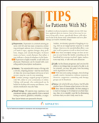
Oral Drug Reduces Relapse Rates in Multiple Sclerosis
TORONTO—Annualized relapse rates in patients with relapsing-remitting multiple sclerosis (MS) were more than 50% lower in subjects taking oral fingolimod (FTY720) than in those taking a placebo, according to research presented at the 62nd Annual Meeting of the American Academy of Neurology. The medication also reduced the risk of disability progression by 30% and significantly decreased the number of new or enlarged T2 lesions and gadolinium-enhancing lesions, per 24-month results of the phase III FTY720 Research Evaluating Effects of Daily Oral therapy in Multiple Sclerosis (FREEDOMS) trial.
Oral fingolimod, a sphingosine 1-phosphate receptor (S1PR) modulator, is the first in a novel class of drugs under evaluation for the treatment of relapsing and progressive forms of MS. S1PR modulators work to retain circulating lymphocytes in the lymph nodes, thereby reducing the recirculation of autoreactive lymphocytes and preventing penetration of these lymphocytes into the CNS.
Fingolimod Versus Placebo
The double-blind, placebo-controlled multicenter FREEDOMS study randomized 1,083 patients (mean age, 40.5) to receive once-daily doses of either 1.25-mg fingolimod, 0.5-mg fingolimod, or a placebo. For the primary end point of annualized relapse rate, both doses of fingolimod were superior to placebo, with a 54% reduction in relapses for the lower dose and a 60% reduction in the higher dose. For the key secondary end point of time to three-month confirmed disability progression, measured by a 1-point increase in the Expanded Disability Status Scale (EDSS), a 30% risk reduction was reported in the 0.5-mg dose, and 32% in the 1.25-mg dose, compared with placebo.
“Overall, 1.25-mg fingolimod did not offer any advantages regarding efficacy, and it had a slightly higher array of side effects; therefore, 0.5 mg is the dosage that we will submit an application for [FDA] approval at this time,” announced Ludwig Kappos, MD, of the Department of Neurology, University Hospital, University of Basel, Switzerland.
The second phase of the study, which was presented in 2006, showed a “clear cut effect on inflammatory outcomes on MRI,” Dr. Kappos noted. “Five years of follow-up support these effects…. Approximately 99.5% of patients remaining in the study do not show MRI activity and have a low annualized relapse rate at approximately 0.2, [which is equivalent to] one relapse every five years.”
The phase III, 24-month MRI inflammatory activity results favored fingolimod over placebo, with 50.5% of the 0.5-mg fingolimod group and 51.9% of the 1.25-mg group free from new or newly enlarged T2 lesions, compared with 21.2% of the placebo group, reported Ernst-Wilhelm Radue, MD, Director of Medical Image Analysis Center, University Hospital Basel, Switzerland. In addition, more patients were free from gadolinium-enhanced lesions with fingolimod (89.7% and 89.8%), compared with placebo (65.1%).
Fingolimod Versus Interferon
A second study, the Trial Assessing Injectable Interferon versus FTY720 Oral in Relapsing-Remitting Multiple Sclerosis (TRANSFORMS), compared both doses of fingolimod to intramuscular interferon beta-1a and found that the oral medicine reduced annualized relapse rates by up to 50%, compared with injectable interferon.
The 12-month phase III study of 1,153 patients also found that fewer patients experienced relapses requiring steroid treatment and/or hospitalization when treated with fingolimod than when treated with interferon beta-1a. Patients in both fingolimod groups also had significantly less deterioration of the ability to perform daily activities, based on the Patient-Reported Indices for Multiple Sclerosis (PRIMUS)–Activities scores, compared with the interferon group.
A 24-month optional extension study to assess the safety and efficacy—both clinically and on MRI—is under way and involves 89% of the patients who completed the core TRANSFORMS study. Patients who were taking weekly interferon beta-1a injections have been switched to daily oral fingolimod, and those who took fingolimod in the earlier phase of the trial have continued on the medication. Following the results of the FREEDOMS II trial, all subjects in the TRANSFORMS extension study are taking the 0.5-mg dose.
Safety and Tolerability
Of the 1,272 patients initially enrolled in the FREEDOMS trial, 81% completed the study. Those who discontinued the trial were proportionately lower in the 0.5-mg fingolimod group (19%), compared with the 1.25-mg dose (31%) or placebo (28%) groups.
The percentages of patients reporting any adverse events were similar for both fingolimod groups (94%) and placebo (93%). Serious adverse events were reported in 10% of patients taking 0.5-mg fingolimod, 12% of those taking 1.25-mg fingolimod, and 13% of the placebo group. Serious infectious adverse events occurred in 1.6% of the 0.5-mg group, 2.6% of the 1.25-mg group, and 1.9% of the placebo group.
Malignant neoplasms were reported in 10 patients in the placebo group and four patients in each active group. Seven cases of macular edema occurred, all in patients on the 1.25-mg dose. A transient heart rate decrease at the beginning of treatment, minor increases in blood pressure, and increases in liver enzymes were also observed.
“These results confirm that fingolimod is generally well tolerated,” the researchers reported. “The 0.5-mg dose was generally better tolerated than the 1.25-mg dose, [and was] associated with fewer bradycardia and serious adverse events and no cases of macular edema.”
—Rebecca K. Abma
Suggested Reading
Cohen JA, Barkhof F, Comi G, et al; TRANSFORMS Study Group. Oral fingolimod or intramuscular interferon for relapsing multiple sclerosis. N Engl J Med. 2010;362(5):402-415.
Kappos L, Radue EW, O’Connor P, et al; FREEDOMS Study Group. A placebo-controlled trial of oral fingolimod in relapsing multiple sclerosis. N Engl J Med. 2010;362(5):387-401.
More Oral MS Drugs in the Pipeline
TORONTO—A number of other oral MS drugs are also in development and were reported on at the 62nd Annual Meeting of the American Academy of Neurology.
Cladribine, an adenosine deaminase-resistant purine nucleoside that targets CD4+ T cells and spares B cells and natural killer cells, is taken as short cycles (once daily for five days), which are then repeated monthly for a total of two (3.5-mg/kg) or four (5.25-mg/kg) cycles in the first year and two cycles in the second year. In the 96-month Cladribine Tablets Treating Multiple Sclerosis Orally (CLARITY) study of 1,326 patients with relapsing-remitting MS, Gavin Giovannoni, PhD, Chair of Neurology, Blizard Institute of Cell and Molecular Science, Barts and The London School of Medicine and Dentistry and the Department of Neurology, Barts and The London NHS Trust, reported that treatment with cladribine in either 3.5-mg/kg or 5.25-mg/kg doses offered significant treatment benefits over placebo, with consistent reductions in the annualized relapse rate and decreases in health care costs. When examining the data for complete disease remission, the investigators found that 71.8% of patients taking 3.5 mg and 70.4% of those taking 5.25 mg were relapse- and disease-progression free, compared with 52.6% of the placebo group. When the third end point of MRI-activity was added, 43.0% of the 3.5-mg group and 44.3% of the 5.25-mg group were disease-activity free, compared with 16% of the placebo group. Cladribine developer Merck Serono SA, Geneva, Switzerland, was issued a refuse to file letter from the FDA late last year.
Teriflunomide, an FDA-approved arthritis treatment from Sanofi-Aventis, Bridgewater, New Jersey, is currently in phase III trials for relapsing-remitting MS. Research presented at the conference noted, “the immunomodulatory effects of teriflunomide are associated with a reduction in the infiltration of macrophages, B cells, and T cells, and an increased survival of oligodendrocytes in the spinal cord. Therefore, teriflunomide treatment could potentially delay neurodegeneration in MS by preventing the loss of myelin and oligodendrocytes.” A six-month, placebo-controlled study of teriflunomide combined with glatiramer acetate found that both 7-mg and 14-mg daily doses improved disease control as measured by MRI scans compared to placebo.
BG-12 is oral dimethyl fumarate taken two to three times a day. Researchers suspect that it may act via the nuclear factor-E2–related factor 2 (Nrf2) transcription pathway. Biogen Idec, Inc, Cambridge, Massachusetts, is conducting two large, phase III trials: Determination of the Efficacy and Safety of Oral Fumarate in Relapsing Remitting MS (DEFINE), a randomized, double-blind, placebo-controlled study of 1,237 patients; and Comparator and an Oral Fumarate in Relapsing Remitting Multiple Sclerosis (CONFIRM), a randomized, double-blind, placebo-controlled and active reference (glatiramer acetate) trial of 1,431 patients.
BAF312, a second-generation S1P receptor modulator, from Novartis Pharma, Basel, Switzerland, selectively targets S1P1 and S1P5 receptors, and is currently in phase II trials. Researchers reported that BAF312 “reverses chronic neurologic paralysis and reduces inflammation and demyelination in EAE mice, and thus may represent a promising treatment modality for inflammatory autoimmune diseases like MS.”
—Rebecca K. Abma
Suggested Reading
Rammohan KW, Shoemaker J. Emerging multiple sclerosis oral therapies. Neurology. 2010;74(Suppl 1):S47-S53.