User login
Remyelination May Continue Despite Age in Patients With MS
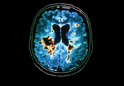
NEW ORLEANS—Despite a consensus that the repair capacity of the adult human brain decreases with age, cortical remyelination appears to continue in patients with multiple sclerosis (MS) into the eighth decade of life, according to research presented at the 2013 Annual Meeting of the American Neurological Association. Depending on the region of the brain, the environment may affect remyelination, said Bruce Trapp, PhD, Chair of the Department of Neurosciences at the Lerner Research Institute of the Cleveland Clinic. Sulfate proteoglycans inhibit the differentiation of oligodendrocyte progenitor cells (OPCs) and are highly expressed in chronic white-matter lesions, but not in gray-matter lesions.
These findings have “therapeutic implications because we’re at the cusp of designing clinical trials for remyelinating therapies,” said Dr. Trapp. “Therapeutic enhancement of white-matter remyelination may be more efficacious for acute lesions in early stages of MS. Cortical remyelination should be considered as a primary outcome measure in clinical trials that test remyelination because it may be a more amenable target for enhancement,” he added.
Oligodendrocytes Were More Common in Gray Matter
Cortical lesions are not inflamed to the same extent as white-matter lesions and consequently are difficult to detect by standard MRI techniques in living patients. Active white-matter lesions, however, can be identified by an abundance of immune cells within the lesion. Immune cells may be involved in cortical lesions, too, albeit in significantly lower numbers, said Dr. Trapp.
He and his colleagues studied 19 brains from patients with MS to determine whether remyelination occurred in cortical lesions. Patients’ age ranged between 27 and 77, and the investigators examined 147 subpial lesions and 65 leukocortical lesions. Approximately 75% of the subpial lesions had evidence of remyelination, and about 25% of them had no remyelination. “We were really quite surprised by how often remyelination was occurring in these subpial lesions,” said Dr. Trapp.
Confocal microscopy indicated that processes sent by oligodendrocytes attached to the ends of inner nodes, not to the center of the internodes. The finding likely will change myelin biologists’ understanding of how remyelination occurs, said Dr. Trapp.
The researchers also identified 65 leukocortical lesions in the study, seven of which were active and had no remyelination in the white or gray matter. Of 58 chronic lesions, 27 had active remyelination, and 24 of these had significantly greater remyelination in the gray matter than in the white matter.
The investigators found 37 remyelinating oligodendrocytes/mm2 of tissue in the gray-matter portion of the leukocoritcal lesions, compared with four oligodendrocytes/mm2 of tissue in the white-matter portion. The oligodendrocytes in the gray-matter lesions were forming 10 to 30 myelin sheaths each, while the oligodendrocytes in the demyelinated white matter were forming few myelin internodes. Two patients with extensive, ongoing cortical remyelination were age 77. The data provide “compelling evidence that remyelination in the cortex is much more apparent than it is in the white matter in aged patients,” said Dr. Trapp.
Hyaluronan May Inhibit Remyelination in White Matter
One reason that cortical remyelination continues throughout a patient’s life may be that little inflammation occurs during demyelination of gray matter. “This inflammatory response sets up certain aspects of the microenvironment of the brain in white matter, but not in gray matter,” said Dr. Trapp.
Another possible explanation is the fact that cortical lesions have normal densities of OPCs, which make new remyelinating oligodendrocytes. White-matter lesions have reduced densities of OPCs. Investigators also suggest that white-matter lesions have molecules that inhibit remyelination, while gray matter does not have these molecules. One such molecule, hyaluronan, inhibits oligodendrocyte differentiation in vitro and remyelination in vivo.
To test whether these molecules are present in white-matter lesions, Dr. Trapp and colleagues examined leukocortical lesions that contain both gray- and white-matter demyelination. They observed that astrocytes in the white-matter portion of leukocoritcal lesions, but not in gray-matter portions, significantly upregulated CD44, hyaluranon, and versican. The group also found a “significant” increase in glial fibrillary acidic protein in the white-matter lesion, but not in the gray-matter lesion.
“These molecules form complexes that can reduce oligodendrocyte maturation and remyelination,” said Dr. Trapp. “These [molecules] are similar to molecules that inhibit axon regeneration in spinal cord injury, so there’s a huge amount of literature on them. Reducing or disrupting this complex may increase remyelination for white-matter lesions.”
—Erik Greb
Suggested Reading
Chang A, Staugaitis SM, Dutta R, et al. Cortical remyelination: a new target for repair therapies in multiple sclerosis. Ann Neurol. 2012;72(6):918-926.
Fancy SP, Glasgow SM, Finley M, et al. Evidence that nuclear factor IA inhibits repair after white matter injury. Ann Neurol. 2012;72(2):224-233.
Fischer MT, Wimmer I, Höftberger R, et al. Disease-specific molecular events in cortical multiple sclerosis lesions. Brain. 2013;136(pt 6):1799-1815.
NEW ORLEANS—Despite a consensus that the repair capacity of the adult human brain decreases with age, cortical remyelination appears to continue in patients with multiple sclerosis (MS) into the eighth decade of life, according to research presented at the 2013 Annual Meeting of the American Neurological Association. Depending on the region of the brain, the environment may affect remyelination, said Bruce Trapp, PhD, Chair of the Department of Neurosciences at the Lerner Research Institute of the Cleveland Clinic. Sulfate proteoglycans inhibit the differentiation of oligodendrocyte progenitor cells (OPCs) and are highly expressed in chronic white-matter lesions, but not in gray-matter lesions.
These findings have “therapeutic implications because we’re at the cusp of designing clinical trials for remyelinating therapies,” said Dr. Trapp. “Therapeutic enhancement of white-matter remyelination may be more efficacious for acute lesions in early stages of MS. Cortical remyelination should be considered as a primary outcome measure in clinical trials that test remyelination because it may be a more amenable target for enhancement,” he added.
Oligodendrocytes Were More Common in Gray Matter
Cortical lesions are not inflamed to the same extent as white-matter lesions and consequently are difficult to detect by standard MRI techniques in living patients. Active white-matter lesions, however, can be identified by an abundance of immune cells within the lesion. Immune cells may be involved in cortical lesions, too, albeit in significantly lower numbers, said Dr. Trapp.
He and his colleagues studied 19 brains from patients with MS to determine whether remyelination occurred in cortical lesions. Patients’ age ranged between 27 and 77, and the investigators examined 147 subpial lesions and 65 leukocortical lesions. Approximately 75% of the subpial lesions had evidence of remyelination, and about 25% of them had no remyelination. “We were really quite surprised by how often remyelination was occurring in these subpial lesions,” said Dr. Trapp.
Confocal microscopy indicated that processes sent by oligodendrocytes attached to the ends of inner nodes, not to the center of the internodes. The finding likely will change myelin biologists’ understanding of how remyelination occurs, said Dr. Trapp.
The researchers also identified 65 leukocortical lesions in the study, seven of which were active and had no remyelination in the white or gray matter. Of 58 chronic lesions, 27 had active remyelination, and 24 of these had significantly greater remyelination in the gray matter than in the white matter.
The investigators found 37 remyelinating oligodendrocytes/mm2 of tissue in the gray-matter portion of the leukocoritcal lesions, compared with four oligodendrocytes/mm2 of tissue in the white-matter portion. The oligodendrocytes in the gray-matter lesions were forming 10 to 30 myelin sheaths each, while the oligodendrocytes in the demyelinated white matter were forming few myelin internodes. Two patients with extensive, ongoing cortical remyelination were age 77. The data provide “compelling evidence that remyelination in the cortex is much more apparent than it is in the white matter in aged patients,” said Dr. Trapp.
Hyaluronan May Inhibit Remyelination in White Matter
One reason that cortical remyelination continues throughout a patient’s life may be that little inflammation occurs during demyelination of gray matter. “This inflammatory response sets up certain aspects of the microenvironment of the brain in white matter, but not in gray matter,” said Dr. Trapp.
Another possible explanation is the fact that cortical lesions have normal densities of OPCs, which make new remyelinating oligodendrocytes. White-matter lesions have reduced densities of OPCs. Investigators also suggest that white-matter lesions have molecules that inhibit remyelination, while gray matter does not have these molecules. One such molecule, hyaluronan, inhibits oligodendrocyte differentiation in vitro and remyelination in vivo.
To test whether these molecules are present in white-matter lesions, Dr. Trapp and colleagues examined leukocortical lesions that contain both gray- and white-matter demyelination. They observed that astrocytes in the white-matter portion of leukocoritcal lesions, but not in gray-matter portions, significantly upregulated CD44, hyaluranon, and versican. The group also found a “significant” increase in glial fibrillary acidic protein in the white-matter lesion, but not in the gray-matter lesion.
“These molecules form complexes that can reduce oligodendrocyte maturation and remyelination,” said Dr. Trapp. “These [molecules] are similar to molecules that inhibit axon regeneration in spinal cord injury, so there’s a huge amount of literature on them. Reducing or disrupting this complex may increase remyelination for white-matter lesions.”
—Erik Greb
Suggested Reading
Chang A, Staugaitis SM, Dutta R, et al. Cortical remyelination: a new target for repair therapies in multiple sclerosis. Ann Neurol. 2012;72(6):918-926.
Fancy SP, Glasgow SM, Finley M, et al. Evidence that nuclear factor IA inhibits repair after white matter injury. Ann Neurol. 2012;72(2):224-233.
Fischer MT, Wimmer I, Höftberger R, et al. Disease-specific molecular events in cortical multiple sclerosis lesions. Brain. 2013;136(pt 6):1799-1815.
Risk Stratification May Mitigate Adverse Events Associated With Newer MS Drugs
COPENHAGEN—Risk stratification can help neurologists determine whether to prescribe one of the newer multiple sclerosis (MS) drugs for their patients, according to a lecture given at the 29th Congress of the European Committee for Treatment and Research in MS. Natalizumab, fingolimod, and alemtuzumab have potentially serious side effects, but their benefits outweigh their risks, said Per Soelberg Sørensen, MD, Head of the Danish MS Center at Copenhagen University Hospital.
“MS is a serious disease, particularly active MS,” said Dr. Sørensen. “Therefore, people need these drugs to control the disease.”
JCV Antibody Status Influences Treatment With Natalizumab
Before treating a patient with natalizumab, a neurologist should administer a blood test and an MRI, according to the European Medicines Agency. Because natalizumab entails the risk of progressive multifocal leukoencephalopathy (PML), the neurologist should determine whether the patient has antibodies to the John Cunningham virus (JCV). Patients with no JCV antibodies have almost no risk of PML, said Dr. Sørensen. Patients who have the antibodies and have used immunosuppressants are at considerable risk for PML and should not take natalizumab, he added. For patients with JCV antibodies who have not used immunosuppressants, the risk of PML is low within the first 24 months of treatment with natalizumab, but it increases during the following 12 months.
Treatment with natalizumab should be stopped if a patient develops hypersensitivity reactions or antibodies against natalizumab, according to Dr. Sørensen, who added that neurologists should monitor liver function and perform annual MRIs for all patients taking the drug. In addition, he advised neurologists to test for JCV antibodies every six months and noted that JCV antibody titer may increase for patients who begin treatment with a low titer. If the neurologist suspects that the patient is developing PML, he or she should stop natalizumab treatment, perform MRI, and perform CSF tests for JCV, said Dr. Sørensen.
Antiarrhythmics and Antineoplastics May Interfere With Fingolimod
Fingolimod, a sphingosine-1 phosphate receptor modulator, helps patients by acting on lymphocytes but may cause macular edema or restrict respiratory flow. The drug is contraindicated in patients who are taking antiarrhythmic, antineoplastic, or immunosuppressant drugs. Neurologists should be cautious when prescribing fingolimod to patients receiving drugs that lower the heart rate (eg, calcium channel blockers and beta-blockers) or medicines that inhibit CYP3A4-mediated metabolism, said Dr. Sørensen. Fingolimod initially decreases a patient’s white blood cell count, and it takes more than four weeks for the count to return to normal. Patients on fingolimod therefore should not receive attenuated live vaccines. Neurologists also should be vigilant for unknown safety signals, added Dr. Sørensen.
Blood and liver function tests are recommended before initiating treatment with fingolimod, and high-risk patients should undergo ophthalmologic exams. Neurologists should observe all patients for six hours during treatment initiation. Because of a risk of infection, clinicians should monitor patients for two months following treatment initiation, said Dr. Sørensen. During treatment, patients should report symptoms of infection and use contraception, and their blood pressure and liver function should be monitored regularly as well, he added.
Alemtuzumab Entails a Risk of Thyroid Disorder
Alemtuzumab is associated with many side effects, most of which are related to the infusion. Approximately 36% of patients develop a thyroid disorder while taking alemtuzumab, and the peak incidence occurs during the second or third year of treatment. About 1% of patients develop immune thrombocytopenia, which, like thyroid disorder, typically occurs during the second or third year of treatment. Diagnosis is important because immune thrombocytopenia responds to standard medical treatment, said Dr. Sørensen. In addition, some patients have developed neuropathy on alemtuzumab, and early detection can improve the patient’s prognosis, he added.
To maximize safety, neurologists should perform a blood test and screen for tuberculosis or HIV before initiating alemtuzumab. Patients who are anti-thyroid peroxidase positive at baseline are more likely to have thyroid-related adverse events on alemtuzumab, said Dr. Sørensen.
To prevent infusion-related reactions, neurologists can administer corticosteroids, antihistamines, and antipyretics during the first three days of treatment. Patients’ pulse and blood pressure should be monitored, and patients should receive an oral antiherpes medication for at least one month after receiving alemtuzumab, noted Dr. Sørensen. Neurologists should perform blood tests and monitor thyroid function regularly, he added. “This [monitoring] has to continue for four years after the last dose of alemtuzumab,” said Dr. Sørensen.
Monthly tests for blood count and urinalysis and thrice-monthly tests for thyroid function are recommended until four years after the last dose of alemtuzumab, although this regimen may pose logistic problems, he noted. Patients on alemtuzumab also should use contraceptives and report symptoms of infections and signs of bleeding, Dr. Sørensen concluded.
—Erik Greb
Senior Associate Editor
Suggested Reading
Perumal J, Khan O. Emerging disease-modifying therapies in multiple sclerosis. Curr Treat Options Neurol. 2012;14(3):256-263.
Rudick R, Polman C, Clifford D, et al. Natalizumab: bench to bedside and beyond. JAMA Neurol. 2013;70(2):172-182.
COPENHAGEN—Risk stratification can help neurologists determine whether to prescribe one of the newer multiple sclerosis (MS) drugs for their patients, according to a lecture given at the 29th Congress of the European Committee for Treatment and Research in MS. Natalizumab, fingolimod, and alemtuzumab have potentially serious side effects, but their benefits outweigh their risks, said Per Soelberg Sørensen, MD, Head of the Danish MS Center at Copenhagen University Hospital.
“MS is a serious disease, particularly active MS,” said Dr. Sørensen. “Therefore, people need these drugs to control the disease.”
JCV Antibody Status Influences Treatment With Natalizumab
Before treating a patient with natalizumab, a neurologist should administer a blood test and an MRI, according to the European Medicines Agency. Because natalizumab entails the risk of progressive multifocal leukoencephalopathy (PML), the neurologist should determine whether the patient has antibodies to the John Cunningham virus (JCV). Patients with no JCV antibodies have almost no risk of PML, said Dr. Sørensen. Patients who have the antibodies and have used immunosuppressants are at considerable risk for PML and should not take natalizumab, he added. For patients with JCV antibodies who have not used immunosuppressants, the risk of PML is low within the first 24 months of treatment with natalizumab, but it increases during the following 12 months.
Treatment with natalizumab should be stopped if a patient develops hypersensitivity reactions or antibodies against natalizumab, according to Dr. Sørensen, who added that neurologists should monitor liver function and perform annual MRIs for all patients taking the drug. In addition, he advised neurologists to test for JCV antibodies every six months and noted that JCV antibody titer may increase for patients who begin treatment with a low titer. If the neurologist suspects that the patient is developing PML, he or she should stop natalizumab treatment, perform MRI, and perform CSF tests for JCV, said Dr. Sørensen.
Antiarrhythmics and Antineoplastics May Interfere With Fingolimod
Fingolimod, a sphingosine-1 phosphate receptor modulator, helps patients by acting on lymphocytes but may cause macular edema or restrict respiratory flow. The drug is contraindicated in patients who are taking antiarrhythmic, antineoplastic, or immunosuppressant drugs. Neurologists should be cautious when prescribing fingolimod to patients receiving drugs that lower the heart rate (eg, calcium channel blockers and beta-blockers) or medicines that inhibit CYP3A4-mediated metabolism, said Dr. Sørensen. Fingolimod initially decreases a patient’s white blood cell count, and it takes more than four weeks for the count to return to normal. Patients on fingolimod therefore should not receive attenuated live vaccines. Neurologists also should be vigilant for unknown safety signals, added Dr. Sørensen.
Blood and liver function tests are recommended before initiating treatment with fingolimod, and high-risk patients should undergo ophthalmologic exams. Neurologists should observe all patients for six hours during treatment initiation. Because of a risk of infection, clinicians should monitor patients for two months following treatment initiation, said Dr. Sørensen. During treatment, patients should report symptoms of infection and use contraception, and their blood pressure and liver function should be monitored regularly as well, he added.
Alemtuzumab Entails a Risk of Thyroid Disorder
Alemtuzumab is associated with many side effects, most of which are related to the infusion. Approximately 36% of patients develop a thyroid disorder while taking alemtuzumab, and the peak incidence occurs during the second or third year of treatment. About 1% of patients develop immune thrombocytopenia, which, like thyroid disorder, typically occurs during the second or third year of treatment. Diagnosis is important because immune thrombocytopenia responds to standard medical treatment, said Dr. Sørensen. In addition, some patients have developed neuropathy on alemtuzumab, and early detection can improve the patient’s prognosis, he added.
To maximize safety, neurologists should perform a blood test and screen for tuberculosis or HIV before initiating alemtuzumab. Patients who are anti-thyroid peroxidase positive at baseline are more likely to have thyroid-related adverse events on alemtuzumab, said Dr. Sørensen.
To prevent infusion-related reactions, neurologists can administer corticosteroids, antihistamines, and antipyretics during the first three days of treatment. Patients’ pulse and blood pressure should be monitored, and patients should receive an oral antiherpes medication for at least one month after receiving alemtuzumab, noted Dr. Sørensen. Neurologists should perform blood tests and monitor thyroid function regularly, he added. “This [monitoring] has to continue for four years after the last dose of alemtuzumab,” said Dr. Sørensen.
Monthly tests for blood count and urinalysis and thrice-monthly tests for thyroid function are recommended until four years after the last dose of alemtuzumab, although this regimen may pose logistic problems, he noted. Patients on alemtuzumab also should use contraceptives and report symptoms of infections and signs of bleeding, Dr. Sørensen concluded.
—Erik Greb
Senior Associate Editor
Suggested Reading
Perumal J, Khan O. Emerging disease-modifying therapies in multiple sclerosis. Curr Treat Options Neurol. 2012;14(3):256-263.
Rudick R, Polman C, Clifford D, et al. Natalizumab: bench to bedside and beyond. JAMA Neurol. 2013;70(2):172-182.
Suggested Reading
Perumal J, Khan O. Emerging disease-modifying therapies in multiple sclerosis. Curr Treat Options Neurol. 2012;14(3):256-263.
Rudick R, Polman C, Clifford D, et al. Natalizumab: bench to bedside and beyond. JAMA Neurol. 2013;70(2):172-182.

Repetitive TMS May Reduce Depression and Fatigue in Patients With MS
COPENHAGEN—When applied to the motor cortex, repetitive transcranial magnetic stimulation (rTMS) may ameliorate depression and fatigue related to multiple sclerosis (MS), according to a study presented at the 29th Congress of the European Committee for Treatment and Research in MS (ECTRIMS).
The effects of rTMS may persist for as long as six weeks after the end of stimulation. To a lesser extent, rTMS of the left prefrontal cortex also may reduce depression and fatigue for patients with MS, said Sven Schippling, MD, deputy head of the Neuroimmunology and MS Research Section at University Hospital Zurich.
Results from the study of patients with MS suggest that the technique is safe and well tolerated. The most frequent side effects included headache on the day of or the day after stimulation and paresthesia in the lower or upper limbs.
Technicians Administered Stimulation With an H-Coil
Dr. Schippling and Friedemann Paul, MD, consultant neurologist at Charité University Medical Center in Berlin, administered rTMS with an H-coil that was jointly developed by the Weizmann Institute of Science in Rehovot, Israel, the NIH, and the Jerusalem-based company Brainsway. Compared with the conventional figure-eight coil, the H-coil directly stimulates deeper brain regions (ie, more than 1 cm below the scalp) over a less focused area.
The investigators randomly assigned 28 patients with MS (26 with relapsing-remitting MS and two with secondary progressive MS, female-to-male ratio 4:1, median EDSS 3.0) to rTMS of the motor cortex, rTMS of the left prefrontal cortex, or sham stimulation of the left prefrontal cortex. Patients who received treatment of the motor cortex did not know whether stimulation was real or sham, but technicians and investigators were not blinded for this condition. Rating physicians, who had been blinded to patients’ assignment, recorded clinical ratings and administered questionnaires. Stimulation of the left prefrontal cortex was performed in a double-blinded fashion. After randomization, participants underwent three stimulation sessions per week during a six-week treatment period. Follow-up lasted for six additional weeks and consisted of three biweekly visits.
The researchers assessed fatigue with the Fatigue Severity Scale (FSS), the Modified Fatigue Impact Scale, and a visual analog scale that the patient completed. Depression was evaluated using the Beck Depression Inventory (BDI) and the 18-item Hamilton Rating Scale for Depression.
Eligible participants were relapse-free for more than 30 days before inclusion and had taken immunomodulatory or immunosuppressant treatment for more than three months before inclusion. To be included, patients had to have an FSS score of 4 or higher or a BDI score of 12 or higher.
Depression Scores Were Not Equally Distributed at Baseline
Approximately one-third of patients had headache on the day of stimulation, and fewer patients had headache on the day after stimulation. Several patients had paresthesia in the lower or upper limbs. Both headache and paresthesia were similar in frequency in the sham and the true stimulation conditions. The investigators observed no severe events in the patients that received true stimulation, and the types and frequency of adverse events were comparable between the three treatment groups.
Baseline BDI scores were not equally distributed between the groups, said Dr. Schippling. Patients who received rTMS of the prefrontal cortex had higher baseline BDI scores, compared with patients who received rTMS of the motor cortex. FSS and BDI scores decreased significantly in patients who received rTMS of the motor cortex. Motor cortex stimulation significantly lowered the rate of fatigue in all the fatigue and depression scores, including the visual analog scale, said Dr. Schippling.
A clear limitation of the study is its small size, said Dr. Schippling. “However, we find the results promising, and we want to embark on a phase II trial because further evidence is needed to draw firm conclusions about what might be an effective treatment. This would be highly desirable, as treatment options in MS-related fatigue are limited,” he concluded.
—Erik Greb
Senior Associate Editor
Suggested Reading
Speer AM, Wassermann EM, Benson BE, et al. Antidepressant efficacy of high and low frequency rTMS at 110% of motor threshold versus sham stimulation over left prefrontal cortex. Brain Stimul. 2013 Jul 29 [Epub ahead of print].
Yusuf A, Koski L. A qualitative review of the neurophysiological underpinnings of fatigue in multiple sclerosis. J Neurol Sci. 2013;330(1-2):4-9.
COPENHAGEN—When applied to the motor cortex, repetitive transcranial magnetic stimulation (rTMS) may ameliorate depression and fatigue related to multiple sclerosis (MS), according to a study presented at the 29th Congress of the European Committee for Treatment and Research in MS (ECTRIMS).
The effects of rTMS may persist for as long as six weeks after the end of stimulation. To a lesser extent, rTMS of the left prefrontal cortex also may reduce depression and fatigue for patients with MS, said Sven Schippling, MD, deputy head of the Neuroimmunology and MS Research Section at University Hospital Zurich.
Results from the study of patients with MS suggest that the technique is safe and well tolerated. The most frequent side effects included headache on the day of or the day after stimulation and paresthesia in the lower or upper limbs.
Technicians Administered Stimulation With an H-Coil
Dr. Schippling and Friedemann Paul, MD, consultant neurologist at Charité University Medical Center in Berlin, administered rTMS with an H-coil that was jointly developed by the Weizmann Institute of Science in Rehovot, Israel, the NIH, and the Jerusalem-based company Brainsway. Compared with the conventional figure-eight coil, the H-coil directly stimulates deeper brain regions (ie, more than 1 cm below the scalp) over a less focused area.
The investigators randomly assigned 28 patients with MS (26 with relapsing-remitting MS and two with secondary progressive MS, female-to-male ratio 4:1, median EDSS 3.0) to rTMS of the motor cortex, rTMS of the left prefrontal cortex, or sham stimulation of the left prefrontal cortex. Patients who received treatment of the motor cortex did not know whether stimulation was real or sham, but technicians and investigators were not blinded for this condition. Rating physicians, who had been blinded to patients’ assignment, recorded clinical ratings and administered questionnaires. Stimulation of the left prefrontal cortex was performed in a double-blinded fashion. After randomization, participants underwent three stimulation sessions per week during a six-week treatment period. Follow-up lasted for six additional weeks and consisted of three biweekly visits.
The researchers assessed fatigue with the Fatigue Severity Scale (FSS), the Modified Fatigue Impact Scale, and a visual analog scale that the patient completed. Depression was evaluated using the Beck Depression Inventory (BDI) and the 18-item Hamilton Rating Scale for Depression.
Eligible participants were relapse-free for more than 30 days before inclusion and had taken immunomodulatory or immunosuppressant treatment for more than three months before inclusion. To be included, patients had to have an FSS score of 4 or higher or a BDI score of 12 or higher.
Depression Scores Were Not Equally Distributed at Baseline
Approximately one-third of patients had headache on the day of stimulation, and fewer patients had headache on the day after stimulation. Several patients had paresthesia in the lower or upper limbs. Both headache and paresthesia were similar in frequency in the sham and the true stimulation conditions. The investigators observed no severe events in the patients that received true stimulation, and the types and frequency of adverse events were comparable between the three treatment groups.
Baseline BDI scores were not equally distributed between the groups, said Dr. Schippling. Patients who received rTMS of the prefrontal cortex had higher baseline BDI scores, compared with patients who received rTMS of the motor cortex. FSS and BDI scores decreased significantly in patients who received rTMS of the motor cortex. Motor cortex stimulation significantly lowered the rate of fatigue in all the fatigue and depression scores, including the visual analog scale, said Dr. Schippling.
A clear limitation of the study is its small size, said Dr. Schippling. “However, we find the results promising, and we want to embark on a phase II trial because further evidence is needed to draw firm conclusions about what might be an effective treatment. This would be highly desirable, as treatment options in MS-related fatigue are limited,” he concluded.
—Erik Greb
Senior Associate Editor
Suggested Reading
Speer AM, Wassermann EM, Benson BE, et al. Antidepressant efficacy of high and low frequency rTMS at 110% of motor threshold versus sham stimulation over left prefrontal cortex. Brain Stimul. 2013 Jul 29 [Epub ahead of print].
Yusuf A, Koski L. A qualitative review of the neurophysiological underpinnings of fatigue in multiple sclerosis. J Neurol Sci. 2013;330(1-2):4-9.

Teriflunomide Is Safe and Effective for Patients With CIS—Main Results From the TOPIC Study
COPENHAGEN—Teriflunomide (14 mg and 7 mg) is safe and effective in the treatment of patients with clinically isolated syndrome, according to TOPIC trial data presented at the 29th Congress of the European Committee for Treatment and Research in Multiple Sclerosis (ECTRIMS).
“These findings highlight the ability of early intervention with teriflunomide to delay onset of MS symptoms,” said Aaron E. Miller, MD, Professor of Neurology at the Icahn School of Medicine at Mount Sinai, New York, and his study collaborators. “To date, all teriflunomide phase III studies have shown consistent safety and efficacy, and greater efficacy for a 14-mg dose.”
Teriflunomide is a novel, once-daily, oral immunomodulator approved in the United States, Argentina, and Australia for the treatment of relapsing-remitting MS. Prior clinical studies of teriflunomide in patients with relapsing-remitting MS (TEMSO and TOWER) showed consistent efficacy across key clinical measures and a well-characterized safety profile. TOPIC was a phase III clinical trial conducted to assess the efficacy and safety of teriflunomide in patients with a first clinical episode suggestive of MS (clinically isolated syndrome).
In TOPIC, a double-blind, placebo-controlled, parallel-group study, 618 patients with clinically isolated syndrome were enrolled from February 2008 to September 2012 (projected end date, December 2012) and randomized to placebo, teriflunomide 7 mg, or teriflunomide 14 mg. TOPIC ended three months early as revised diagnostic criteria enabled earlier diagnosis of MS. The primary and key secondary end points were conversion to clinically definite MS and occurrence of a new clinical relapse or MRI lesion. Other efficacy end points and safety and tolerability were also assessed.
Baseline characteristics were generally well balanced among the treatment groups. Of the randomized population (n = 618), 59.1% had monofocal and 40.9% had multifocal lesion presentation, and 31.4% had one or more gadolinium-enhancing lesions. Median time since first neurologic event was two months.
Compared with placebo, teriflunomide 14 mg reduced the risk of conversion to clinically definite MS by 42.6%, with a probability of conversion to clinically definite MS at 108 weeks of 24.0% (probability for placebo group, 35.9%). Teriflunomide 14 mg also reduced the risk of occurrence of new relapse or new MRI lesion by 34.9%. As measured by MRI over the two-year study period, patients treated with teriflunomide 14 mg had a 5% increase in total lesion volume, compared with a 28% increase among patients treated with placebo. In addition, patients treated with teriflunomide 14 mg had a 59% reduction in gadolinium-enhancing T1 lesions, compared with patients receiving placebo.
Teriflunomide 7 mg reduced the risk of conversion to clinically definite MS by 37.2% (108-week probability, 27.6%) and the risk of occurrence of new relapse or new MRI lesion by 31.4%. The occurrence of adverse events was similar across treatment groups. The most common adverse events reported more frequently in the teriflunomide arms included alanine aminotransferase elevation, headache, hair thinning (14 mg only), diarrhea, and paresthesia.
—Glenn S. Williams
Vice President/Group Editor
New and Noteworthy Information—December 2013
Mild traumatic brain injury (TBI) may be associated with increased cortical fractional anisotropy, but not with cortical or subcortical atrophy, according to research published online ahead of print November 20 in Neurology. Investigators evaluated 50 patients and 50 sex-, age-, and education-matched controls with a clinical and neuroimaging battery approximately 14 days after TBI. A total of 26 patients returned for follow-up four months after injury. Patients had increased fractional anisotropy in the bilateral superior frontal cortex during the semiacute phase of injury. Fractional anisotropy in the left superior frontal cortex remained elevated at four months after injury. The researchers found no significant differences between patients and matched controls on neuropsychologic testing or measures of gray matter atrophy or mean diffusivity at either time point.
Researchers detailed the early clinical course, morbidity, and mortality of the 2012 outbreak of fungal infections associated with methylprednisolone injections in two articles published October 24, 2013, in the New England Journal of Medicine. As of July 1, 2013, a total of 749 cases of infection had been reported in 20 states, including 61 deaths. Of 728 patients for whom data were available, 31% had meningitis and no other documented infection. Of 328 patients without peripheral joint infection who were included in one investigation, 81% had CNS infection, and 19% had non-CNS infections only. The investigators found evidence of Exserohilum rostratum in 36% of patients for whom samples were available. Patients’ median age was 64, and the median incubation period was 47 days. Forty patients had a stroke.
An algorithm may accurately predict time to death, institutionalization, and need for full-time care in patients with Alzheimer’s disease, according to an article published online ahead of print September 24 in the Journal of Alzheimer’s Disease. Investigators followed two study cohorts with mild Alzheimer’s disease for 10 years. The first cohort included 252 patients, and the second included 254 patients. Participants underwent semiannual assessments that included cognition, functional capacity, and medical, psychiatric, and neurologic information. For each of the three outcome measures, the predicted survival curves were well within the 95% confidence intervals of the observed survival curves. The actual and predicted survival curves were statistically equivalent. The algorithm can be adapted to predict other important disease end points, according to the researchers.
High pulse pressure may be associated with increased CSF phosphorylated tau and decreased β-amyloid 1–42 (Aβ1–42) in cognitively normal older adults, according to research published online ahead of print November 13 in Neurology. A total of 177 cognitively normal, stroke-free older adults underwent blood pressure assessment for determination of pulse pressure, as well as lumbar puncture for measurement of CSF Aβ1–42 and phosphorylated tau. High pulse pressure was associated with increased phosphorylated tau, reduced Aβ1–42, and increased phosphorylated tau to Aβ1–42 ratio. After controlling for covariates, the investigators found that pulse pressure remained associated with phosphorylated tau and phosphorylated tau to Aβ1–42 ratio, but was no longer associated with Aβ1–42. The relationship between pulse pressure and CSF biomarkers is age-dependent, said the researchers.
Acute stroke care in hospitals with neurology residency programs may be associated with an increased use of thrombolytics, investigators reported online ahead of print November 1 in Neurology. The disparities between the thrombolysis rates in hospitals with neurology residency programs and those in other teaching hospitals and nonteaching hospitals may be greater among elderly patients. Researchers retrospectively studied a nationally representative sample of patients with ischemic stroke. A total of 712,433 individuals from 6,839 hospital samples were included. Of these patients, 10.1%, 29.1%, and 60.8% were treated in hospitals with neurology residency programs, other teaching hospitals, and nonteaching hospitals, respectively. Patients in hospitals with neurology residency programs received thrombolysis more frequently (3.74%) than those in other teaching hospitals (2.28%) and those in nonteaching hospitals (1.44%).
The FDA has approved Aptiom (eslicarbazepine acetate) as an add-on medication to treat partial-onset seizures associated with epilepsy. In three large, phase III safety and efficacy trials that included more than 1,400 patients with inadequately controlled partial-onset seizures, eslicarbazepine acetate was associated with statistically significant reductions in standardized seizure frequency, compared with placebo. Significantly more patients who received eslicarbazepine acetate had a reduction in seizure frequency of 50% or more, compared with controls. The most common side effects include dizziness, somnolence, nausea, headache, diplopia, vomiting, fatigue, vertigo, ataxia, and blurred vision. Eslicarbazepine acetate will not be classified as a controlled substance. Sunovion (Marlborough, Massachusetts) markets the drug and expects it to be available in the US during the second quarter of 2014.
The FDA has approved the NeuroPace RNS System, a device intended to reduce the frequency of seizures in patients with epilepsy who have not responded well to medications. The device consists of a small neurostimulator implanted within the skull. The neurostimulator is connected to one or two electrodes that are placed where the seizures are suspected to originate within the brain or on the surface of the brain. When it detects abnormal electrical activity, the neurostimulator delivers electrical stimulation to normalize brain activity and prevent seizures. In a randomized study of 191 patients, the average number of seizures per month was reduced by approximately 38% at three months in patients in whom the device was turned on. The RNS System is manufactured by NeuroPace (Mountainview, California).
Reducing blood pressure with antihypertensive medications may not decrease the likelihood of death and major disability among patients with acute ischemic stroke, according to a study published online ahead of print November 17 in JAMA. Researchers studied 4,071 patients with nonthrombolyzed ischemic stroke within 48 hours of onset and elevated systolic blood pressure. Patients were randomized to receive antihypertensive treatment or to discontinue all antihypertensive medications during hospitalization. Mean systolic blood pressure was reduced from 166.7 mm Hg to 144.7 mm Hg within 24 hours in the antihypertensive treatment group and from 165.6 mm Hg to 152.9 mm Hg in the control group within 24 hours after randomization. The researchers found no difference in the rates of death and major disability between the treatment groups.
Persons with high urinary concentrations of tungsten may have an increased risk of stroke, according to a study published November 11 in PLOS One. Investigators analyzed associations between tungsten, commonly used in mobile phones and computers, and cardiovascular disease or stroke using crude and adjusted logistic regression models in a cohort of 8,614 adults (ages 18 to 74) with 193 reported stroke diagnoses and 428 reported diagnoses of cardiovascular disease. The researchers also stratified the data to characterize associations in a subset of individuals between ages 18 and 50. Elevated tungsten concentrations were strongly associated with an increase in the prevalence of stroke, independent of typical risk factors (odds ratio: 1.66). The association between tungsten and stroke in the young age category was still evident (odds ratio: 2.17).
Traumatic brain injury (TBI) may be associated with increased amyloid deposition, according to research published online ahead of print November 11 in JAMA Neurology. Investigators used carbon 11-labeled Pittsburgh Compound B ([11C]PiB) PET to image amyloid deposition in 11 controls and 15 patients between one and 361 days after TBI. Compared with the controls, the patients with TBI had significantly increased [11C]PiB distribution volume ratios in cortical gray matter and the striatum, but not in the thalamus or white matter. The investigators observed increases in [11C]PiB distribution volume ratios in patients with TBI across most cortical subregions. The increases were replicated using comparisons of standardized uptake value ratios and could not be accounted for by methodologic confounders.
Compared with persons who speak only one language, bilingual individuals may have a delayed onset of dementia, according to a study published online ahead of print November 6 in Neurology. Investigators reviewed case records of 648 patients with dementia (391 bilinguals) diagnosed in a specialist clinic. They compared age at onset of first symptoms between monolingual and bilingual groups and examined the influence of the number of languages spoken, education, occupation, and other potentially interacting variables. Bilingual patients developed dementia 4.5 years later than the monolingual patients. The researchers found a significant difference in age at onset of Alzheimer’s disease dementia, frontotemporal dementia, and vascular dementia. The age difference was also observed in illiterate patients. The investigators found no additional benefit to speaking more than two languages.
Temporal lobe epilepsy (TLE) may entail altered structural connectivity in the brain, according to a study published online ahead of print November 8 in Radiology. Investigators analyzed 60-direction diffusion-tensor imaging and magnetization-prepared rapid acquisition gradient-echo (MP-RAGE) MRI volumes for 24 patients with left TLE and 24 healthy control subjects. MP-RAGE volumes were segmented into 1,015 regions of interest that spanned the entire brain. Patients with TLE had 22% to 45% reduced distant connectivity in the medial orbitofrontal cortex, temporal cortex, posterior cingulate cortex, and precuneus, compared with healthy subjects. Local connectivity, as measured by means of network efficiency, was increased by 85% to 270% in the medial and lateral frontal cortices, insular cortex, posterior cingulate cortex, precuneus, and occipital cortex in patients with TLE, compared with healthy subjects.
Gray matter damage may be a key factor associated with long-term accumulation of disability and cognitive impairment in multiple sclerosis (MS), according to research published November 12 in Neurology. Investigators obtained conventional and magnetization transfer (MT) MRI brain scans at baseline and at 12 months for 73 patients with MS, who were followed prospectively with clinical visits and rating of the Expanded Disability Status Scale (EDSS) score and the MS severity score for a median of 13.3 years. At 13-year follow-up, 66% of patients had significant worsening of disability, and 37% had worse cognition. The multivariable model identified baseline gray matter fraction as the only predictor of disability worsening. Baseline disease duration and average gray matter lesion MT ratio were independent variables associated with cognitive deterioration.
—Erik Greb
Senior Associate Editor
What Is the Future of Disease-Modifying Therapies for MS?
COPENHAGEN—The many drugs in development for multiple sclerosis (MS) have various modes of action and could increase patients’ treatment options, according to an overview presented at the 29th Congress of the European Committee for Treatment and Research in MS. If these drugs reach the market, they may make it difficult for neurologists to stratify patients to various treatment regimens, but they will open new possibilities such as combination therapies, said Tobias Derfuss, MD.
Laquinimod Reduced Disability Progression
Two recently completed phase III trials found that laquinimod, a drug that appeared to promote remyelination in animal models, reduced patients’ annualized relapse rate by 20%. The drug also was associated with a “surprising” 46% reduction in disability progression, said Dr. Derfuss, Cohead of the Outpatient Care Unit and the Neuroimmunological Research Group at the University Hospital Basel. “You expect this effect on disability progression only if you also have a strong effect on the inflammatory components or [if] the drug has an impact on neuroprotection,” he said.
In addition, laquinimod reduced patients’ rate of brain atrophy by 30% during a 24-month period, which indicated that the drug might have a neuroprotective effect. New trials are required to identify the optimal dose and determine whether the drug could be part of a combination therapy.
Siponimod Decreased the Number of Unique Active Lesions
Siponimod is an oral compound in development that acts on the sphingosine-1-phosphate receptor. A phase II trial that compared siponimod to placebo in patients with relapsing-remitting MS suggested that 2 mg was the most effective dose of the drug. Compared with placebo, siponimod reduced unique active lesions at three months.
Investigators are recruiting participants for a phase III trial of siponimod in patients with secondary progressive MS. The trial will continue until a certain number of patients have three-month confirmed disability progression, which is the primary end point. When patients have six-month confirmed disability progression, they will be given the option of switching to open-label treatment. “This trial is a very good option for patients with secondary progressive MS,” said Dr. Derfuss.
Simvastatin May Reduce Brain Atrophy
Researchers have investigated the efficacy of statins in treating relapsing-remitting MS, and a double blind placebo-controlled trial in 2012 evaluated the efficacy of 80 mg/day of simvastatin in patients with secondary progressive MS. The drug was associated with significant reduction in brain atrophy, as well as with reductions in Expanded Disability Status Scale score and the MS Impact Scale, a patient-related outcome.
The results are surprising, in light of the trial’s small patient population, said Dr. Derfuss. In all, 140 patients participated in the trial, which lasted for two years. “We know from other trials in secondary progressive MS [that] it might take longer to see the effect,” said Dr. Derfuss. But because simvastatin no longer has patent protection, he added, pharmaceutical companies may be unwilling to conduct larger phase III trials, which means that MS societies and independent research organizations may have to provide the necessary funding.
Monoclonal Antibodies May Improve Patients’ Outcomes
Several monoclonal antibodies in development also show promise for the treatment of MS. Investigators recently completed a phase II trial that tested two doses of daclizumab, an IL-2 receptor antagonist, for one year. Compared with placebo, daclizumab reduced patients’ annualized relapse rate by more than 50%. An extension study and another phase IIb trial have supported the treatment’s efficacy. Daclizumab is a “promising drug” that has an “impressive effect on disability progression” within a short time, said Dr. Derfuss.
Secukinumab is a fully humanized antibody against the IL-17A cytokine. Patients in a small trial received 10 mg/kg/day of secukinumab. Despite the trial’s brief duration, researchers observed a significant reduction in new gadolinium-enhancing lesions among patients receiving secukinumab. Patients in the treatment arm also had a reduction in relapse rates, but it was not statistically significant. Investigators are recruiting patients for a second, larger phase II trial.
Ocrelizumab, a third antibody, depletes the body’s B-cells. A recent phase II trial compared ocrelizumab with interferon beta-1a and placebo. After eight weeks, patients who received ocrelizumab had a significant reduction in gadolinium-enhancing lesions, compared with patients receiving placebo and those receiving interferon. Ocrelizumab also reduced relapse rates. Two phase III trials are studying ocrelizumab for primary progressive MS, and one trial is evaluating ocrelizumab as a treatment for relapsing-remitting MS.
—Erik Greb
Senior Associate Editor
Suggested Reading
He D, Han K, Gao X, et al. Laquinimod for multiple sclerosis. Cochrane Database Syst Rev. 2013 Aug 6;8:CD010475.
Oh J, Calabresi PA. Emerging injectable therapies for multiple sclerosis. Lancet Neurol. 2013;12(11):1115-1126.
Selmaj K, Li DK, Hartung HP, et al. Siponimod for patients with relapsing-remitting multiple sclerosis (BOLD): an adaptive, dose-ranging, randomised, phase 2 study. Lancet Neurol. 2013;12(8):756-767.
Wang J, Xiao Y, Luo M, Luo H. Statins for multiple sclerosis. Cochrane Database Syst Rev. 2011 Dec 7;(12):CD008386.
COPENHAGEN—The many drugs in development for multiple sclerosis (MS) have various modes of action and could increase patients’ treatment options, according to an overview presented at the 29th Congress of the European Committee for Treatment and Research in MS. If these drugs reach the market, they may make it difficult for neurologists to stratify patients to various treatment regimens, but they will open new possibilities such as combination therapies, said Tobias Derfuss, MD.
Laquinimod Reduced Disability Progression
Two recently completed phase III trials found that laquinimod, a drug that appeared to promote remyelination in animal models, reduced patients’ annualized relapse rate by 20%. The drug also was associated with a “surprising” 46% reduction in disability progression, said Dr. Derfuss, Cohead of the Outpatient Care Unit and the Neuroimmunological Research Group at the University Hospital Basel. “You expect this effect on disability progression only if you also have a strong effect on the inflammatory components or [if] the drug has an impact on neuroprotection,” he said.
In addition, laquinimod reduced patients’ rate of brain atrophy by 30% during a 24-month period, which indicated that the drug might have a neuroprotective effect. New trials are required to identify the optimal dose and determine whether the drug could be part of a combination therapy.
Siponimod Decreased the Number of Unique Active Lesions
Siponimod is an oral compound in development that acts on the sphingosine-1-phosphate receptor. A phase II trial that compared siponimod to placebo in patients with relapsing-remitting MS suggested that 2 mg was the most effective dose of the drug. Compared with placebo, siponimod reduced unique active lesions at three months.
Investigators are recruiting participants for a phase III trial of siponimod in patients with secondary progressive MS. The trial will continue until a certain number of patients have three-month confirmed disability progression, which is the primary end point. When patients have six-month confirmed disability progression, they will be given the option of switching to open-label treatment. “This trial is a very good option for patients with secondary progressive MS,” said Dr. Derfuss.
Simvastatin May Reduce Brain Atrophy
Researchers have investigated the efficacy of statins in treating relapsing-remitting MS, and a double blind placebo-controlled trial in 2012 evaluated the efficacy of 80 mg/day of simvastatin in patients with secondary progressive MS. The drug was associated with significant reduction in brain atrophy, as well as with reductions in Expanded Disability Status Scale score and the MS Impact Scale, a patient-related outcome.
The results are surprising, in light of the trial’s small patient population, said Dr. Derfuss. In all, 140 patients participated in the trial, which lasted for two years. “We know from other trials in secondary progressive MS [that] it might take longer to see the effect,” said Dr. Derfuss. But because simvastatin no longer has patent protection, he added, pharmaceutical companies may be unwilling to conduct larger phase III trials, which means that MS societies and independent research organizations may have to provide the necessary funding.
Monoclonal Antibodies May Improve Patients’ Outcomes
Several monoclonal antibodies in development also show promise for the treatment of MS. Investigators recently completed a phase II trial that tested two doses of daclizumab, an IL-2 receptor antagonist, for one year. Compared with placebo, daclizumab reduced patients’ annualized relapse rate by more than 50%. An extension study and another phase IIb trial have supported the treatment’s efficacy. Daclizumab is a “promising drug” that has an “impressive effect on disability progression” within a short time, said Dr. Derfuss.
Secukinumab is a fully humanized antibody against the IL-17A cytokine. Patients in a small trial received 10 mg/kg/day of secukinumab. Despite the trial’s brief duration, researchers observed a significant reduction in new gadolinium-enhancing lesions among patients receiving secukinumab. Patients in the treatment arm also had a reduction in relapse rates, but it was not statistically significant. Investigators are recruiting patients for a second, larger phase II trial.
Ocrelizumab, a third antibody, depletes the body’s B-cells. A recent phase II trial compared ocrelizumab with interferon beta-1a and placebo. After eight weeks, patients who received ocrelizumab had a significant reduction in gadolinium-enhancing lesions, compared with patients receiving placebo and those receiving interferon. Ocrelizumab also reduced relapse rates. Two phase III trials are studying ocrelizumab for primary progressive MS, and one trial is evaluating ocrelizumab as a treatment for relapsing-remitting MS.
—Erik Greb
Senior Associate Editor
Suggested Reading
He D, Han K, Gao X, et al. Laquinimod for multiple sclerosis. Cochrane Database Syst Rev. 2013 Aug 6;8:CD010475.
Oh J, Calabresi PA. Emerging injectable therapies for multiple sclerosis. Lancet Neurol. 2013;12(11):1115-1126.
Selmaj K, Li DK, Hartung HP, et al. Siponimod for patients with relapsing-remitting multiple sclerosis (BOLD): an adaptive, dose-ranging, randomised, phase 2 study. Lancet Neurol. 2013;12(8):756-767.
Wang J, Xiao Y, Luo M, Luo H. Statins for multiple sclerosis. Cochrane Database Syst Rev. 2011 Dec 7;(12):CD008386.

Fine-Tuning the Ketogenic Diet May Benefit Children With Epilepsy—And Other News From the Child Neurology Society Meeting
AUSTIN—Adjusting the ketogenic diet is helpful in one of five children with epilepsy, researchers reported at the 2013 Annual Meeting of the Child Neurology Society.
“Medication adjustment and dietary modification lead to similar outcomes,” stated Eric Kossoff, MD, Assistant Professor of Neurology and Pediatrics at Johns Hopkins Children’s Center in Baltimore, and colleagues. “Therefore, both [tactics] can be tried if the ketogenic diet is not meeting seizure-control expectations.”
The findings are based on a retrospective review of 200 children with epilepsy who had begun the ketogenic diet at the Johns Hopkins Hospital between October 2007 and June 2013. Dr. Kossoff’s group identified 10 commonly implemented dietary and supplement changes among the children. The investigators also reviewed medication adjustments in the children, as well as patient records during the course of adherence to the ketogenic diet for as many as four interventions per child. Greater than 50% seizure reduction after a change was defined as success, according to the study authors.
A total of 156 children (78%) had at least one intervention, and 391 distinct and occasionally concurrent interventions were recorded, of which 265 were made specifically for seizure control, noted Dr. Kossoff.
“Overall, there was an 18% chance that any intervention would be successful, but only a 3% chance of resultant seizure freedom,” reported the researchers. “The likelihood of success did not decrease with each subsequent intervention. There was a trend toward medication adjustments being more successful than dietary modifications (24% vs 15%, respectively).”
Analgesic Overuse May Worsen Chronic Post-Traumatic Headaches in Adolescents With Concussion
Excessive use of analgesics may exacerbate post-traumatic headaches among adolescents who have had a concussion, investigators reported.
“Analgesics should be minimized or discontinued when headaches continue several weeks following concussion,” recommended Geoffrey L. Heyer, MD, of the Division of Pediatric Neurology, Nationwide Children’s Hospital in Columbus, Ohio, and colleagues.
The researchers conducted a retrospective review of 104 adolescents with concussion who had been referred to a pediatric headache clinic for chronic post-traumatic headaches of three to 12 months in duration. The study authors compared concussion symptoms, headache symptoms before and after concussion, demographic data, and headache outcomes between patients who had probable medication-overuse headache and those who did not have probable medication-overuse headache.
A total of 77 patients had chronic post-traumatic headache between three and 12 months, and 54 patients (70%) met the criteria for probable medication-overuse headache. The investigators found that patients with medication overuse were more likely to have had daily headaches, to be female, to have had nausea or throbbing associated with their headaches, to have had increased irritability following concussion, and to have had a longer interval between injury and neurologic evaluation.
“Of the patients with medication overuse, 37 (68.5%) had resolution of headaches or improvements to preconcussion headache patterns after discontinuing analgesics, seven (13%) had no change in headaches or worsening of headaches after discontinuing analgesics, and 10 (18.5%) did not discontinue analgesics or were lost to follow-up,” stated Dr. Heyer.
2010 McDonald Criteria Are Effective for Early Diagnosis of Pediatric MS
The 2010 Revised McDonald Diagnostic Criteria for Multiple Sclerosis (MS) may have a higher sensitivity for diagnosing pediatric MS, compared with the 2005 criteria. The revised criteria thus could allow for earlier initiation of disease-modifying therapy, researchers reported.
Mitchel T. Williams, MD, Pediatric Neurologist at the Children’s Hospital of Michigan in Detroit, and colleagues conducted a retrospective study of 25 children who had been diagnosed with MS at the Children’s Hospital of Michigan from 2005 through early 2013.
The researchers applied the 2005 and 2010 McDonald criteria based on neuroimaging findings as well as initial clinical presentation. The investigators also analyzed demographic data and compared those data with those of previous pediatric MS cohorts.
The median age at presentation, sex ratio, clinical symptoms, and relapse rate among the participants were comparable to those in previously published data, except for a high rate of African Americans (64%) among the 25 children.
The researchers found that the initial MS diagnosis rate based on the 2005 McDonald criteria was 32%, compared with a rate of 92% using the 2010 McDonald criteria. The mean time after initial symptom presentation until the 2005 criteria for MS were met was 5.0 months, compared with 0.7 months for the 2010 McDonald criteria.
“[Our findings] suggest that the 2010 McDonald criteria are a more appropriate tool for the timely diagnosis of pediatric MS,” concluded the researchers.
—Colby Stong
Editor
Conference News Update—Society for Neuroscience
Dot Test May Gauge the Progression of Dopamine Loss in Parkinson’s Disease
A simple dot test could help gauge the extent of dopamine loss in individuals with Parkinson’s disease, according to a pilot study by a multidisciplinary team of investigators. “It is difficult now to assess the extent of dopamine loss—a hallmark of Parkinson’s disease—in people with the disease,” said Katherine R. Gamble, a doctoral student in psychology at Georgetown University in Washington, DC. “This test, called the Triplets Learning Task (TLT), may provide some help for physicians who treat people with Parkinson’s disease, but we still have much work to do to better understand its utility,” she added.
The TLT tests implicit learning, which occurs without awareness or intent and relies on the caudate nucleus, an area of the brain affected by loss of dopamine. The test is a sequential learning task that does not require complex motor skills. In the TLT, participants see four open circles, and then two red dots appear. Patients are asked to respond when they see a green dot appear. Unknown to them, the location of the first red dot predicts the location of the green dot. Participants learn implicitly where the green dot will appear, and their responses become faster and more accurate.
Previous studies have shown that the caudate region in the brain underlies implicit learning. In the study, participants with Parkinson’s disease implicitly learned the dot pattern with training, but a loss of dopamine appears to negatively affect that learning. “Their performance began to decline toward the end of training, suggesting that people with Parkinson’s disease lack the neural resources in the caudate, such as dopamine, to complete the learning task,” said Ms. Gamble. The current study included 27 patients with Parkinson’s disease. The research team is now testing how implicit learning may differ by different stages of Parkinson’s disease and drug doses.
The Eyes May Help Diagnose Alzheimer’s Disease
The loss of a particular layer of retinal cells may reveal the presence of Alzheimer’s disease and provide a new way to track disease progression, according to an international team of researchers. The investigators examined retinas from the eyes of mice genetically engineered to develop Alzheimer’s disease.
“The retina is an extension of the brain, so it makes sense to see if the same pathologic processes found in an Alzheimer’s brain are also found in the eye,” said R. Scott Turner, MD, PhD, Director of the Memory Disorders Program at Georgetown University Medical Center in Washington, DC. “We know there’s an association between glaucoma and Alzheimer’s disease, in that both are characterized by loss of neurons, but the mechanisms are not clear.” Many researchers increasingly view glaucoma as a neurodegenerative disorder similar to Alzheimer’s disease, he added.
Most of the research to date examining the relationship between glaucoma and Alzheimer’s disease focused on the retinal ganglion cell layer, which transmits visual information via the optic nerve into the brain. Before that transmission happens, though, the retinal ganglion cells receive information from another layer in the retina—the inner nuclear layer.
The researchers examined the thickness of the retina, including the inner nuclear layer (which had not been studied previously in this setting) and the retinal ganglion cell layer. They found a significant loss of thickness in both layers. The inner nuclear layer had a 37% loss of neurons, and the retinal ganglion cell layer had a 49% loss of neurons, compared with healthy, age-matched control mice.
In humans, the structure and thickness of the retina can be readily measured using optical coherence tomography. This new tool is finding an increasing number of applications in research and clinical care, said Dr. Turner.
“This study suggests another path forward in understanding the disease process and could lead to new ways to diagnose or predict Alzheimer’s that could be as simple as looking into the eyes,” said Dr. Turner. “Parallel disease mechanisms suggest that new treatments developed for Alzheimer’s disease may also be useful for glaucoma.”
Research Suggests Warning Signs and Potential Treatments for MS
Recent research is increasing clinicians’ understanding of multiple sclerosis (MS) and may lead to new treatments and management strategies, said investigators. One study has shed light on the way in which antibodies in the bloodstream break through the blood–brain barrier to attack the optic nerves, spinal cord, and brain, causing the symptoms of neuromyelitis optica. Understanding how the antibodies bypass the blood–brain barrier could provide new approaches to treating the disease.
A protein involved in blood clotting might serve as an early biomarker for MS before symptoms occur, said Katerina Akassoglou, PhD, Professor in Residence of Neurology at the University of California, San Francisco School of Medicine. Early detection of the disease could lead to more effective early treatments.
In another study, low levels of a cholesterol protein correlated with the severity of MS. The finding suggests that the protein, which protects against inflammation, may protect against developing MS and may help regenerate damaged neurons, said Lidia Gardner, PhD, Assistant Professor of Neurology at the University of Tennessee in Memphis. Cholesterol drugs thus may be potential treatments for MS.
Researchers found that a type of immune system cell directly targets and damages nerve cell axons, just as MS does. This cell may be a target for new disease-modifying therapies. In addition, researchers have found that when exosomes—tiny, naturally occurring nanovesicles—are produced by dendritic cells and applied to the brain, they can deliver a mixture of proteins and RNAs that promote the regeneration of protective myelin sheaths and guard against MS symptoms.

Tobias Derfuss, MD, Part 1


Tobias Derfuss, MD, Part 2

