User login
Rituximab and vemurafenib could challenge frontline chemotherapy for HCL
AMSTERDAM – A combination of rituximab and the BRAF inhibitor vemurafenib could be the one-two punch needed for relapsed or refractory hairy cell leukemia (HCL), according to investigators.
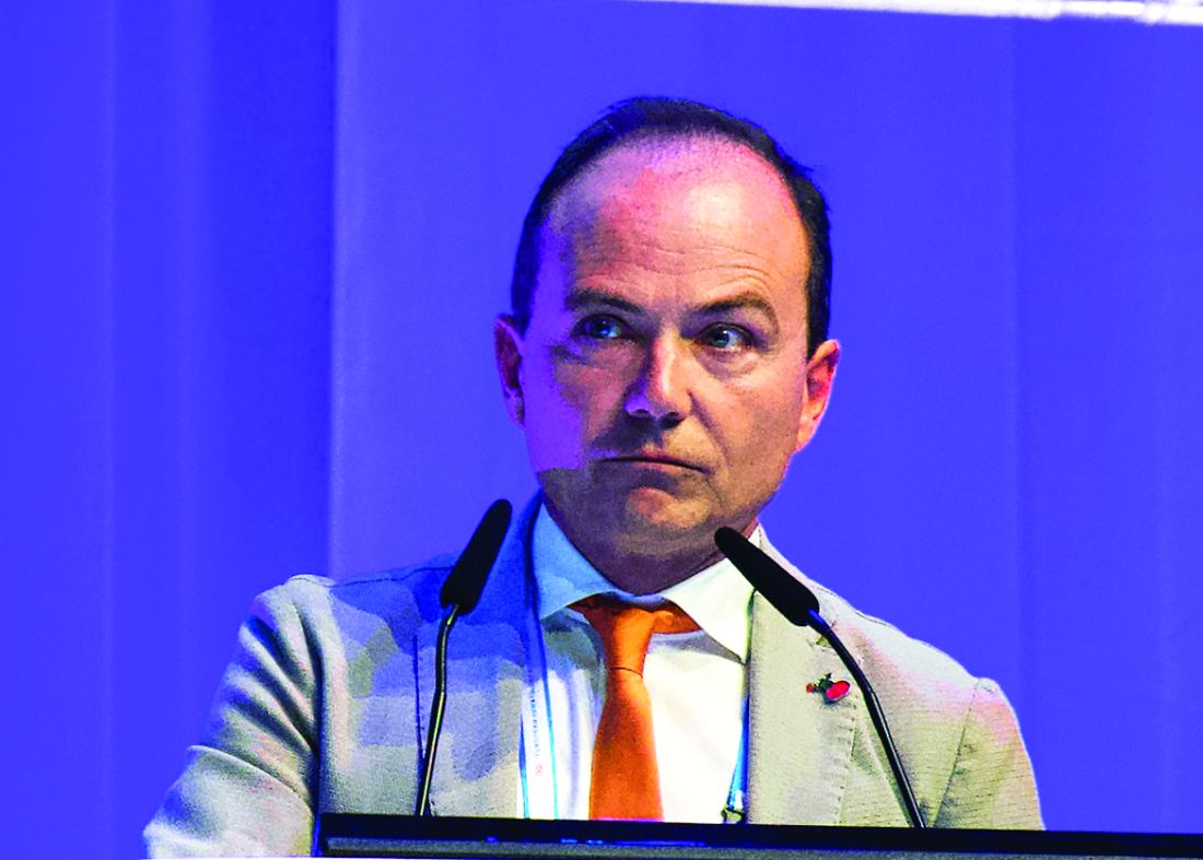
Among evaluable patients treated with this combination, 96% achieved complete remission, reported lead author, Enrico Tiacci, MD, of the University and Hospital of Perugia, Italy.
This level of efficacy is “clearly superior to historical results with either agent alone,” Dr. Tiacci said during a presentation at the annual congress of the European Hematology Association, citing previous complete response rates with vemurafenib alone of 35%-40%. “[This combination] has potential for challenging chemotherapy in the frontline setting,” he said.
The phase 2 trial involved 31 patients with relapsed or refractory HCL who had received a median of three previous therapies. Eight of the patients (26%) had primary refractory disease. Patients received vemurafenib 960 mg, twice daily for 8 weeks and rituximab 375 mg/m2, every 2 weeks. After finishing vemurafenib, patients received rituximab four more times, keeping the interval of 2 weeks. Complete remission was defined as a normal blood count, no leukemic cells in bone marrow biopsies and blood smears, and no palpable splenomegaly.
Out of 31 patients, 27 were evaluable at data cutoff. Of these, 26 (96%) achieved complete remission. The investigators noted that two complete responders had incomplete platelet recovery at the end of treatment that resolved soon after, and two patients had persistent splenomegaly, but were considered to be in complete remission at 22.5 and 25 months after finishing therapy.
All of the complete responders had previously received purine analogs, while a few had been refractory to a prior BRAF inhibitor (n = 7) and/or rituximab (n = 5).
The investigators also pointed out that 15 out of 24 evaluable patients (63%) achieved complete remission just 4 weeks after starting the trial regimen. Almost two-thirds of patients (65%) were negative for minimal residual disease (MRD). The rate of progression-free survival at a median follow-up of 29.5 months was 83%. Disease progression occurred exclusively in patients who were MRD positive.
The combination was well tolerated; most adverse events were of grade 1 or 2, overlapping with the safety profile of each agent alone.
Reflecting on the study findings, Dr. Tiacci suggested that the combination could be most effective if delivered immediately, instead of after BRAF failure.
“Interestingly,” he said, “the relapse-free survival in patients naive to a BRAF inhibitor remained significantly longer than the relapse-free interval that patients previously exposed to a BRAF inhibitor enjoyed, both following monotherapy with a BRAF inhibitor and following subsequent combination with rituximab, potentially suggesting that vemurafenib should be used directly in combination with rituximab rather than being delivered first as a monotherapy and then added to rituximab at relapse.”
Randomized testing of the combination against the chemotherapy-based standard of care in the frontline setting is warranted, the investigators concluded.
Dr. Tiacci reported financial relationships with Roche, AbbVie, and Shire.
SOURCE: Tiacci E et al. EHA Congress, Abstract S104.
AMSTERDAM – A combination of rituximab and the BRAF inhibitor vemurafenib could be the one-two punch needed for relapsed or refractory hairy cell leukemia (HCL), according to investigators.
Among evaluable patients treated with this combination, 96% achieved complete remission, reported lead author, Enrico Tiacci, MD, of the University and Hospital of Perugia, Italy.
This level of efficacy is “clearly superior to historical results with either agent alone,” Dr. Tiacci said during a presentation at the annual congress of the European Hematology Association, citing previous complete response rates with vemurafenib alone of 35%-40%. “[This combination] has potential for challenging chemotherapy in the frontline setting,” he said.
The phase 2 trial involved 31 patients with relapsed or refractory HCL who had received a median of three previous therapies. Eight of the patients (26%) had primary refractory disease. Patients received vemurafenib 960 mg, twice daily for 8 weeks and rituximab 375 mg/m2, every 2 weeks. After finishing vemurafenib, patients received rituximab four more times, keeping the interval of 2 weeks. Complete remission was defined as a normal blood count, no leukemic cells in bone marrow biopsies and blood smears, and no palpable splenomegaly.
Out of 31 patients, 27 were evaluable at data cutoff. Of these, 26 (96%) achieved complete remission. The investigators noted that two complete responders had incomplete platelet recovery at the end of treatment that resolved soon after, and two patients had persistent splenomegaly, but were considered to be in complete remission at 22.5 and 25 months after finishing therapy.
All of the complete responders had previously received purine analogs, while a few had been refractory to a prior BRAF inhibitor (n = 7) and/or rituximab (n = 5).
The investigators also pointed out that 15 out of 24 evaluable patients (63%) achieved complete remission just 4 weeks after starting the trial regimen. Almost two-thirds of patients (65%) were negative for minimal residual disease (MRD). The rate of progression-free survival at a median follow-up of 29.5 months was 83%. Disease progression occurred exclusively in patients who were MRD positive.
The combination was well tolerated; most adverse events were of grade 1 or 2, overlapping with the safety profile of each agent alone.
Reflecting on the study findings, Dr. Tiacci suggested that the combination could be most effective if delivered immediately, instead of after BRAF failure.
“Interestingly,” he said, “the relapse-free survival in patients naive to a BRAF inhibitor remained significantly longer than the relapse-free interval that patients previously exposed to a BRAF inhibitor enjoyed, both following monotherapy with a BRAF inhibitor and following subsequent combination with rituximab, potentially suggesting that vemurafenib should be used directly in combination with rituximab rather than being delivered first as a monotherapy and then added to rituximab at relapse.”
Randomized testing of the combination against the chemotherapy-based standard of care in the frontline setting is warranted, the investigators concluded.
Dr. Tiacci reported financial relationships with Roche, AbbVie, and Shire.
SOURCE: Tiacci E et al. EHA Congress, Abstract S104.
REPORTING FROM EHA CONGRESS
Risk model could help predict VTE in acute leukemia
AMSTERDAM – A new clinical prediction model can determine the risk of venous thromboembolism in patients with leukemia, according to investigators.
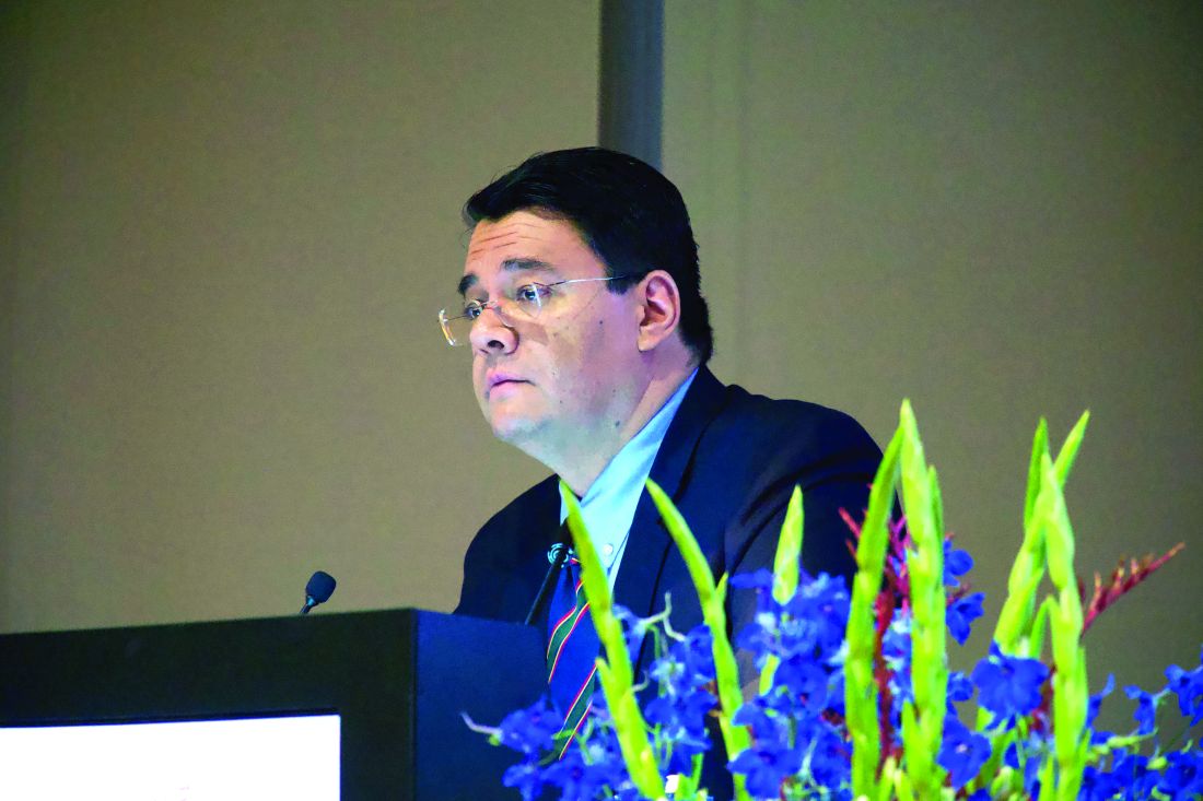
The scoring system, which incorporates historical, morphological, and cytologic factors, was internally validated at multiple time points over the course of a year, reported lead author, Alejandro Lazo-Langner, MD, of the University of Western Ontario, London.
“It is important that we can predict or anticipate which patients [with acute leukemia] will develop venous thrombosis so that we can develop preventions and aim for better surveillance strategies,” Dr. Lazo-Langner said at the annual congress of the European Hematology Association. Venous thromboembolism (VTE) risk modeling is available for patients with solid tumors, but a similar prognostic tool for leukemia patients has been missing.
To fill this practice gap, Dr. Lazo-Langner and colleagues conducted a retrospective cohort study involving 501 patients with acute leukemia who were diagnosed between 2006 and 2017. Of these patients, 427 (85.2%) had myeloid lineage and 74 (14.8%) had lymphoblastic disease. VTE outcomes of interest included proximal lower- and upper-extremity deep vein thrombosis; pulmonary embolism; and thrombosis of unusual sites, such as splanchnic and cerebral. Patients were followed until last follow-up, VTE, or death. Single variable and multiple variable logistic regression were used sequentially to evaluate and confirm potential predictive factors, with nonparametric bootstrapping for internal validation.
After last follow-up, 77 patients (15.3%) had developed VTE; specifically, 44 patients had upper-extremity deep vein thrombosis, 28 had lower-extremity deep vein thrombosis or pulmonary embolism, and 5 had cerebral vein thrombosis. The median time from leukemia diagnosis to VTE was approximately 2 months (64 days). Out of 20 possible predictive factors, 7 were included in the multivariable model, and 3 constitute the final model. These three factors are platelet count greater than 50 x 109/L at time of diagnosis (1 point), lymphoblastic leukemia (2 points), and previous history of venous thromboembolism (3 points).
Dr. Lazo-Langner explained that leukemia patients at high risk of VTE are those with a score of 3 or more points. Using this risk threshold, the investigators found that the overall cumulative incidence of VTE in the high-risk group was 44.0%, compared with 10.5% in the low-risk group. Temporal analysis showed a widening disparity between the two groups, from 3 months (28.8% vs. 6.3%), to 6 months (41.1% vs. 7.9%), and 12 months (42.5% vs. 9.3%).
When asked if treatment type was evaluated, Dr. Lazo-Langner said that treatment type was evaluated but proved unfruitful for the model, which is designed for universal use in leukemia.
“We did include a number of different chemotherapy regimens,” he said. “The problem is, because we included both AML [acute myeloid leukemia] and ALL [acute lymphoblastic leukemia] lineage, and the cornerstone of treatment is different for both lineages. It’s difficult to actually include what kind of chemotherapy [patients had]. For instance, it is known that anthracyclines increase risk of thrombosis, but in both lineages, you use anthracyclines, so you really cannot use that as a predictor.”
Looking to the future, the next step will be validation in other cohorts. If this is successful, then Dr. Lazo-Langner speculated that clinicians could use the scoring system to direct monitoring and treatment. For example, patients with high scores and low platelet counts could receive earlier transfusional support, while all high-risk patients could be placed under more intensive surveillance and given additional education about thrombosis.
“I think recognizing symptoms early is important,” Dr. Lazo-Langner said, “and that would be training not only clinicians, but also nursing personnel and the patients themselves to be aware of the symptoms, so they can actually recognize them sooner.”
The study was funded by the Canadian Institutes of Health Research. Dr. Lazo-Langner is an investigator with the Canadian Venous Thromboembolism Clinical Trials and Outcomes Research (CanVECTOR) Network.
SOURCE: Lazo-Langner A et al. EHA 2019, Abstract S1642.
AMSTERDAM – A new clinical prediction model can determine the risk of venous thromboembolism in patients with leukemia, according to investigators.
The scoring system, which incorporates historical, morphological, and cytologic factors, was internally validated at multiple time points over the course of a year, reported lead author, Alejandro Lazo-Langner, MD, of the University of Western Ontario, London.
“It is important that we can predict or anticipate which patients [with acute leukemia] will develop venous thrombosis so that we can develop preventions and aim for better surveillance strategies,” Dr. Lazo-Langner said at the annual congress of the European Hematology Association. Venous thromboembolism (VTE) risk modeling is available for patients with solid tumors, but a similar prognostic tool for leukemia patients has been missing.
To fill this practice gap, Dr. Lazo-Langner and colleagues conducted a retrospective cohort study involving 501 patients with acute leukemia who were diagnosed between 2006 and 2017. Of these patients, 427 (85.2%) had myeloid lineage and 74 (14.8%) had lymphoblastic disease. VTE outcomes of interest included proximal lower- and upper-extremity deep vein thrombosis; pulmonary embolism; and thrombosis of unusual sites, such as splanchnic and cerebral. Patients were followed until last follow-up, VTE, or death. Single variable and multiple variable logistic regression were used sequentially to evaluate and confirm potential predictive factors, with nonparametric bootstrapping for internal validation.
After last follow-up, 77 patients (15.3%) had developed VTE; specifically, 44 patients had upper-extremity deep vein thrombosis, 28 had lower-extremity deep vein thrombosis or pulmonary embolism, and 5 had cerebral vein thrombosis. The median time from leukemia diagnosis to VTE was approximately 2 months (64 days). Out of 20 possible predictive factors, 7 were included in the multivariable model, and 3 constitute the final model. These three factors are platelet count greater than 50 x 109/L at time of diagnosis (1 point), lymphoblastic leukemia (2 points), and previous history of venous thromboembolism (3 points).
Dr. Lazo-Langner explained that leukemia patients at high risk of VTE are those with a score of 3 or more points. Using this risk threshold, the investigators found that the overall cumulative incidence of VTE in the high-risk group was 44.0%, compared with 10.5% in the low-risk group. Temporal analysis showed a widening disparity between the two groups, from 3 months (28.8% vs. 6.3%), to 6 months (41.1% vs. 7.9%), and 12 months (42.5% vs. 9.3%).
When asked if treatment type was evaluated, Dr. Lazo-Langner said that treatment type was evaluated but proved unfruitful for the model, which is designed for universal use in leukemia.
“We did include a number of different chemotherapy regimens,” he said. “The problem is, because we included both AML [acute myeloid leukemia] and ALL [acute lymphoblastic leukemia] lineage, and the cornerstone of treatment is different for both lineages. It’s difficult to actually include what kind of chemotherapy [patients had]. For instance, it is known that anthracyclines increase risk of thrombosis, but in both lineages, you use anthracyclines, so you really cannot use that as a predictor.”
Looking to the future, the next step will be validation in other cohorts. If this is successful, then Dr. Lazo-Langner speculated that clinicians could use the scoring system to direct monitoring and treatment. For example, patients with high scores and low platelet counts could receive earlier transfusional support, while all high-risk patients could be placed under more intensive surveillance and given additional education about thrombosis.
“I think recognizing symptoms early is important,” Dr. Lazo-Langner said, “and that would be training not only clinicians, but also nursing personnel and the patients themselves to be aware of the symptoms, so they can actually recognize them sooner.”
The study was funded by the Canadian Institutes of Health Research. Dr. Lazo-Langner is an investigator with the Canadian Venous Thromboembolism Clinical Trials and Outcomes Research (CanVECTOR) Network.
SOURCE: Lazo-Langner A et al. EHA 2019, Abstract S1642.
REPORTING FROM EHA CONGRESS
Cell count ratios appear to predict thromboembolism in lymphoma
AMSTERDAM – When predicting the risk of thromboembolism in lymphoma patients receiving chemotherapy, clinicians can rely on a routine diagnostic tool: complete blood count, investigators reported.
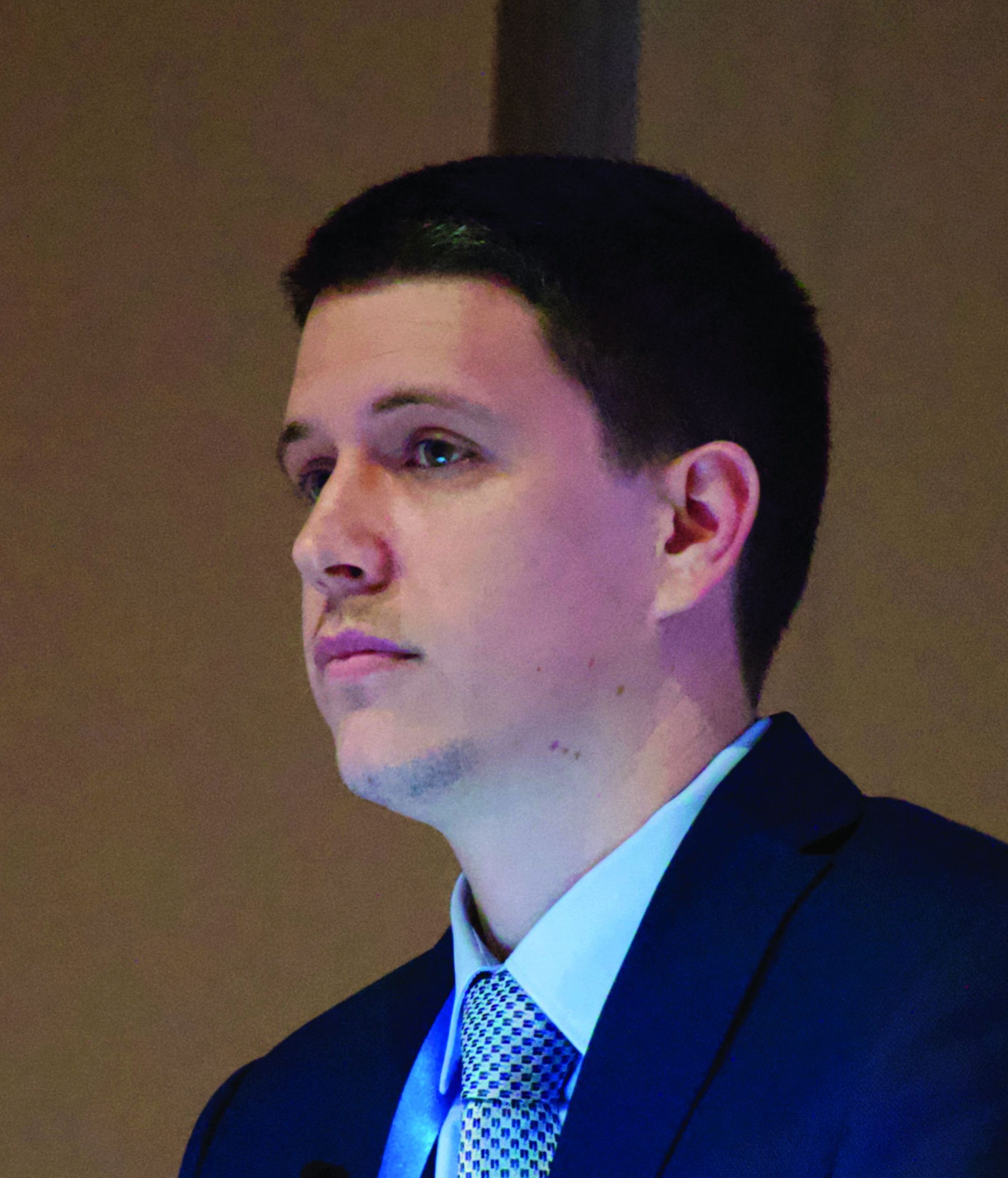
A recent study found that high neutrophil to lymphocyte (NLR) and platelet to lymphocyte (PLR) ratios were prognostic for thromboembolism in this setting, reported lead author Vladimir Otasevic, MD, of the Clinical Centre of Serbia in Belgrade.
“Because of the presence of a broad spectrum of risk factors [in patients with lymphoma undergoing chemotherapy], some authors have published risk-assessment models for prediction of thromboembolism,” Dr. Otasevic said during a presentation at the annual congress of the European Hematology Association. While the underlying pathophysiology that precedes thromboembolism is complex, Dr. Otasevic suggested that risk prediction may not have to be, noting that NLR and PLR were recently proposed as risk biomarkers.
To test the utility of these potential biomarkers, Dr. Otasevic and his colleagues retrospectively analyzed data from 484 patients with non-Hodgkin and Hodgkin lymphoma who had undergone at least one cycle of chemotherapy at the Clinic for Hematology, Clinical Centre of Serbia. Patients were followed for venous and arterial thromboembolic events from the time of diagnosis to 3 months beyond their final cycle of chemotherapy. NLR and PLR ratios were calculated from complete blood count. Thromboembolism was diagnosed by radiography, clinical exam, and laboratory evaluation, with probable diagnoses reviewed by an internist and radiologist.
The median patient age was 53 years with a range from 18 to 89 years. Most patients were recently diagnosed with advanced disease (21.1% stage III and 42.5% stage IV). Half of the population had high-grade non-Hodgkin lymphoma (50.0%) and slightly more than a quarter had low-grade non-Hodgkin lymphoma (28.3%). Low-grade Hodgkin lymphoma was less common (17.4%) and followed distantly by other forms (4.3%).
Thirty-five patients (7.2%) developed thromboembolic events; of these, 30 had venous thromboembolism (6.2%), 6 had arterial thromboembolism (1.2%), and 1 had both. Patients who experienced thromboembolic events had significantly higher NLR and PLR than patients without thromboembolism, and both ratios were significantly associated with one another.
A positive NLR, defined as a ratio of 3.1 or more, was associated with a relative risk of 4.1 for thromboembolism (P less than .001), while a positive PLR, defined as a ratio of 10 or more, was associated with a relative risk of 2.9 (P = .008). Using a multivariate model, a positive NLR was associated with an even higher relative risk (RR = 4.5; P less than .001).
“NLR and PLR demonstrated significant powerfulness in prediction of future risk of [thromboembolism] in lymphoma patients,” the investigators concluded. “Simplicity, effectiveness, modesty, and practicability qualify these new tools for routine [thromboembolism] prognostic assessment.”
Dr. Otasevic said that he and his colleagues have plans to build on these findings with further analysis involving progression-free and overall survival.
The investigators reported no disclosures.
SOURCE: Otasevic V et al. EHA Congress, Abstract S1645.
AMSTERDAM – When predicting the risk of thromboembolism in lymphoma patients receiving chemotherapy, clinicians can rely on a routine diagnostic tool: complete blood count, investigators reported.
A recent study found that high neutrophil to lymphocyte (NLR) and platelet to lymphocyte (PLR) ratios were prognostic for thromboembolism in this setting, reported lead author Vladimir Otasevic, MD, of the Clinical Centre of Serbia in Belgrade.
“Because of the presence of a broad spectrum of risk factors [in patients with lymphoma undergoing chemotherapy], some authors have published risk-assessment models for prediction of thromboembolism,” Dr. Otasevic said during a presentation at the annual congress of the European Hematology Association. While the underlying pathophysiology that precedes thromboembolism is complex, Dr. Otasevic suggested that risk prediction may not have to be, noting that NLR and PLR were recently proposed as risk biomarkers.
To test the utility of these potential biomarkers, Dr. Otasevic and his colleagues retrospectively analyzed data from 484 patients with non-Hodgkin and Hodgkin lymphoma who had undergone at least one cycle of chemotherapy at the Clinic for Hematology, Clinical Centre of Serbia. Patients were followed for venous and arterial thromboembolic events from the time of diagnosis to 3 months beyond their final cycle of chemotherapy. NLR and PLR ratios were calculated from complete blood count. Thromboembolism was diagnosed by radiography, clinical exam, and laboratory evaluation, with probable diagnoses reviewed by an internist and radiologist.
The median patient age was 53 years with a range from 18 to 89 years. Most patients were recently diagnosed with advanced disease (21.1% stage III and 42.5% stage IV). Half of the population had high-grade non-Hodgkin lymphoma (50.0%) and slightly more than a quarter had low-grade non-Hodgkin lymphoma (28.3%). Low-grade Hodgkin lymphoma was less common (17.4%) and followed distantly by other forms (4.3%).
Thirty-five patients (7.2%) developed thromboembolic events; of these, 30 had venous thromboembolism (6.2%), 6 had arterial thromboembolism (1.2%), and 1 had both. Patients who experienced thromboembolic events had significantly higher NLR and PLR than patients without thromboembolism, and both ratios were significantly associated with one another.
A positive NLR, defined as a ratio of 3.1 or more, was associated with a relative risk of 4.1 for thromboembolism (P less than .001), while a positive PLR, defined as a ratio of 10 or more, was associated with a relative risk of 2.9 (P = .008). Using a multivariate model, a positive NLR was associated with an even higher relative risk (RR = 4.5; P less than .001).
“NLR and PLR demonstrated significant powerfulness in prediction of future risk of [thromboembolism] in lymphoma patients,” the investigators concluded. “Simplicity, effectiveness, modesty, and practicability qualify these new tools for routine [thromboembolism] prognostic assessment.”
Dr. Otasevic said that he and his colleagues have plans to build on these findings with further analysis involving progression-free and overall survival.
The investigators reported no disclosures.
SOURCE: Otasevic V et al. EHA Congress, Abstract S1645.
REPORTING FROM EHA CONGRESS
For tough AML, half respond to selinexor plus chemotherapy
AMSTERDAM – Patients with relapsed or refractory acute myeloid leukemia (AML) may be more likely to respond when selinexor is added to standard chemotherapy, according to investigators.
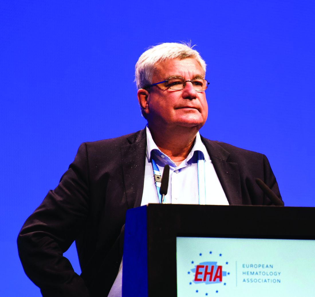
In a recent phase 2 trial, selinexor given with cytarabine and idarubicin led to a 50% overall response rate, reported lead author Walter Fiedler, MD, of University Medical Center Hamburg-Eppendorf (Germany). This response rate is at the upper end of what has been seen in published studies, Dr. Fiedler said at the annual congress of the European Hematology Association.
He also noted that giving a flat dose of selinexor improved tolerability in the trial, a significant finding in light of common adverse events and recent concerns from the Food and Drug Administration about the safety of selinexor for patients with multiple myeloma.
“The rationale to employ selinexor in this study is that there is a synergy between anthracyclines and selinexor,” Dr. Fiedler said, which may restore anthracycline sensitivity in relapsed or refractory patients. “Secondly, there is a c-myc reduction pathway that leads to a reduction of DNA damage repair genes such as Rad51 and Chk1, and this might result in inhibition of homologous recombination.”
The study involved 44 patients with relapsed or refractory AML, of whom 17 (39%) had previously received stem cell transplantation and 11 (25%) exhibited therapy-induced or secondary disease. The median patient age was 59.5 years.
Patients were given idarubicin 10 mg/m2 on days 1, 3, and 5, and cytarabine 100 mg/m2 on days 1-7. Initially, selinexor was given at a dose of 40 mg/m2 twice per week for 4 weeks, but this led to high rates of febrile neutropenia and grade 3 or higher diarrhea, along with prolonged aplasia. In response to this issue, after the first 27 patients, the dose was reduced to a flat amount of 60 mg, given twice weekly for 3 weeks.
For patients not undergoing transplantation after the first or second induction cycle, selinexor maintenance monotherapy was offered for up to 1 year.
The primary endpoint was overall remission rate, reported as complete remission, complete remission with incomplete blood count recovery, and morphological leukemia-free status. Secondary endpoints included the rate of partial remissions, percentage of patients being transplanted after induction, early death rate, overall survival, event-free survival, and relapse-free survival.
The efficacy analysis revealed an overall response rate of 50%. A total of 9 patients had complete remission (21.4%), 11 achieved complete remission with incomplete blood count recovery (26.2%), and 1 exhibited morphological leukemia-free status (2.4%). Of note, almost half of the patients (47%) who had relapsed after previous stem cell transplantation responded, as did three-quarters who tested positive for an NPM1 mutation. After a median follow-up of 8.2 months, the median overall survival was 8.2 months, relapse-free survival was 17.7 months, and event-free survival was 4.9 months.
Adverse events occurred frequently, with a majority of patients experiencing nausea (86%), diarrhea (83%), vomiting (74%), decreased appetite (71%), febrile neutropenia (67%), fatigue (64%), leukopenia (62%), thrombocytopenia (62%), or anemia (60%).
Grade 3 or higher adverse events were almost as common, and included febrile neutropenia (67%), leukopenia (62%), thrombocytopenia (62%), anemia (57%), and diarrhea (50%). Reducing the dose did improve tolerability, with notable drops in the rate of severe diarrhea (56% vs. 40%) and febrile neutropenia (85% vs. 33%). In total, 19% of patients discontinued treatment because of adverse events.
A total of 25 patients (60%) died during the study, with about half dying from disease progression (n = 12), and fewer succumbing to infectious complications, graft-versus-host disease, multiorgan failure, multiple brain infarct, or asystole. Two deaths, one from suspected hemophagocytosis and another from systemic inflammatory response syndrome, were considered possibly related to selinexor.
“The results should be further evaluated in a phase 3 study,” Dr. Fiedler said. However, plans for this are not yet underway, he said, adding that Karyopharm Therapeutics will be focusing its efforts on selinexor for myeloma first.
The study was funded by Karyopharm. Dr. Fielder reported financial relationships with Amgen, Pfizer, Jazz Pharmaceuticals, and other companies.
SOURCE: Fiedler W et al. EHA Congress, Abstract S880.
AMSTERDAM – Patients with relapsed or refractory acute myeloid leukemia (AML) may be more likely to respond when selinexor is added to standard chemotherapy, according to investigators.
In a recent phase 2 trial, selinexor given with cytarabine and idarubicin led to a 50% overall response rate, reported lead author Walter Fiedler, MD, of University Medical Center Hamburg-Eppendorf (Germany). This response rate is at the upper end of what has been seen in published studies, Dr. Fiedler said at the annual congress of the European Hematology Association.
He also noted that giving a flat dose of selinexor improved tolerability in the trial, a significant finding in light of common adverse events and recent concerns from the Food and Drug Administration about the safety of selinexor for patients with multiple myeloma.
“The rationale to employ selinexor in this study is that there is a synergy between anthracyclines and selinexor,” Dr. Fiedler said, which may restore anthracycline sensitivity in relapsed or refractory patients. “Secondly, there is a c-myc reduction pathway that leads to a reduction of DNA damage repair genes such as Rad51 and Chk1, and this might result in inhibition of homologous recombination.”
The study involved 44 patients with relapsed or refractory AML, of whom 17 (39%) had previously received stem cell transplantation and 11 (25%) exhibited therapy-induced or secondary disease. The median patient age was 59.5 years.
Patients were given idarubicin 10 mg/m2 on days 1, 3, and 5, and cytarabine 100 mg/m2 on days 1-7. Initially, selinexor was given at a dose of 40 mg/m2 twice per week for 4 weeks, but this led to high rates of febrile neutropenia and grade 3 or higher diarrhea, along with prolonged aplasia. In response to this issue, after the first 27 patients, the dose was reduced to a flat amount of 60 mg, given twice weekly for 3 weeks.
For patients not undergoing transplantation after the first or second induction cycle, selinexor maintenance monotherapy was offered for up to 1 year.
The primary endpoint was overall remission rate, reported as complete remission, complete remission with incomplete blood count recovery, and morphological leukemia-free status. Secondary endpoints included the rate of partial remissions, percentage of patients being transplanted after induction, early death rate, overall survival, event-free survival, and relapse-free survival.
The efficacy analysis revealed an overall response rate of 50%. A total of 9 patients had complete remission (21.4%), 11 achieved complete remission with incomplete blood count recovery (26.2%), and 1 exhibited morphological leukemia-free status (2.4%). Of note, almost half of the patients (47%) who had relapsed after previous stem cell transplantation responded, as did three-quarters who tested positive for an NPM1 mutation. After a median follow-up of 8.2 months, the median overall survival was 8.2 months, relapse-free survival was 17.7 months, and event-free survival was 4.9 months.
Adverse events occurred frequently, with a majority of patients experiencing nausea (86%), diarrhea (83%), vomiting (74%), decreased appetite (71%), febrile neutropenia (67%), fatigue (64%), leukopenia (62%), thrombocytopenia (62%), or anemia (60%).
Grade 3 or higher adverse events were almost as common, and included febrile neutropenia (67%), leukopenia (62%), thrombocytopenia (62%), anemia (57%), and diarrhea (50%). Reducing the dose did improve tolerability, with notable drops in the rate of severe diarrhea (56% vs. 40%) and febrile neutropenia (85% vs. 33%). In total, 19% of patients discontinued treatment because of adverse events.
A total of 25 patients (60%) died during the study, with about half dying from disease progression (n = 12), and fewer succumbing to infectious complications, graft-versus-host disease, multiorgan failure, multiple brain infarct, or asystole. Two deaths, one from suspected hemophagocytosis and another from systemic inflammatory response syndrome, were considered possibly related to selinexor.
“The results should be further evaluated in a phase 3 study,” Dr. Fiedler said. However, plans for this are not yet underway, he said, adding that Karyopharm Therapeutics will be focusing its efforts on selinexor for myeloma first.
The study was funded by Karyopharm. Dr. Fielder reported financial relationships with Amgen, Pfizer, Jazz Pharmaceuticals, and other companies.
SOURCE: Fiedler W et al. EHA Congress, Abstract S880.
REPORTING FROM EHA CONGRESS
Two trials support shorter DAPT without aspirin after stent
An entire year of dual-antiplatelet therapy may be no better at limiting ischemic events or death than a shorter course for patients who have undergone percutaneous coronary intervention with a drug-eluting stent.
The two trials, which tested dual-antiplatelet therapy (DAPT) regimens of 3 months and 1 month, are also noteworthy for giving a P2Y12 inhibitor after DAPT instead of aspirin monotherapy, which is a more common approach. Each randomized about 3,000 patients.
According to lead author Joo-Yong Hahn, MD, of Sungkyunkwan University in Seoul, South Korea, and colleagues, who conducted the first trial (SMART-CHOICE), both shorter and longer DAPT regimens with aspirin have been associated with shortcomings.
Specifically, shorter duration DAPT with subsequent aspirin monotherapy carries increased risks of MI and stent thrombosis, the investigators wrote. “Conversely, prolonged DAPT increases the risk of bleeding, which offsets the benefit from reducing recurrent ischemic events. Therefore, neither prolonged DAPT nor short-duration DAPT followed by aspirin monotherapy is fully satisfactory.” Because of these shortcomings, the investigators suggested that developing novel strategies “is of paramount importance.”
SMART-CHOICE
The multicenter trial by Dr. Hahn and colleagues, conducted in South Korea, involved 2,993 patients undergoing percutaneous coronary intervention with drug-eluting stents. Patients were randomized to receive either standard DAPT with aspirin and a P2Y12 inhibitor for 12 months, or aspirin plus a P2Y12 inhibitor for 3 months followed by 9 months of P2Y12 monotherapy. Patients were stratified by enrolling center, clinical presentation, type of stent, and type of P2Y12 therapy. Stents were limited to those eluting cobalt-chromium everolimus (Xience Prime, Xience Expedition, or Xience Alpine; Abbott Vascular), platinum-chromium everolimus (Promus Element, Promus Premier, or SYNERGY; Boston Scientific), or sirolimus (Orsiro; Biotronik). Acceptable P2Y12 therapies were clopidogrel, ticagrelor, and prasugrel. The primary endpoint was a composite of major adverse cerebrovascular and cardiac events, including stroke, MI, or all-cause death, at 12 months after percutaneous coronary intervention. A number of secondary endpoints were also evaluated, such as bleeding rate, stent thrombosis, and the individual components of the primary endpoint.
Almost all patients (95%) in the DAPT group adhered to the study protocol, while a smaller proportion (79%) followed P2Y12 monotherapy as described. Still, for both groups, more than 97% of patients completed 1-year follow-up. Primary endpoint analysis showed that the cumulative rate of major adverse cerebrovascular and cardiac events was similar between both groups, at 2.9% in the P2Y12 group versus 2.5% in the DAPT group, which was statistically significant for noninferiority (P = .007). Per-protocol analysis supported this finding.
Similarly, the components of the primary endpoint – stroke, MI, or all-cause death – did not vary significantly between groups. No significant difference was detected for the risk of stent thrombosis. Although the major bleeding rate was comparable between groups, the overall bleeding rate was significantly lower in the P2Y12 inhibitor group than the DAPT group (2.0% vs. 3.4%; P = .02); this finding also was supported by per-protocol analysis (1.8% vs. 3.1%; P = .04).
The investigators proposed several explanations for the results. “First, aspirin might provide little additional inhibition of platelet aggregation in the presence of a P2Y12 inhibitor. … Second, the risk of bleeding was significantly lower with P2Y12 inhibitor monotherapy than with DAPT in the present study.”
They noted that second-generation drug-eluting stents were used, which have been shown to significantly reduce MI and stent thrombosis, compared with first-generation products.
STOPDAPT-2
This study, led by Hirotoshi Watanabe, MD, of Kyoto University, and colleagues, followed a similar design, but with an even shorter duration of DAPT in the treatment arm, at 1 month, and stricter criteria for the stent, which was limited to one cobalt-chromium everolimus-eluting model (Xience Series; Abbott Vascular). During the first month of the trial, all patients received aspirin plus either clopidogrel or prasugrel; thereafter, patients in the 12-month group received aspirin and clopidogrel while the 1-month group was given clopidogrel alone.
The primary endpoint was a composite of cardiovascular and bleeding events, including MI, stent thrombosis, cardiovascular death, stroke, and major or minor bleeding. Secondary endpoints included these components individually, as well as a list of other cardiovascular and bleeding measures.
Similarly to the first trial, Dr. Watanabe and colleagues found that the shorter DAPT protocol was noninferior to standard DAPT and associated with a lower rate of bleeding events. The primary endpoint occurred in 2.4% of the 1-month DAPT group, compared with 3.7% of the 12-month DAPT group, thereby meeting noninferiority criteria (P less than .001). This finding was confirmed in the per-protocol population. The 1-month DAPT regimen was significantly associated with fewer major bleeding events than the 12-month protocol (0.41% vs. 1.54%), demonstrating superiority (P = .004). In addition, seven other measures of bleeding frequency were lower in the 1-month DAPT group than the standard DAPT group, including Bleeding Academic Research Consortium type 3 or 5 criteria, and Global Use of Strategies to Open Occluded Arteries moderate or severe criteria.
Dr. Watanabe and colleagues provided some insight into these findings and described clinical implications. “The benefit [of the 1-month DAPT regimen] was driven by a significant reduction of bleeding events without an increase in cardiovascular events,” they wrote. “Therefore, the very short DAPT duration of 1 month would be a potential option even in patients without high bleeding risk. Given the very low rates of stent thrombosis in studies using contemporary drug-eluting stents, avoiding bleeding with de-escalation of antiplatelet therapy may be more important than attempting further reduction of stent thrombosis with intensive antiplatelet therapy.”
SMART-CHOICE was funded by the Korean Society of Interventional Cardiology, Biotronik, Abbott Vascular, and Boston Scientific. Dr. Hahn and colleagues reported receiving additional financial relationships with AstraZeneca, Daiichi Sankyo, Sanofi-Aventis, and others. STOPDAPT-2 was funded by Abbott Vascular. Dr. Watanabe and colleagues reported receiving additional funding from Daiichi Sankyo, Otsuka Pharmaceutical, Kowa Pharmaceuticals, and others.
SOURCES: Watanabe H et al. JAMA. 2019 Jun 25. doi: 10.1001/jama.2019.8145; Hahn J-Y et al. JAMA. 2019 Jun 25. doi: 10.1001/jama.2019.8146.
These two studies evaluated shorter-duration dual-antiplatelet therapy (DAPT) with a novel variation: Instead of discontinuing the P2Y12 inhibitor, which is a more common approach, the regimens discontinued aspirin. Although the studies had slightly different 1-year endpoints, both found that shorter DAPT with continued P2Y12 monotherapy reduced bleeding complications without increasing risk of ischemic events or death.
Based on these findings, and those from other trials, shorter DAPT will likely gain support, particularly when used with atherosclerosis risk factor reduction, newer implantation techniques, and contemporary stents. However, clinicians considering shorter DAPT for their patients should do so in light of baseline ischemic complication risk and clinical presentation. Furthermore, it remains unclear whether P2Y12 or aspirin monotherapy should be given after shorter DAPT. Until more evidence is available, it is too soon to abandon aspirin monotherapy or traditional DAPT.
Khaled M. Ziada, MD, and David J. Moliterno, MD, are with the department of cardiovascular medicine at the University of Kentucky, Lexington. Dr. Moliterno has received grants from AstraZeneca. No other disclosures were reported. Their remarks are adapted from an accompanying editorial (JAMA. 2019 June 25. doi: 10.1001/jama.2019.7025).
An entire year of dual-antiplatelet therapy may be no better at limiting ischemic events or death than a shorter course for patients who have undergone percutaneous coronary intervention with a drug-eluting stent.
The two trials, which tested dual-antiplatelet therapy (DAPT) regimens of 3 months and 1 month, are also noteworthy for giving a P2Y12 inhibitor after DAPT instead of aspirin monotherapy, which is a more common approach. Each randomized about 3,000 patients.
According to lead author Joo-Yong Hahn, MD, of Sungkyunkwan University in Seoul, South Korea, and colleagues, who conducted the first trial (SMART-CHOICE), both shorter and longer DAPT regimens with aspirin have been associated with shortcomings.
Specifically, shorter duration DAPT with subsequent aspirin monotherapy carries increased risks of MI and stent thrombosis, the investigators wrote. “Conversely, prolonged DAPT increases the risk of bleeding, which offsets the benefit from reducing recurrent ischemic events. Therefore, neither prolonged DAPT nor short-duration DAPT followed by aspirin monotherapy is fully satisfactory.” Because of these shortcomings, the investigators suggested that developing novel strategies “is of paramount importance.”
SMART-CHOICE
The multicenter trial by Dr. Hahn and colleagues, conducted in South Korea, involved 2,993 patients undergoing percutaneous coronary intervention with drug-eluting stents. Patients were randomized to receive either standard DAPT with aspirin and a P2Y12 inhibitor for 12 months, or aspirin plus a P2Y12 inhibitor for 3 months followed by 9 months of P2Y12 monotherapy. Patients were stratified by enrolling center, clinical presentation, type of stent, and type of P2Y12 therapy. Stents were limited to those eluting cobalt-chromium everolimus (Xience Prime, Xience Expedition, or Xience Alpine; Abbott Vascular), platinum-chromium everolimus (Promus Element, Promus Premier, or SYNERGY; Boston Scientific), or sirolimus (Orsiro; Biotronik). Acceptable P2Y12 therapies were clopidogrel, ticagrelor, and prasugrel. The primary endpoint was a composite of major adverse cerebrovascular and cardiac events, including stroke, MI, or all-cause death, at 12 months after percutaneous coronary intervention. A number of secondary endpoints were also evaluated, such as bleeding rate, stent thrombosis, and the individual components of the primary endpoint.
Almost all patients (95%) in the DAPT group adhered to the study protocol, while a smaller proportion (79%) followed P2Y12 monotherapy as described. Still, for both groups, more than 97% of patients completed 1-year follow-up. Primary endpoint analysis showed that the cumulative rate of major adverse cerebrovascular and cardiac events was similar between both groups, at 2.9% in the P2Y12 group versus 2.5% in the DAPT group, which was statistically significant for noninferiority (P = .007). Per-protocol analysis supported this finding.
Similarly, the components of the primary endpoint – stroke, MI, or all-cause death – did not vary significantly between groups. No significant difference was detected for the risk of stent thrombosis. Although the major bleeding rate was comparable between groups, the overall bleeding rate was significantly lower in the P2Y12 inhibitor group than the DAPT group (2.0% vs. 3.4%; P = .02); this finding also was supported by per-protocol analysis (1.8% vs. 3.1%; P = .04).
The investigators proposed several explanations for the results. “First, aspirin might provide little additional inhibition of platelet aggregation in the presence of a P2Y12 inhibitor. … Second, the risk of bleeding was significantly lower with P2Y12 inhibitor monotherapy than with DAPT in the present study.”
They noted that second-generation drug-eluting stents were used, which have been shown to significantly reduce MI and stent thrombosis, compared with first-generation products.
STOPDAPT-2
This study, led by Hirotoshi Watanabe, MD, of Kyoto University, and colleagues, followed a similar design, but with an even shorter duration of DAPT in the treatment arm, at 1 month, and stricter criteria for the stent, which was limited to one cobalt-chromium everolimus-eluting model (Xience Series; Abbott Vascular). During the first month of the trial, all patients received aspirin plus either clopidogrel or prasugrel; thereafter, patients in the 12-month group received aspirin and clopidogrel while the 1-month group was given clopidogrel alone.
The primary endpoint was a composite of cardiovascular and bleeding events, including MI, stent thrombosis, cardiovascular death, stroke, and major or minor bleeding. Secondary endpoints included these components individually, as well as a list of other cardiovascular and bleeding measures.
Similarly to the first trial, Dr. Watanabe and colleagues found that the shorter DAPT protocol was noninferior to standard DAPT and associated with a lower rate of bleeding events. The primary endpoint occurred in 2.4% of the 1-month DAPT group, compared with 3.7% of the 12-month DAPT group, thereby meeting noninferiority criteria (P less than .001). This finding was confirmed in the per-protocol population. The 1-month DAPT regimen was significantly associated with fewer major bleeding events than the 12-month protocol (0.41% vs. 1.54%), demonstrating superiority (P = .004). In addition, seven other measures of bleeding frequency were lower in the 1-month DAPT group than the standard DAPT group, including Bleeding Academic Research Consortium type 3 or 5 criteria, and Global Use of Strategies to Open Occluded Arteries moderate or severe criteria.
Dr. Watanabe and colleagues provided some insight into these findings and described clinical implications. “The benefit [of the 1-month DAPT regimen] was driven by a significant reduction of bleeding events without an increase in cardiovascular events,” they wrote. “Therefore, the very short DAPT duration of 1 month would be a potential option even in patients without high bleeding risk. Given the very low rates of stent thrombosis in studies using contemporary drug-eluting stents, avoiding bleeding with de-escalation of antiplatelet therapy may be more important than attempting further reduction of stent thrombosis with intensive antiplatelet therapy.”
SMART-CHOICE was funded by the Korean Society of Interventional Cardiology, Biotronik, Abbott Vascular, and Boston Scientific. Dr. Hahn and colleagues reported receiving additional financial relationships with AstraZeneca, Daiichi Sankyo, Sanofi-Aventis, and others. STOPDAPT-2 was funded by Abbott Vascular. Dr. Watanabe and colleagues reported receiving additional funding from Daiichi Sankyo, Otsuka Pharmaceutical, Kowa Pharmaceuticals, and others.
SOURCES: Watanabe H et al. JAMA. 2019 Jun 25. doi: 10.1001/jama.2019.8145; Hahn J-Y et al. JAMA. 2019 Jun 25. doi: 10.1001/jama.2019.8146.
FROM JAMA
Hedgehog signaling offers prognostic, therapeutic potential in CLL
Activation of hedgehog (Hh) signaling could predict early disease progression in chronic lymphocytic leukemia (CLL) and offer a new therapeutic target, according to investigators.
Approximately 11% of treatment-naive patients had mutations associated with Hh signaling, reported lead author Emanuela M. Ghia, PhD, of the University of California, San Diego, and colleagues. In addition to early progression, Hh signaling was associated with expression of GLI1, which could be a future therapeutic target. In support of this possibility, in vitro experimentation with a GLI1 inhibitor showed high cytotoxicity for GLI1-positive CLL cells.
“Targeting GLI1 could block ligand-independent and ligand-dependent Hh pathway activation and perhaps overcome the apparent resistance to SMO inhibitors,” the investigators wrote in Blood.
Using the HALT Pan-Leukemia Gene Panel, which includes 103 genes, the investigators tested for mutations in cell samples from 841 patients with treatment-naive CLL. Specifically, the investigators focused on mutations that did not map to seven well-known signaling/metabolic pathways, such as Wnt and Notch.
This strategy revealed that 89 patients (11%) had nonsynonymous mutations in genes that drive Hh signaling. These mutations were highly associated with GLI1 expression (x2 test; P less than .0001), which stands to reason, as GLI1 is the main effector of the Hh signaling pathway. Of note, 62 of the 161 patients (38%) who did not test positive for an Hh pathway mutation still tested positive for GLI1, suggesting that they had Hh pathway activation, albeit without an identifiable mutational cause.
These findings are clinically significant, as GLI1 overexpression has been linked to numerous types of cancer and is an adverse prognostic indicator for acute myeloid leukemia and several carcinomas, the investigators wrote.
Considering these associations with other cancer types, the investigators looked for a relationship between GLI1 expression and outcomes in CLL. Comparing 103 patients with GLI1-positive disease with 107 GLI-negative patients revealed that GLI1 positivity was significantly associated with shorter median treatment-free survival (4.7 vs. 6.4 years; P = .002). Additional analysis showed that this prognostic relationship was present regardless of IgVH mutational status.
Based on these findings, the investigators concluded that “[a]ctivation of the Hh pathway can strongly influence disease progression in CLL.”
Two additional tests showed that GLI1-positivity was associated with cell survival. First, silencing GLI1 with a GLI1-specific small interfering RNA led to decreased cell viability. Second, GANT61, a small molecule inhibitor of GLI1, was highly cytotoxic for GLI1-positive cells. According to the investigators, these findings suggest that GLI1 could be a future therapeutic target.
“[T]his report shows that the Hh pathway frequently is activated in CLL and associated with relatively rapid disease progression, while identifying a new avenue for therapeutic intervention for patients with this disease,” they concluded.
The study was funded by the National Institutes of Health, the Cancer Stem Cell Consortium, the Canadian Institutes of Health Research, the Ontario Institute for Cancer Research, and other groups. The investigators reported having no competing financial interests.
SOURCE: Ghia EM et al. Blood. 2019 Jun 20;133(25):2651-63.
FROM BLOOD
Rozanolixizumab may offer new treatment paradigm for ITP
AMSTERDAM – Rozanolixizumab, a subcutaneous antibody for the human neonatal Fc receptor, provides clinically meaningful improvements in platelet count for patients with primary immune thrombocytopenia, according to results from a recent phase 2 trial.

Rozanolixizumab was well tolerated across all dose groups, with higher doses delivering faster responses, reported lead author Tadeusz Robak, MD, PhD, of the Medical University of Lodz (Poland).
Targeting the Fc receptor interrupts recirculation of IgG, a key autoantibody in immune thrombocytopenia (ITP) pathogenesis, Dr. Robak explained during a presentation at the annual congress of the European Hematology Association. This approach represents an emerging treatment paradigm, he said, noting that rozanolixizumab is also being studied for the treatment of other IgG-driven autoimmune diseases, such as myasthenia gravis and chronic inflammatory demyelinating polyneuropathy.
The present open-label, dose-escalation study involved 54 adult patients with primary ITP of at least 3 months duration and platelet counts of less than 30 x 109/L at screening and 35 x 109/L at baseline. Eligibility required a previous response to ITP therapy. Enrolled patients were randomized into four dose groups: 4 mg/kg (five doses), 7 mg/kg (three doses), 10 mg/kg (two doses), or 15 mg/kg (one dose). After dosing, patients were followed for 8 weeks. Clinically relevant efficacy was defined as a platelet count of at least 50 x 109/L. Decreases in IgG were also reported.
A safety analysis showed that the regimen was well tolerated across all dose groups. In total, 20.4% of patients experienced at least one treatment-related adverse event. The most common adverse events were headache (31.5%), diarrhea (11.1%), and vomiting (3.7%); all of which were mild or moderate. Headache appeared to be dose related, as 42% of patients in the 15-mg/kg group reported headache, compared with 8% in the 10-mg/kg group, 7% in the 7-mg/kg group, and none in the 4-mg/kg group. Out of four reported serious adverse events, none were considered treatment related.
Concerning efficacy, higher doses were associated with higher response rates and faster response times. In the 4-mg/kg group, 33% of patients achieved a platelet count of at least 50 x 109/L, compared with 33% of the 7-mg/kg group, 50% of the 10-mg/kg group, and 67% of the 15-mg/kg group. Of the patients that achieved clinically meaningful responses, 20% of the 4-mg/kg group did so within 8 days, compared with 40% of 7-mg/kg responders, 50% of 10-mg/kg responders, and 87.5% of 15-mg/kg responders. Additional observations included dose-dependent decreases in IgG titer and longer response durations after multiple lower doses.
“Data from this study indicate that we can achieve effective increases in platelet levels, we can observe decreasing IgG levels, and the treatment was safe for the patients,” Dr. Robak said.
When asked about the intended clinical application of rozanolixizumab, Dr. Robak suggested that the agent may have a role in the postacute care setting. “We should develop a method of prolonged administration of [rozanolixizumab], as we saw that lower, multiple doses gave longer response durations.”
Still, he noted that more research is needed, since responses in diverse patient populations remain unknown. “We do not know how the drug will be active in truly refractory patients and we need this response before we establish the indication for the drug.”
The investigators reported financial relationships with Celgene, Roche, GlaxoSmithKline, Amgen, AbbVie, and other companies.
SOURCE: Robak T et al. EHA Congress, Abstract S850.
AMSTERDAM – Rozanolixizumab, a subcutaneous antibody for the human neonatal Fc receptor, provides clinically meaningful improvements in platelet count for patients with primary immune thrombocytopenia, according to results from a recent phase 2 trial.
Rozanolixizumab was well tolerated across all dose groups, with higher doses delivering faster responses, reported lead author Tadeusz Robak, MD, PhD, of the Medical University of Lodz (Poland).
Targeting the Fc receptor interrupts recirculation of IgG, a key autoantibody in immune thrombocytopenia (ITP) pathogenesis, Dr. Robak explained during a presentation at the annual congress of the European Hematology Association. This approach represents an emerging treatment paradigm, he said, noting that rozanolixizumab is also being studied for the treatment of other IgG-driven autoimmune diseases, such as myasthenia gravis and chronic inflammatory demyelinating polyneuropathy.
The present open-label, dose-escalation study involved 54 adult patients with primary ITP of at least 3 months duration and platelet counts of less than 30 x 109/L at screening and 35 x 109/L at baseline. Eligibility required a previous response to ITP therapy. Enrolled patients were randomized into four dose groups: 4 mg/kg (five doses), 7 mg/kg (three doses), 10 mg/kg (two doses), or 15 mg/kg (one dose). After dosing, patients were followed for 8 weeks. Clinically relevant efficacy was defined as a platelet count of at least 50 x 109/L. Decreases in IgG were also reported.
A safety analysis showed that the regimen was well tolerated across all dose groups. In total, 20.4% of patients experienced at least one treatment-related adverse event. The most common adverse events were headache (31.5%), diarrhea (11.1%), and vomiting (3.7%); all of which were mild or moderate. Headache appeared to be dose related, as 42% of patients in the 15-mg/kg group reported headache, compared with 8% in the 10-mg/kg group, 7% in the 7-mg/kg group, and none in the 4-mg/kg group. Out of four reported serious adverse events, none were considered treatment related.
Concerning efficacy, higher doses were associated with higher response rates and faster response times. In the 4-mg/kg group, 33% of patients achieved a platelet count of at least 50 x 109/L, compared with 33% of the 7-mg/kg group, 50% of the 10-mg/kg group, and 67% of the 15-mg/kg group. Of the patients that achieved clinically meaningful responses, 20% of the 4-mg/kg group did so within 8 days, compared with 40% of 7-mg/kg responders, 50% of 10-mg/kg responders, and 87.5% of 15-mg/kg responders. Additional observations included dose-dependent decreases in IgG titer and longer response durations after multiple lower doses.
“Data from this study indicate that we can achieve effective increases in platelet levels, we can observe decreasing IgG levels, and the treatment was safe for the patients,” Dr. Robak said.
When asked about the intended clinical application of rozanolixizumab, Dr. Robak suggested that the agent may have a role in the postacute care setting. “We should develop a method of prolonged administration of [rozanolixizumab], as we saw that lower, multiple doses gave longer response durations.”
Still, he noted that more research is needed, since responses in diverse patient populations remain unknown. “We do not know how the drug will be active in truly refractory patients and we need this response before we establish the indication for the drug.”
The investigators reported financial relationships with Celgene, Roche, GlaxoSmithKline, Amgen, AbbVie, and other companies.
SOURCE: Robak T et al. EHA Congress, Abstract S850.
REPORTING FROM EHA CONGRESS
Platelet glycoprotein genotypes predict responses to ITP therapy
AMSTERDAM – For patients with immune thrombocytopenia (ITP), platelet glycoprotein genotypes may predict effectiveness of specific therapies, according to investigators.
Significant relationships were detected between allelic polymorphisms of glycoprotein genes and durable responses to corticosteroids, thrombopoietin receptor agonists (aTPOr), and splenectomy, reported lead author Irina Zotova, MD, of the Russian Research Institute of Hematology and Transfusiology, Federal Biomedical Agency, in Saint Petersburg and colleagues.
These findings could guide clinical decision making in ITP, according to Dr. Zotova.
“Currently, the choice of therapeutic approach to ITP treatment, especially the second-line, is empirical, and often based on the experience of the clinician,” Dr. Zotova said during her presentation at the annual congress of the European Hematology Association. “Recent studies have shown that ... genes coding for glycoprotein are involved in platelet function and are associated with different responses to ITP treatment. This provides an opportunity for an individual approach and personalization of ITP therapy in accordance with the genetic status of the patient.”
In the present study, the investigators used PCR-RFLP (polymerase chain reaction–restriction fragment length polymorphism) to determine GPIa A1648G and GPIIb 2622TG gene status in 81 patients with primary ITP and compared these genotypes with clinical responses.
All patients received first-line corticosteroid therapy. If necessary, second-line treatment was delivered. In total, 37 patients (46%) received aTPOr, while 22 patients (27%) underwent splenectomy. Responses were classified as durable response, nondurable response, or no response.
Analysis showed that a significantly greater proportion of patients who had durable responses to corticosteroid therapy exhibited a heterozygous GPIIb 2622TG genotype, compared with patients who had nondurable responses (72.2% vs. 30.9%; odds ratio, 5.8; P = .005).
For second-line responses, patients who had durable response to aTPOr were much more likely to be homozygous for the GPIa A1648G than nonresponders (87.5% vs. 20.0%; OR, 28.0; P = .005). More strikingly, all patients who achieved durable responses to splenectomy had the GPIa A1648G genotype, compared with just 44% of nonresponders (OR, 33.0; P = .005).
“Gentoyping for the GPIIb T2622G and GPIa A1648G polymorphisms allows [clinicians] to predict the response to first-line and second-line treatment, giving the possibility to stratify patients to groups with favorable and unfavorable courses of the disease,” Dr. Zotova concluded.
Session moderator János Kappelmayer, MD, of the University of Debrecen (Hungary) highlighted a key opportunity presented by the study. “I think that one important aspect of these findings could be that splenectomy is an irreversible procedure, so one can exclude those patients who will not benefit from splenectomy,” he said. “I think this is a real advantage.”
The investigators reported no study funding or conflicts of interest.
SOURCE: Zotova I et al. EHA Congress, Abstract S848.
REPORTING FROM EHA CONGRESS
Obinutuzumab provides strong early responses in untreated MCL
Amsterdam – For patients with untreated mantle cell lymphoma (MCL), the anti-CD20 monoclonal antibody obinutuzumab may one day offer an alternative to rituximab, according to investigators.
Patients in the LYMA-101 trial were given four cycles of obinutuzumab in combination with dexamethasone, high-dose aracytine, and platinum chemotherapy (O-DHAP), followed by autologous stem cell transplantation (ASCT) and maintenance obinutuzumab. After a median follow-up of 14.6 months, ranging from 3.8 to 24.4 months, 85% of evaluable patients had achieved minimal residual disease (MRD) in bone marrow, reported lead author Steven Le Gouill, MD, PhD, of the University Hospital of Nantes, and his colleagues.
In this disease population, an MRD rate of 85% is “unprecedented,” Dr. Le Gouill said during his presentation at the annual congress of the European Hematology Association. Based on findings from LYMA-101 and preclinical data, Dr. Le Gouill suggested that obinutuzumab may become an alternative to rituximab, the current standard anti-CD20 antibody.
“There are few data of interest for obinutuzumab in MCL, but there is a strong rationale in the lab as obinutuzumab has a different mechanism of action against tumor cells [than rituximab], with more efficacy against MCL cells,” Dr. Le Gouill said.
Data from the ongoing phase 2 trial were drawn from 85 patients with untreated MCL who were 65 years or younger at the time of enrollment. More specifically, median patient age was 55.5 years and 17.4% of patients had blastoid disease. All patients were given the O-DHAP/ASCT/obinutuzumab protocol, with a maintenance period of 3 years. Thereafter, MRD-positive patients may receive obinutuzumab on-demand.
The primary endpoint was MRD in bone marrow after induction therapy, measured by quantitative PCR (qPCR) and droplet digital PCR (ddPCR). Secondary endpoints included response rates, survival measures, incidence of stem cell collection failure after O-DHAP, and MRD rates at additional therapeutic time points.
Owing to the ongoing nature of the study, Dr. Le Gouill focused on the primary endpoint during his presentation.
Analysis showed that 75% and 85% of evaluable patients had achieved negative MRD in bone marrow after induction, according to qPCR and ddPCR, respectively.
These early findings give “a flavor of the results in terms of efficacy,” Dr. Le Gouill said, noting that “the median follow-up is pretty short.”
Still, 1-year findings were “very promising,” he said, with a progression-free survival of 93.4% and overall survival of 96%.
Twelve patients stopped treatment before ASCT, three prior to maintenance, and nine during maintenance. Of these 24 patients, 13 stopped treatment because of adverse events. The remaining 11 patients halted therapy because of disease progression, other malignancies, or death.
From the original 85 patients, 3 patients died and 3 progressed. Considering all of these findings, and that no major toxicities were encountered, the investigators concluded that the regimen was safe.
Overall, the results suggest that further research is needed, Dr. Le Gouill concluded. “Maybe this is where obinutuzumab may have stronger efficacy in MCL, as compared to rituximab,” he said.
The study is sponsored by the Lymphoma Academic Research Organisation. The investigators reported relationships with Roche, Janssen-Cilag, Gilead, Servier, and Novartis.
SOURCE: Le Gouill S et al. EHA Congress, Abstract S103.
REPORTING FROM EHA CONGRESS
Key clinical point:
Major finding: Out of 73 patients, 62 (85%) achieved negative minimal residual disease (MRD) in bone marrow based on ddPCR.
Study details: LYMA-101 is an ongoing phase 2 trial involving 85 patients with untreated mantle cell lymphoma.
Disclosures: The investigators reported relationships with Roche, Janssen-Cilag, Gilead, Servier, and Novartis.
Source: Le Gouill S et al. EHA Congress, Abstract S103.
Early TIPS shows superiority to standard care for advanced cirrhosis with acute variceal bleeding
Early TIPS “should therefore be preferred to the current standard of care,” reported lead author Yong Lv, MD, of the Fourth Military Medical University in Xi’an, China, and colleagues, referring to standard pharmaceutical and endoscopic therapy.
“[The current standard] approach has improved patient outcomes,” the investigators wrote in the Lancet Gastroenterology & Hepatology. “However, up to 10%-20% of patients still experience treatment failure, requiring further intensive management. In such patients, [TIPS] is successful in achieving hemostasis in 90%-100% of patients. However, 6-week mortality remains high [35%-55%]. This is probably because the severity of the underlying liver disease has worsened and additional organ dysfunction may have occurred after several failed endoscopic therapy attempts.”
Previous studies have explored earlier intervention with TIPS; however, according to the investigators, these trials were inconclusive for various reasons. For example, uncovered stents and an out-of-date control therapy were employed in a trial by Monescillo et al., while a study by Garcia-Pagan et al. lacked a primary survival endpoint and has been criticized for selection bias. “Thus, whether early TIPS confers a survival benefit in a broader population remains to be assessed,” the investigators wrote.
To this end, the investigators screened 373 patients with advanced cirrhosis (Child-Pugh class B or C) and acute variceal bleeding. Of these, 132 were eligible for inclusion based on age, disease severity, willingness to participate, comorbidities, and other factors. Patients were randomized 2:1 to receive either early TIPS or standard therapy. Within 12 hours of hospital admission for the initial bleeding episode, all patients received vasoactive drugs or endoscopic band ligation and prophylactic antibiotics. Control patients continued vasoactive drugs for up to 5 days, followed by propranolol, which was titrated to reduce resting heart rate by 25% but not less than 55 beats per minute. Elective endoscopic band ligation was performed within 1-2 weeks of initial endoscopic treatment, then approximately every 2 weeks until variceal eradication, and additionally if varices reappeared. TIPS was allowed as rescue therapy. In contrast, patients in the TIPS group underwent the procedure with conscious sedation and local anesthesia within 72 hours of diagnostic endoscopy, followed by approximately 1 week of antibiotics and vasoactive drugs. TIPS revision with angioplasty or another stent placement was performed in the event of shunt dysfunction or reemergence of portal hypertensive complications. The final dataset contained 127 patients, as 3 were excluded after randomization because of exclusionary diagnoses, 1 withdrew consent, and 1 died before TIPS placement.
The primary endpoint was transplantation-free survival. The secondary endpoints were new or worsening ascites based on ultrasound score or sustained ascites necessitating paracentesis, failure to control bleeding or rebleeding, overt hepatic encephalopathy, other complications of portal hypertension, and adverse events.
After a median follow-up of 24 months, data analysis showed a survival benefit associated with early TIPS based on multiple measures. Out of 84 patients in the TIPS group, 15 (18%) died during follow-up, compared with 15 (33%) in the control group. Actuarial transplantation-free survival was also better with TIPS instead of standard therapy at 6 weeks (99% vs. 84%), 1 year (86% vs. 73%), and 2 years (79% vs. 64%). The hazard ratio for transplantation-free survival was 0.50 in favor of TIPS (P = .04). These survival advantages were maintained regardless of hepatitis B virus status or Child-Pugh/Model for End-Stage Liver Disease score.
Similarly to transplantation-free survival, patients treated with TIPS were more likely to be free of uncontrolled bleeding or rebleeding at 1 year (89% vs. 66%) and 2 years (86% vs. 57%). The associated hazard ratio for this outcome favored early TIPS (HR, 0.26; P less than .0001), and univariate and multivariate analysis confirmed an independent protective role. In further support of superiority over standard therapy, patients treated with TIPS were more likely than those in the control group to be free of new or worsening ascites at 1 year (89% vs. 57%) and 2 years (81% vs. 54%).
No significant intergroup differences were found for rates of overt hepatic encephalopathy, hepatic hydrothorax, hepatorenal syndrome, spontaneous bacterial peritonitis, hepatocellular carcinoma, serious adverse events, or nonserious adverse events. At 1 and 3 months, patients in the TIPS group had a slight increase of median bilirubin concentrations and median international normalized ratio; however, these values normalized after 6 months. A similar temporal pattern was observed in early TIPS patients with regard to median Model for End-Stage Liver Disease score.
“[The transplantation-free survival benefit of early TIPS] was probably related to better control of factors contributing to death, such as failure to control bleeding or rebleeding or new or worsening ascites, without increasing the frequency and severity of overt hepatic encephalopathy and other adverse events,” the investigators concluded. “This study provides direct evidence and greater confidence in the recommendations of current guidelines that early TIPS should be performed in high-risk patients without contraindications.
“Future studies addressing whether early TIPS can be equally recommended in Child-Pugh B and C patients are warranted,” the investigators added.
The study was funded by the National Key Technology R&D Program, Boost Program of Xijing Hospital, Optimized Overall Project of Shaanxi Province, and National Natural Science Foundation of China. The investigators reported no conflicts of interest.
SOURCE: Lv Y et al. Lancet Gastroenterol Hepatol. 2019 May 29. doi: 10.1016/S2468-1253(19)30090-1.
Although the paper published by Lv et al. supports early transjugular intrahepatic portosystemic shunt (TIPS) for some patients with cirrhosis and variceal bleeding, Dominique Thabut, MD, and Marika Rudler, MD, pointed out that this conclusion cannot be applied to all patients.
“First ... the landscape of cirrhosis with acute variceal bleeding in China is different from that in Europe,” they wrote. “Second, the authors chose to include patients with Child-Pugh B disease without active bleeding at endoscopy [the largest group of patients in this trial]; such patients are not often seen in Europe. Last, a survival benefit was only observed when the Child-Pugh B and Child-Pugh C patients were combined, with and without active bleeding. Hence, this study does not permit conclusions to be made for patients with Child-Pugh B disease.”
“Overall, the authors should be congratulated for tackling the much debated issue of preemptive TIPS,” Dr. Thabut and Dr. Rudler wrote. “There is now no doubt about the benefit of preemptive TIPS in patients with Child-Pugh C disease. The beneficial effects of preemptive TIPS on ascites should push us to consider this approach in all patients, in the absence of contraindication.”
Dr. Tabut and Dr. Rudler, of the Institute of Cardiometabolism and Nutrition, Paris, made their remarks in an accompanying editorial (Lancet Gastroenterol Hepatol. 2019 May 29. doi: 10.1016/S2468-1253[19]30172-4). They reported no conflicts of interest.
Early TIPS “should therefore be preferred to the current standard of care,” reported lead author Yong Lv, MD, of the Fourth Military Medical University in Xi’an, China, and colleagues, referring to standard pharmaceutical and endoscopic therapy.
“[The current standard] approach has improved patient outcomes,” the investigators wrote in the Lancet Gastroenterology & Hepatology. “However, up to 10%-20% of patients still experience treatment failure, requiring further intensive management. In such patients, [TIPS] is successful in achieving hemostasis in 90%-100% of patients. However, 6-week mortality remains high [35%-55%]. This is probably because the severity of the underlying liver disease has worsened and additional organ dysfunction may have occurred after several failed endoscopic therapy attempts.”
Previous studies have explored earlier intervention with TIPS; however, according to the investigators, these trials were inconclusive for various reasons. For example, uncovered stents and an out-of-date control therapy were employed in a trial by Monescillo et al., while a study by Garcia-Pagan et al. lacked a primary survival endpoint and has been criticized for selection bias. “Thus, whether early TIPS confers a survival benefit in a broader population remains to be assessed,” the investigators wrote.
To this end, the investigators screened 373 patients with advanced cirrhosis (Child-Pugh class B or C) and acute variceal bleeding. Of these, 132 were eligible for inclusion based on age, disease severity, willingness to participate, comorbidities, and other factors. Patients were randomized 2:1 to receive either early TIPS or standard therapy. Within 12 hours of hospital admission for the initial bleeding episode, all patients received vasoactive drugs or endoscopic band ligation and prophylactic antibiotics. Control patients continued vasoactive drugs for up to 5 days, followed by propranolol, which was titrated to reduce resting heart rate by 25% but not less than 55 beats per minute. Elective endoscopic band ligation was performed within 1-2 weeks of initial endoscopic treatment, then approximately every 2 weeks until variceal eradication, and additionally if varices reappeared. TIPS was allowed as rescue therapy. In contrast, patients in the TIPS group underwent the procedure with conscious sedation and local anesthesia within 72 hours of diagnostic endoscopy, followed by approximately 1 week of antibiotics and vasoactive drugs. TIPS revision with angioplasty or another stent placement was performed in the event of shunt dysfunction or reemergence of portal hypertensive complications. The final dataset contained 127 patients, as 3 were excluded after randomization because of exclusionary diagnoses, 1 withdrew consent, and 1 died before TIPS placement.
The primary endpoint was transplantation-free survival. The secondary endpoints were new or worsening ascites based on ultrasound score or sustained ascites necessitating paracentesis, failure to control bleeding or rebleeding, overt hepatic encephalopathy, other complications of portal hypertension, and adverse events.
After a median follow-up of 24 months, data analysis showed a survival benefit associated with early TIPS based on multiple measures. Out of 84 patients in the TIPS group, 15 (18%) died during follow-up, compared with 15 (33%) in the control group. Actuarial transplantation-free survival was also better with TIPS instead of standard therapy at 6 weeks (99% vs. 84%), 1 year (86% vs. 73%), and 2 years (79% vs. 64%). The hazard ratio for transplantation-free survival was 0.50 in favor of TIPS (P = .04). These survival advantages were maintained regardless of hepatitis B virus status or Child-Pugh/Model for End-Stage Liver Disease score.
Similarly to transplantation-free survival, patients treated with TIPS were more likely to be free of uncontrolled bleeding or rebleeding at 1 year (89% vs. 66%) and 2 years (86% vs. 57%). The associated hazard ratio for this outcome favored early TIPS (HR, 0.26; P less than .0001), and univariate and multivariate analysis confirmed an independent protective role. In further support of superiority over standard therapy, patients treated with TIPS were more likely than those in the control group to be free of new or worsening ascites at 1 year (89% vs. 57%) and 2 years (81% vs. 54%).
No significant intergroup differences were found for rates of overt hepatic encephalopathy, hepatic hydrothorax, hepatorenal syndrome, spontaneous bacterial peritonitis, hepatocellular carcinoma, serious adverse events, or nonserious adverse events. At 1 and 3 months, patients in the TIPS group had a slight increase of median bilirubin concentrations and median international normalized ratio; however, these values normalized after 6 months. A similar temporal pattern was observed in early TIPS patients with regard to median Model for End-Stage Liver Disease score.
“[The transplantation-free survival benefit of early TIPS] was probably related to better control of factors contributing to death, such as failure to control bleeding or rebleeding or new or worsening ascites, without increasing the frequency and severity of overt hepatic encephalopathy and other adverse events,” the investigators concluded. “This study provides direct evidence and greater confidence in the recommendations of current guidelines that early TIPS should be performed in high-risk patients without contraindications.
“Future studies addressing whether early TIPS can be equally recommended in Child-Pugh B and C patients are warranted,” the investigators added.
The study was funded by the National Key Technology R&D Program, Boost Program of Xijing Hospital, Optimized Overall Project of Shaanxi Province, and National Natural Science Foundation of China. The investigators reported no conflicts of interest.
SOURCE: Lv Y et al. Lancet Gastroenterol Hepatol. 2019 May 29. doi: 10.1016/S2468-1253(19)30090-1.
FROM THE LANCET GASTROENTEROLOGY & HEPATOLOGY