User login
Serious hypoglycemic events doubled dementia risk in diabetes
In older adults who have diabetes, clinically significant hypoglycemic events are associated with a doubling of the risk for developing dementia, according to a report published online June 10 in JAMA Internal Medicine.
In addition, older adults with diabetes who already have cognitive impairment are more likely than others to experience severe hypoglycemic events, and these can further compromise their cognition, said Dr. Kristine Yaffe of the department of psychiatry, University of California, San Francisco, and her associates.
The findings "provide evidence for a reciprocal association between hypoglycemia and dementia," which can devolve into "a detrimental cycle" for some older patients, the investigators said (JAMA Intern. Med. 2013 June 10 [doi:10.1001/jamainternmed.2013.6176]).

"Our findings emphasize the importance of cognitive function in the clinical management of older adults with diabetes," they noted. "Certain medications known to carry a higher risk for hypoglycemia, such as insulin secretagogues and some sulfonylureas, may be inappropriate for older patients with or at risk for cognitive impairment."
Physicians "should also consider the implications for the management and care of patients with lesser, subclinical levels of cognitive dysfunction," Dr. Yaffe and her associates said. And caretakers should be educated about the symptoms and treatment of hypoglycemia.
The researchers examined the association between hypoglycemia and cognitive impairment because the few studies that have assessed the issue produced conflicting results. They used data collected in a large, prospective cohort study involving a random sample of 3,075 people aged 70-79 years who lived in Memphis or Pittsburgh at the study’s inception in 1997.
For their analysis, Dr. Yaffe and her colleagues included 783 of the study subjects who had diabetes at baseline or developed it during the 12-year follow-up. Approximately half the study population was black and half was white; approximately half of the patients were men and half were women.
During follow-up, 61 (7.8%) of the older adults with diabetes reported having at least one hypoglycemic event that required a hospital visit. Twenty-one of those patients had more than one such event.
Overall, 148 (18.9%) of the study patients developed dementia during follow-up.
The study subjects who were hospitalized for a hypoglycemic event were approximately twice as likely to develop dementia (21 of 61 patients, or 34.4%), compared with the 127 of 722 patients (17.6%) who did not have a hypoglycemic event.
After the data were adjusted to account for differences in age, sex, education level, insulin use, race/ethnicity, apo E–epsilon 4 allele status, baseline scores on the Modified Mini-Mental State Examination, and glycated hemoglobin level at baseline, analysis of the data produced similar results. Additional adjustment for MI, stroke, and hypertension did not change the results appreciably.
The association between hypoglycemia and dementia risk also remained significant in a sensitivity analysis, the investigators said.
"Hypoglycemia may contribute to the pathogenesis of dementia through several possible mechanisms," they noted. Hypoglycemia can directly induce neuronal damage, preferentially affecting the cerebral cortex and hippocampus. It also can act indirectly by causing a loss of ionic hemostasis or raising the number of reactive oxygen species, which in turn induce neuronal death.
Hypoglycemia also may contribute to the production of amyloid precursor protein, disruption of the metabolism of amyloid and tau proteins, an increase in inflammatory markers, and exacerbation of oxidative stress. In addition, it may precipitate microinfarcts in the brain, the researchers said.
This study was supported by the National Institute on Aging, the National Institute of Nursing Research, and the American Health Assistance Foundation. Dr. Yaffe reported ties to Medivation, Novartis, Pfizer, and Takeda.
Clinicians and patients should consider setting higher HbA1c targets and changing their antihyperglycemic approach to diabetes management to diminish the risk of hypoglycemia, said Dr. Kasia J. Lipska and Dr. Victor M. Montori.
Hypoglycemia is common with attempted tight control of diabetes, but many older patients are "unlikely to experience more benefit than harm from targeting an HbA1c level below 7%," they said.
Less-intensive management would have the added benefit of simplifying the treatment regimen for older patients, which would in turn reduce the chance that they would inadvertently induce hypoglycemia and keep the vicious circle going.
Dr. Lipska is in the endocrinology section of the department of medicine at Yale University, New Haven. Dr. Montori is in the knowledge and evaluation research unit at the Mayo Clinic, Rochester, Minn. They reported no financial conflicts of interest. These remarks were taken from their invited commentary accompanying Dr. Yaffe’s report (JAMA Intern. Med. 2013 June 10 [doi:10.1001/jamainternmed.2013.6189]).
In older adults who have diabetes, clinically significant hypoglycemic events are associated with a doubling of the risk for developing dementia, according to a report published online June 10 in JAMA Internal Medicine.
In addition, older adults with diabetes who already have cognitive impairment are more likely than others to experience severe hypoglycemic events, and these can further compromise their cognition, said Dr. Kristine Yaffe of the department of psychiatry, University of California, San Francisco, and her associates.
The findings "provide evidence for a reciprocal association between hypoglycemia and dementia," which can devolve into "a detrimental cycle" for some older patients, the investigators said (JAMA Intern. Med. 2013 June 10 [doi:10.1001/jamainternmed.2013.6176]).
"Our findings emphasize the importance of cognitive function in the clinical management of older adults with diabetes," they noted. "Certain medications known to carry a higher risk for hypoglycemia, such as insulin secretagogues and some sulfonylureas, may be inappropriate for older patients with or at risk for cognitive impairment."
Physicians "should also consider the implications for the management and care of patients with lesser, subclinical levels of cognitive dysfunction," Dr. Yaffe and her associates said. And caretakers should be educated about the symptoms and treatment of hypoglycemia.
The researchers examined the association between hypoglycemia and cognitive impairment because the few studies that have assessed the issue produced conflicting results. They used data collected in a large, prospective cohort study involving a random sample of 3,075 people aged 70-79 years who lived in Memphis or Pittsburgh at the study’s inception in 1997.
For their analysis, Dr. Yaffe and her colleagues included 783 of the study subjects who had diabetes at baseline or developed it during the 12-year follow-up. Approximately half the study population was black and half was white; approximately half of the patients were men and half were women.
During follow-up, 61 (7.8%) of the older adults with diabetes reported having at least one hypoglycemic event that required a hospital visit. Twenty-one of those patients had more than one such event.
Overall, 148 (18.9%) of the study patients developed dementia during follow-up.
The study subjects who were hospitalized for a hypoglycemic event were approximately twice as likely to develop dementia (21 of 61 patients, or 34.4%), compared with the 127 of 722 patients (17.6%) who did not have a hypoglycemic event.
After the data were adjusted to account for differences in age, sex, education level, insulin use, race/ethnicity, apo E–epsilon 4 allele status, baseline scores on the Modified Mini-Mental State Examination, and glycated hemoglobin level at baseline, analysis of the data produced similar results. Additional adjustment for MI, stroke, and hypertension did not change the results appreciably.
The association between hypoglycemia and dementia risk also remained significant in a sensitivity analysis, the investigators said.
"Hypoglycemia may contribute to the pathogenesis of dementia through several possible mechanisms," they noted. Hypoglycemia can directly induce neuronal damage, preferentially affecting the cerebral cortex and hippocampus. It also can act indirectly by causing a loss of ionic hemostasis or raising the number of reactive oxygen species, which in turn induce neuronal death.
Hypoglycemia also may contribute to the production of amyloid precursor protein, disruption of the metabolism of amyloid and tau proteins, an increase in inflammatory markers, and exacerbation of oxidative stress. In addition, it may precipitate microinfarcts in the brain, the researchers said.
This study was supported by the National Institute on Aging, the National Institute of Nursing Research, and the American Health Assistance Foundation. Dr. Yaffe reported ties to Medivation, Novartis, Pfizer, and Takeda.
FROM JAMA INTERNAL MEDICINE
Major finding: Older patients with diabetes who were hospitalized for a hypoglycemic event were approximately twice as likely to develop dementia (34.4% rate) as were patients who did not have a hypoglycemic event (17.6% rate).
Data source: A secondary analysis of data collected in a prospective population-based cohort of 783 men and women with diabetes who were aged 70-79 years at baseline and were followed for the development of dementia for 12 years.
Disclosures: This study was supported by the National Institute on Aging, the National Institute of Nursing Research, and the American Health Assistance Foundation. Dr. Yaffe reported ties to Medivation, Novartis, Pfizer, and Takeda.

Estimated 4,870 future cancers induced by pediatric CT annually
An estimated 4,870 future cancers are induced each year because so many children are exposed to high radiation doses from CT scans, according to a report published online June 10 in JAMA Pediatrics.
Currently, the doses of radiation vary dramatically among radiologists, even for the same type of scan in children of the same age and size. Reducing the highest 25% of radiation doses to the median dose for that type of scan would prevent nearly half of these cancers from developing, said Diana L. Miglioretti, Ph.D., of the biostatistics unit at the Group Health Research Institute and the department of public health sciences at the University of Washington, Seattle, and her associates.
Noting that the ionizing radiation doses delivered by CT are 100-500 times higher than those of conventional radiography and fall within ranges that have been linked to increased cancer risk, Dr. Miglioretti and her colleagues examined time trends in CT imaging of pediatric patients from 1996 to 2010. CT exposure "is especially concerning for children because they are more sensitive to radiation-induced carcinogenesis [than are adults] and have many remaining years of life left for cancer to develop," they noted.
The researchers used data from the HMO Research Network to retrospectively assess randomly selected CT scans in children aged 15 years and younger enrolled in six health care systems covering diverse racial/ethnic and socioeconomic populations across the country. Between 152,419 and 371,095 patients were included for each year, for a total of 4,857,736 child-years of observation.
Radiation doses were calculated for a subset of 744 pediatric CTs of the head, chest, abdomen/pelvis, and spine. These regions together account for more than 95% of all pediatric CT scans. The study population was equally divided among boys and girls, and 29% of the patients were younger than 5 years at the time of their CT scans.
For children aged 5-15 years, the use of CT nearly tripled during the first decade of the study period, from 10.5 scans/1,000 in 1996 to 27.0/1,000 in 2006, then decreased somewhat to 23.9/1,000 in 2010, Dr. Miglioretti and her colleagues reported
The pattern was similar in children aged 0-5 years: CT scanning doubled from 11/1,000 in 1996 to 20/1,000 in 2006, and then dropped somewhat to 15.8/1,000 in 2010. This trend was seen across all six health care systems.
The stabilization and slight decline in pediatric CT scanning may have resulted from increased awareness about the cancer risks from pediatric imaging, particularly given the Image Gently campaign that began in 2007, they said.
Among the anatomic locations for CT scans, increases in the number of scans were greatest for abdominal and pelvic imaging, which happen to deliver the highest doses of radiation. The head was the most commonly scanned region for children of all ages, and head CTs increased by approximately 50% during the study period. Chest CTs also rose by 50%, and the number of spinal scans increased as much as ninefold, depending on the age of the patient.
Thus, the greater use of CT scans overall and the increased use of scans for regions that required higher radiation doses both contributed to the increase in radiation doses to the pediatric population, Dr. Miglioretti and her colleagues said.
However, variability in the radiation dose administered for a given type of scan also accounted for much of the increased exposure, and targeting the highest 25% of doses would yield the largest population benefits, the investigators said.
For example, radiation doses were highest for abdominal/pelvic scans, with a mean effective dose of 14.8 mSv for the oldest and largest children. But, as many as one-fourth of all the children who underwent a single abdominal/pelvic CT scan received a dose of 20 mSv or higher, Dr. Miglioretti and her associates said.
In another example, up to 14% of all head CTs delivered radiation doses of 50 mGy to the brain in a single examination and many children who require head CT undergo multiple such examinations. Reports in the literature cite 50 mGy of exposure as raising the risk of brain cancer by two- to threefold.
The investigators used the data on radiation exposure to estimate the lifetime attributable risks of various cancers nationwide.
One radiation-induced solid cancer was projected to arise from every 300-390 abdominal or pelvic scans among girls and for every 670-760 such scans among boys. For girls, one solid cancer was projected to arise from every 330-480 chest scans and from every 270-800 spinal scans, depending on the age of the child, Dr. Miglioretti and her colleagues said
The projected lifetime attributable risk of leukemia was highest among the youngest children who received head scans, and decreased with increasing age of the patient. For children younger than 5 years who underwent head CT scanning, leukemia was projected to develop in 1.9 patients/10,000 scans, while the rate was only 0.5 cases/10,000 for patients older than 10 years. Abdominal and pelvic CT scans also raised the risk of later leukemia.
"A case of leukemia was projected to result from 1 in 5,250 head scans performed for children younger than 5 years of age and from 1 in 21,160 scans for children 10-14 years of age. The risk of leukemia was 0.8-1.0 cases/10,000 abdomen and pelvic scans and 0.4-0.7 cases/10,000 chest and spine scans," the researchers said.
"Conservatively assuming that 4.25 million pediatric CT scans are performed each year in the United States, 4.0 million CT scans would be of the head, abdomen/pelvis, chest, or spine, based on our observed distribution. If radiation doses from those CT scans parallel our observed dose distributions, approximately 4,870 future cancers could be induced by pediatric CT scans each year," they wrote (JAMA Pediatr. 2013 June 10 [doi:10.1001/jamapediatrics.2013.311]).
"Cases of breast, thyroid, and lung cancers and cases of leukemia account for 68% of projected cancers in exposed girls, whereas cases of brain, lung, and colon cancers and cases of leukemia account for 51% of future cancers in boys."
The number of radiation-induced cancers could be markedly reduced if standard dose-reduction CT protocols were implemented more uniformly across the country. "Reducing the highest 25% of doses within age groups and anatomic regions to the median dose could prevent 2,090 (43%) of these cancers," Dr. Miglioretti and her colleagues said
The benefits of medically necessary CT scans far exceed the increase in cancer risk to a given patient, but CT scans that are not necessary place patients at risk for no reason. Some studies suggest that as many as one-third of pediatric CT scans are not medically necessary and eliminating them would reduce future cancers by another 33%.
"Combining these two strategies could prevent 3,020 (62%) of these cancers," Dr. Miglioretti and her colleagues said.
"It is important for both the referring physician and the radiologist to consider whether the risks of CT exceed the diagnostic value it provides over other tests," they noted.
For example, the indications for most of the abdominal and pelvic scans in this study were pain (40%), possible appendicitis (11%), or possible infection (6%). Ultrasound, which doesn’t use ionizing radiation, is a reasonable alternative for such assessments, with CT reserved for patients whose findings are equivocal or negative on ultrasonography.
Similarly, 23% of the head scans in this study were to evaluate trauma, 22% to assess upper respiratory issues, and 17% to evaluate headache. The use of CT for trauma can be reduced if highly sensitive prediction rules are used to select only the most appropriate patients, and CT has not been established as having value in the pediatric population for assessing headache or sinusitis, the researchers said.
They cautioned that their risk projections "are only estimates based on the best available evidence and are in no way definitive."
This study was supported by the National Cancer Institute. No financial conflicts of interest were reported.
"We can still do more" to decrease the use of unnecessary CT scans in children and to decrease the amount of radiation exposure in those scans that are medically necessary, said Dr. Alan R. Schroeder and Dr. Rita F. Redberg.
"This will require a shift in our culture to become more tolerant of clinical diagnoses without confirmatory imaging, more accepting of ‘watch and wait’ approaches, and less accepting of the ‘another test can’t hurt’ mentality.
"Uncertainty can be unsettling, but it is a small price to pay for protecting ourselves and our children from thousands of preventable cancers," they said.
Dr. Schroeder is in the department of pediatrics at Santa Clara Valley Medical Center, San Jose, Calif. Dr. Redberg is in the department of medicine and women’s cardiovascular services at the University of California, San Francisco. They reported no financial conflicts of interest. These remarks were taken from their editorial accompanying Dr. Miglioretti’s report (JAMA Pediatr. 2013 June 10 [doi:10.1001/jamapediatrics.2013.356]).
An estimated 4,870 future cancers are induced each year because so many children are exposed to high radiation doses from CT scans, according to a report published online June 10 in JAMA Pediatrics.
Currently, the doses of radiation vary dramatically among radiologists, even for the same type of scan in children of the same age and size. Reducing the highest 25% of radiation doses to the median dose for that type of scan would prevent nearly half of these cancers from developing, said Diana L. Miglioretti, Ph.D., of the biostatistics unit at the Group Health Research Institute and the department of public health sciences at the University of Washington, Seattle, and her associates.
Noting that the ionizing radiation doses delivered by CT are 100-500 times higher than those of conventional radiography and fall within ranges that have been linked to increased cancer risk, Dr. Miglioretti and her colleagues examined time trends in CT imaging of pediatric patients from 1996 to 2010. CT exposure "is especially concerning for children because they are more sensitive to radiation-induced carcinogenesis [than are adults] and have many remaining years of life left for cancer to develop," they noted.
The researchers used data from the HMO Research Network to retrospectively assess randomly selected CT scans in children aged 15 years and younger enrolled in six health care systems covering diverse racial/ethnic and socioeconomic populations across the country. Between 152,419 and 371,095 patients were included for each year, for a total of 4,857,736 child-years of observation.
Radiation doses were calculated for a subset of 744 pediatric CTs of the head, chest, abdomen/pelvis, and spine. These regions together account for more than 95% of all pediatric CT scans. The study population was equally divided among boys and girls, and 29% of the patients were younger than 5 years at the time of their CT scans.
For children aged 5-15 years, the use of CT nearly tripled during the first decade of the study period, from 10.5 scans/1,000 in 1996 to 27.0/1,000 in 2006, then decreased somewhat to 23.9/1,000 in 2010, Dr. Miglioretti and her colleagues reported
The pattern was similar in children aged 0-5 years: CT scanning doubled from 11/1,000 in 1996 to 20/1,000 in 2006, and then dropped somewhat to 15.8/1,000 in 2010. This trend was seen across all six health care systems.
The stabilization and slight decline in pediatric CT scanning may have resulted from increased awareness about the cancer risks from pediatric imaging, particularly given the Image Gently campaign that began in 2007, they said.
Among the anatomic locations for CT scans, increases in the number of scans were greatest for abdominal and pelvic imaging, which happen to deliver the highest doses of radiation. The head was the most commonly scanned region for children of all ages, and head CTs increased by approximately 50% during the study period. Chest CTs also rose by 50%, and the number of spinal scans increased as much as ninefold, depending on the age of the patient.
Thus, the greater use of CT scans overall and the increased use of scans for regions that required higher radiation doses both contributed to the increase in radiation doses to the pediatric population, Dr. Miglioretti and her colleagues said.
However, variability in the radiation dose administered for a given type of scan also accounted for much of the increased exposure, and targeting the highest 25% of doses would yield the largest population benefits, the investigators said.
For example, radiation doses were highest for abdominal/pelvic scans, with a mean effective dose of 14.8 mSv for the oldest and largest children. But, as many as one-fourth of all the children who underwent a single abdominal/pelvic CT scan received a dose of 20 mSv or higher, Dr. Miglioretti and her associates said.
In another example, up to 14% of all head CTs delivered radiation doses of 50 mGy to the brain in a single examination and many children who require head CT undergo multiple such examinations. Reports in the literature cite 50 mGy of exposure as raising the risk of brain cancer by two- to threefold.
The investigators used the data on radiation exposure to estimate the lifetime attributable risks of various cancers nationwide.
One radiation-induced solid cancer was projected to arise from every 300-390 abdominal or pelvic scans among girls and for every 670-760 such scans among boys. For girls, one solid cancer was projected to arise from every 330-480 chest scans and from every 270-800 spinal scans, depending on the age of the child, Dr. Miglioretti and her colleagues said
The projected lifetime attributable risk of leukemia was highest among the youngest children who received head scans, and decreased with increasing age of the patient. For children younger than 5 years who underwent head CT scanning, leukemia was projected to develop in 1.9 patients/10,000 scans, while the rate was only 0.5 cases/10,000 for patients older than 10 years. Abdominal and pelvic CT scans also raised the risk of later leukemia.
"A case of leukemia was projected to result from 1 in 5,250 head scans performed for children younger than 5 years of age and from 1 in 21,160 scans for children 10-14 years of age. The risk of leukemia was 0.8-1.0 cases/10,000 abdomen and pelvic scans and 0.4-0.7 cases/10,000 chest and spine scans," the researchers said.
"Conservatively assuming that 4.25 million pediatric CT scans are performed each year in the United States, 4.0 million CT scans would be of the head, abdomen/pelvis, chest, or spine, based on our observed distribution. If radiation doses from those CT scans parallel our observed dose distributions, approximately 4,870 future cancers could be induced by pediatric CT scans each year," they wrote (JAMA Pediatr. 2013 June 10 [doi:10.1001/jamapediatrics.2013.311]).
"Cases of breast, thyroid, and lung cancers and cases of leukemia account for 68% of projected cancers in exposed girls, whereas cases of brain, lung, and colon cancers and cases of leukemia account for 51% of future cancers in boys."
The number of radiation-induced cancers could be markedly reduced if standard dose-reduction CT protocols were implemented more uniformly across the country. "Reducing the highest 25% of doses within age groups and anatomic regions to the median dose could prevent 2,090 (43%) of these cancers," Dr. Miglioretti and her colleagues said
The benefits of medically necessary CT scans far exceed the increase in cancer risk to a given patient, but CT scans that are not necessary place patients at risk for no reason. Some studies suggest that as many as one-third of pediatric CT scans are not medically necessary and eliminating them would reduce future cancers by another 33%.
"Combining these two strategies could prevent 3,020 (62%) of these cancers," Dr. Miglioretti and her colleagues said.
"It is important for both the referring physician and the radiologist to consider whether the risks of CT exceed the diagnostic value it provides over other tests," they noted.
For example, the indications for most of the abdominal and pelvic scans in this study were pain (40%), possible appendicitis (11%), or possible infection (6%). Ultrasound, which doesn’t use ionizing radiation, is a reasonable alternative for such assessments, with CT reserved for patients whose findings are equivocal or negative on ultrasonography.
Similarly, 23% of the head scans in this study were to evaluate trauma, 22% to assess upper respiratory issues, and 17% to evaluate headache. The use of CT for trauma can be reduced if highly sensitive prediction rules are used to select only the most appropriate patients, and CT has not been established as having value in the pediatric population for assessing headache or sinusitis, the researchers said.
They cautioned that their risk projections "are only estimates based on the best available evidence and are in no way definitive."
This study was supported by the National Cancer Institute. No financial conflicts of interest were reported.
FROM JAMA PEDIATRICS
Major finding: Use of CT scans nearly tripled in children aged 5-15 years and doubled in those aged 0-5 years during the first decade of the study period, and then dropped somewhat from 2006 to 2010 in both age groups.
Data source: A retrospective observational study of time trends in CT scanning of up to 372,000 pediatric patients per year during 1996-2010 in six U.S. health care systems.
Disclosures: This study was supported by the National Cancer Institute. No financial conflicts of interest were reported.
Obsessive-compulsive symptoms can manifest through ADHD
In adult patients with obsessive-compulsive disorder who are thought to have comorbid attention-deficit/hyperactivity disorder, the symptoms of inattention, forgetfulness, and impaired executive function might actually be an epiphenomenon of OCD rather than a manifestation of ADHD, a study has shown.
OCD patients’ "continuous and excessive attempts to control behavior and thoughts" may cause a flooding of the executive system, interfering with attention, memory, and other executive processes, said Amitai Abramovitch, Ph.D., of the department of psychiatry, Harvard Medical School, Boston, and his associates.

"We believe that there is a growing convergence of evidence that may, at least in some cases, challenge the diagnostic validity of OCD and ADHD comorbidity." Making a clear distinction between the two disorders is crucial because stimulant medication given to treat ADHD is known to exacerbate OCD symptoms and has even been reported to induce full-blown OCD, the researchers noted in the second of two papers they have written on the topic (J. Obsessive Compuls. Relat. Disord. 2013;2:53-61).
Dr. Abramovitch and his colleagues began with the observation that OCD and ADHD have very different, even directly opposite, clinical presentations. ADHD is characterized primarily by impulsivity, risk taking, and novelty-seeking behavior. "In contrast, the behavioral manifestations of OCD seem to lie on the opposite end of an impulsive-compulsive spectrum," typified by inhibited temperament, avoidance of novel stimuli, increased risk avoidance, and lower than normal impulsivity.
In addition, the hallmark of OCD is performance of repetitive, precise, and accurately timed rituals, which requires extremely focused attention. "It seems highly unlikely that individuals with ADHD would be able to perform such precise and repetitive rituals," wrote Dr. Abramovitch, also with the department of psychiatry, OCD and related disorders program, at Massachusetts General Hospital, Boston.
Another distinction is that ADHD presents early in childhood, while the average age of onset for OCD is 19 years.
Physiologically, both disorders are characterized by abnormal frontostriatal activity, which until now might have been mistaken as a similarity between the two. But the pattern of this activity is very different.
OCD is associated with increased metabolic activity in certain areas of the frontostriatal network such as the orbitofrontal cortex, thalamus, and caudate nucleus, during resting state, performance on some neuropsychological tasks, and under symptom provocation conditions. This is thought to reflect "executive hypercontrol and a preference toward controlled information processing." In contrast, ADHD is associated with frontostriatal hypoactivity during resting state and provocation.
To test their hypothesis that OCD and ADHD are so fundamentally different that they’re unlikely to coexist in the same person, the researchers assessed 30 men with OCD, 30 men with ADHD, and 30 healthy control subjects who had no history of psychiatric, neurologic, developmental, or learning abnormalities. The three groups were well matched for age (mean, approximately 30 years) and level of education (mean, approximately 13 years) (J. Neuropsychology 2012;6:161-91).
All the subjects in the OCD and ADHD groups had their diagnoses validated using the Mini International Neuropsychiatric Interview (MINI), and the absence of mental disorders was verified in the control subjects using the same instrument. All the subjects completed a short computerized battery of neuropsychological tests, personal interviews, and assessments using the Eysenck Impulsiveness-Venturesomeness-Empathy (IVE) scale, an ADHD questionnaire based on DSM-IV criteria, and the Obsessive-Compulsive Inventory-Revised (OCI-R).
The ADHD group scored significantly higher on measures of impulsiveness than both the OCD group and the control subjects. Scores in the latter two groups were similar to each other. In contrast, the OCD group achieved significantly higher scores on the OCI-R than both the ADHD group and the control subjects. However, both the ADHD and OCD groups performed worse than controls on neuropsychological measures.
The ADHD and OCD groups also were significantly different from each other in an assessment of longitudinal behavioral traits from childhood through the present day. The subjects with ADHD showed continuity of inattention, impulsivity, and hyperactivity throughout their life spans.
In contrast, almost all the subjects with OCD reported that they never had symptoms during childhood similar to their current symptoms in adulthood. Only the five subjects with the most severe OCD reported such continuity of symptoms throughout their life spans.
These same five men with OCD scored high on inattention items on the ADHD questionnaire, but not on impulsivity or other items. This suggests that their inattention, which could mistakenly be interpreted as representing ADHD, was actually an epiphenomenon of executive impairment from the OCD, Dr. Abramovitch and his associates said.
"Our model may contribute to clinicians as well as individuals diagnosed with OCD in proposing that neuropsychological impairments may be viewed as a second-order consequence of OC symptoms," they wrote.
Their findings also challenge the "possibility comorbidity between the two disorders in the adult population," they wrote.
The investigators cited several limitations. Among them is that the study included only male participants and should be replicated with both sexes. In addition, the study used self-reported measures, including current and retrospective accounts of ADHD childhood symptoms, the latter of which "are prone to error."
Nevertheless, the authors cites studies demonstrating that self-report ADHD measures have equally sound psychometric properties as clinician administered ones.
The authors conclude that "clinicians ought to pay careful consideration to OCD symptoms in the diagnostic process of individuals suspected of having ADHD, and be mindful that [obsessive-compulsive] symptomatology has the possibility to manifest through ADHD-like symptoms," the investigators said.
No funding was received for this study, and the investigators reported no relevant financial conflicts.
In adult patients with obsessive-compulsive disorder who are thought to have comorbid attention-deficit/hyperactivity disorder, the symptoms of inattention, forgetfulness, and impaired executive function might actually be an epiphenomenon of OCD rather than a manifestation of ADHD, a study has shown.
OCD patients’ "continuous and excessive attempts to control behavior and thoughts" may cause a flooding of the executive system, interfering with attention, memory, and other executive processes, said Amitai Abramovitch, Ph.D., of the department of psychiatry, Harvard Medical School, Boston, and his associates.
"We believe that there is a growing convergence of evidence that may, at least in some cases, challenge the diagnostic validity of OCD and ADHD comorbidity." Making a clear distinction between the two disorders is crucial because stimulant medication given to treat ADHD is known to exacerbate OCD symptoms and has even been reported to induce full-blown OCD, the researchers noted in the second of two papers they have written on the topic (J. Obsessive Compuls. Relat. Disord. 2013;2:53-61).
Dr. Abramovitch and his colleagues began with the observation that OCD and ADHD have very different, even directly opposite, clinical presentations. ADHD is characterized primarily by impulsivity, risk taking, and novelty-seeking behavior. "In contrast, the behavioral manifestations of OCD seem to lie on the opposite end of an impulsive-compulsive spectrum," typified by inhibited temperament, avoidance of novel stimuli, increased risk avoidance, and lower than normal impulsivity.
In addition, the hallmark of OCD is performance of repetitive, precise, and accurately timed rituals, which requires extremely focused attention. "It seems highly unlikely that individuals with ADHD would be able to perform such precise and repetitive rituals," wrote Dr. Abramovitch, also with the department of psychiatry, OCD and related disorders program, at Massachusetts General Hospital, Boston.
Another distinction is that ADHD presents early in childhood, while the average age of onset for OCD is 19 years.
Physiologically, both disorders are characterized by abnormal frontostriatal activity, which until now might have been mistaken as a similarity between the two. But the pattern of this activity is very different.
OCD is associated with increased metabolic activity in certain areas of the frontostriatal network such as the orbitofrontal cortex, thalamus, and caudate nucleus, during resting state, performance on some neuropsychological tasks, and under symptom provocation conditions. This is thought to reflect "executive hypercontrol and a preference toward controlled information processing." In contrast, ADHD is associated with frontostriatal hypoactivity during resting state and provocation.
To test their hypothesis that OCD and ADHD are so fundamentally different that they’re unlikely to coexist in the same person, the researchers assessed 30 men with OCD, 30 men with ADHD, and 30 healthy control subjects who had no history of psychiatric, neurologic, developmental, or learning abnormalities. The three groups were well matched for age (mean, approximately 30 years) and level of education (mean, approximately 13 years) (J. Neuropsychology 2012;6:161-91).
All the subjects in the OCD and ADHD groups had their diagnoses validated using the Mini International Neuropsychiatric Interview (MINI), and the absence of mental disorders was verified in the control subjects using the same instrument. All the subjects completed a short computerized battery of neuropsychological tests, personal interviews, and assessments using the Eysenck Impulsiveness-Venturesomeness-Empathy (IVE) scale, an ADHD questionnaire based on DSM-IV criteria, and the Obsessive-Compulsive Inventory-Revised (OCI-R).
The ADHD group scored significantly higher on measures of impulsiveness than both the OCD group and the control subjects. Scores in the latter two groups were similar to each other. In contrast, the OCD group achieved significantly higher scores on the OCI-R than both the ADHD group and the control subjects. However, both the ADHD and OCD groups performed worse than controls on neuropsychological measures.
The ADHD and OCD groups also were significantly different from each other in an assessment of longitudinal behavioral traits from childhood through the present day. The subjects with ADHD showed continuity of inattention, impulsivity, and hyperactivity throughout their life spans.
In contrast, almost all the subjects with OCD reported that they never had symptoms during childhood similar to their current symptoms in adulthood. Only the five subjects with the most severe OCD reported such continuity of symptoms throughout their life spans.
These same five men with OCD scored high on inattention items on the ADHD questionnaire, but not on impulsivity or other items. This suggests that their inattention, which could mistakenly be interpreted as representing ADHD, was actually an epiphenomenon of executive impairment from the OCD, Dr. Abramovitch and his associates said.
"Our model may contribute to clinicians as well as individuals diagnosed with OCD in proposing that neuropsychological impairments may be viewed as a second-order consequence of OC symptoms," they wrote.
Their findings also challenge the "possibility comorbidity between the two disorders in the adult population," they wrote.
The investigators cited several limitations. Among them is that the study included only male participants and should be replicated with both sexes. In addition, the study used self-reported measures, including current and retrospective accounts of ADHD childhood symptoms, the latter of which "are prone to error."
Nevertheless, the authors cites studies demonstrating that self-report ADHD measures have equally sound psychometric properties as clinician administered ones.
The authors conclude that "clinicians ought to pay careful consideration to OCD symptoms in the diagnostic process of individuals suspected of having ADHD, and be mindful that [obsessive-compulsive] symptomatology has the possibility to manifest through ADHD-like symptoms," the investigators said.
No funding was received for this study, and the investigators reported no relevant financial conflicts.
FROM THE JOURNAL OF OBSESSIVE-COMPULSIVE AND RELATED DISORDERS
Major finding: Men with OCD showed markedly different behaviors and clinical histories than did those with ADHD, suggesting that the two disorders are mutually exclusive.
Data source: An observational study of ADHD and OCD symptoms in 30 men with ADHD, 30 with OCD, and 30 control subjects matched for age and education level.
Disclosures: No funding was received for this study, and the investigators reported no relevant financial conflicts.

Postop care program cuts pneumonia, unplanned intubation
A multidisciplinary postoperative care program of patient and staff education, early patient mobilization, and pulmonary interventions has begun to reduce the excessive rate of postsurgical pulmonary complications at a large urban safety-net hospital, according to a report published online June 5 in JAMA Surgery (formerly Archives of Surgery).
"We are eager to monitor our outcomes over a longer period, and we are stimulated by the possibility that postoperative complications may be diminished by adherence to simple, inexpensive, easily performed patient care strategies," said Dr. Michael R. Cassidy of the department of surgery, Boston University Medical Center, and his associates.
When data collected in the National Surgical Quality Improvement Program (NSQIP) revealed that their center "was a high outlier for all measured postoperative pulmonary complications," the investigators formed a multidisciplinary group to address the problem.
BUMC is the largest safety-net facility in New England. The annual income of more than half of its patients is below $20,420, approximately 25% of its patients do not speak English, and 70% belong to racial or ethnic minorities, the investigators noted.
The committee included representatives from the hospital’s departments of surgery, nursing, and quality improvement, as well as from the units on respiratory therapy, preoperative assessment, infection control, and physical therapy. To devise a program to reduce the incidence of adverse pulmonary complications, these members reviewed the sparse literature regarding prevention of postoperative pneumonia and audited postsurgical pulmonary practices at their medical center.
The audit found that patients received no formal preoperative education about the importance of lung expansion, mobility, and other strategies to prevent pulmonary complications, and that families usually weren’t included in whatever minimal education did take place. In addition, physicians’ orders for nurses regarding postoperative pulmonary care "were irregular or absent."
The program that was then developed was given the acronym "I COUGH" to help physicians, nurses, patients, and families remember its key principles: Incentive spirometry, Coughing and deep breathing, Oral care, Understanding, Getting out of bed frequently, and Head-of-the-bed elevation. It was intended for all patients on the general surgery and vascular surgery services.
I COUGH included brochures, a video, and posters to educate patients, families, nurses, and physicians about the importance of pulmonary care. Proper use of incentive spirometry was demonstrated, the use of mouthwash and toothbrushing was recommended at least twice a day, and elevation of the head of the bed to at least 30 degrees was advocated. All this information was reinforced at preoperative clinic visits and in the preoperative holding area just before surgery. Nursing staff also reiterated the information after the procedures, as did surgeons, attending physicians, and house staff during rounds.
The effort also included standardized electronic physician order sets with "specific and detailed orders for all elements of the I COUGH program." These included instructions for patients to perform deep breathing and coughing every 2 hours; for patients to perform incentive spirometry 10 times every hour while awake; for nurses to document incentive spirometry volume every 4 hours and to ensure that the head of the patient’s bed was elevated to at least 30 degrees; for patients to walk at least once on the day of operation unless contraindicated; and for patients to get out of bed and sit in a chair for a while at least 3 times per day.
Dr. Cassidy and his associates then compared data collected during the year before I COUGH was implemented to that collected during the year afterward.
Before I COUGH, 80% of 250 patients were in bed at the time of the audit, with only 19.6% seated in a chair or walking. After I COUGH, 69.1% of 250 patients were out of bed. Before I COUGH, only 52.8% of patients had an incentive spirometer within reach and an unknown number were using it appropriately, whereas afterward 77.2% of patients had the device within reach and were using it appropriately. Both findings were statistically significant.
The incidence of postoperative pneumonia was 2.6% before I COUGH, which dropped to 1.6% in the year afterward (P = .09). Similarly, the incidence of unplanned intubations was 2.0% before I COUGH, which decreased to 1.2% afterward, the authors reported (JAMA Surg. 2013 June 5 [doi: 10.1001/jamasurg.2013.358]).
These successes are due in part to the involvement of the multidisciplinary team at all stages of development of the I COUGH program, the investigators said.
"We have not imposed a standard of care by mandate, but instead have involved nursing leadership and practicing ward and ICU nurses in the process of redefining the culture.
"We found that involvement of representatives of each discipline significantly increased acceptance of the I COUGH program, and instilled a sense of commitment and pride that could not have been achieved by simply instituting and enforcing a policy without input from all parties involved," Dr. Cassidy and his colleagues added.
While the study had several limitations, including variations in data-gathering techniques and NSQIP reporting protocols, the investigators pointed to "substantial differences in nursing practice documented between the audits before and after I COUGH implementation."
"We believe that a favorable change in practice occurred as a result of our program," they wrote.
The investigators reported having no financial conflicts of interest.
Bruce J. Leavitt, M.D., commented: The reductions in postoperative pneumonia and unplanned intubations fell short of statistical significance in this study, but the investigators still demonstrated many positive outcomes, said Dr. Bruce J. Leavitt.
"Cassidy and his colleagues have shown that creating a multidisciplinary team that implements simple measures involving the pulmonary care of the surgical patient can improve outcomes and lower medical costs," he noted.
Bruce J. Leavitt, M.D., is in the department of surgery at Fletcher Allen Health Care, Burlington, Vt. He reported having no financial conflicts of interest. These remarks were taken from his invited critique accompanying Dr. Cassidy’s report (JAMA Surg. 2013 June 5 [doi: 10.1001/jamasurg.2013.375]).
A multidisciplinary postoperative care program of patient and staff education, early patient mobilization, and pulmonary interventions has begun to reduce the excessive rate of postsurgical pulmonary complications at a large urban safety-net hospital, according to a report published online June 5 in JAMA Surgery (formerly Archives of Surgery).
"We are eager to monitor our outcomes over a longer period, and we are stimulated by the possibility that postoperative complications may be diminished by adherence to simple, inexpensive, easily performed patient care strategies," said Dr. Michael R. Cassidy of the department of surgery, Boston University Medical Center, and his associates.
When data collected in the National Surgical Quality Improvement Program (NSQIP) revealed that their center "was a high outlier for all measured postoperative pulmonary complications," the investigators formed a multidisciplinary group to address the problem.
BUMC is the largest safety-net facility in New England. The annual income of more than half of its patients is below $20,420, approximately 25% of its patients do not speak English, and 70% belong to racial or ethnic minorities, the investigators noted.
The committee included representatives from the hospital’s departments of surgery, nursing, and quality improvement, as well as from the units on respiratory therapy, preoperative assessment, infection control, and physical therapy. To devise a program to reduce the incidence of adverse pulmonary complications, these members reviewed the sparse literature regarding prevention of postoperative pneumonia and audited postsurgical pulmonary practices at their medical center.
The audit found that patients received no formal preoperative education about the importance of lung expansion, mobility, and other strategies to prevent pulmonary complications, and that families usually weren’t included in whatever minimal education did take place. In addition, physicians’ orders for nurses regarding postoperative pulmonary care "were irregular or absent."
The program that was then developed was given the acronym "I COUGH" to help physicians, nurses, patients, and families remember its key principles: Incentive spirometry, Coughing and deep breathing, Oral care, Understanding, Getting out of bed frequently, and Head-of-the-bed elevation. It was intended for all patients on the general surgery and vascular surgery services.
I COUGH included brochures, a video, and posters to educate patients, families, nurses, and physicians about the importance of pulmonary care. Proper use of incentive spirometry was demonstrated, the use of mouthwash and toothbrushing was recommended at least twice a day, and elevation of the head of the bed to at least 30 degrees was advocated. All this information was reinforced at preoperative clinic visits and in the preoperative holding area just before surgery. Nursing staff also reiterated the information after the procedures, as did surgeons, attending physicians, and house staff during rounds.
The effort also included standardized electronic physician order sets with "specific and detailed orders for all elements of the I COUGH program." These included instructions for patients to perform deep breathing and coughing every 2 hours; for patients to perform incentive spirometry 10 times every hour while awake; for nurses to document incentive spirometry volume every 4 hours and to ensure that the head of the patient’s bed was elevated to at least 30 degrees; for patients to walk at least once on the day of operation unless contraindicated; and for patients to get out of bed and sit in a chair for a while at least 3 times per day.
Dr. Cassidy and his associates then compared data collected during the year before I COUGH was implemented to that collected during the year afterward.
Before I COUGH, 80% of 250 patients were in bed at the time of the audit, with only 19.6% seated in a chair or walking. After I COUGH, 69.1% of 250 patients were out of bed. Before I COUGH, only 52.8% of patients had an incentive spirometer within reach and an unknown number were using it appropriately, whereas afterward 77.2% of patients had the device within reach and were using it appropriately. Both findings were statistically significant.
The incidence of postoperative pneumonia was 2.6% before I COUGH, which dropped to 1.6% in the year afterward (P = .09). Similarly, the incidence of unplanned intubations was 2.0% before I COUGH, which decreased to 1.2% afterward, the authors reported (JAMA Surg. 2013 June 5 [doi: 10.1001/jamasurg.2013.358]).
These successes are due in part to the involvement of the multidisciplinary team at all stages of development of the I COUGH program, the investigators said.
"We have not imposed a standard of care by mandate, but instead have involved nursing leadership and practicing ward and ICU nurses in the process of redefining the culture.
"We found that involvement of representatives of each discipline significantly increased acceptance of the I COUGH program, and instilled a sense of commitment and pride that could not have been achieved by simply instituting and enforcing a policy without input from all parties involved," Dr. Cassidy and his colleagues added.
While the study had several limitations, including variations in data-gathering techniques and NSQIP reporting protocols, the investigators pointed to "substantial differences in nursing practice documented between the audits before and after I COUGH implementation."
"We believe that a favorable change in practice occurred as a result of our program," they wrote.
The investigators reported having no financial conflicts of interest.
FROM JAMA SURGERY
Major finding: Before I COUGH, 52.8% of patients had an incentive spirometer within reach; an unknown number were using it appropriately. Afterward, 77.2% had the device within reach and were using it appropriately.
Data source: A comparison of postoperative complications between 250 patients hospitalized before I COUGH was implemented and 250 hospitalized afterward.
Disclosures: The investigators reported having no financial conflicts of interest.
Gastric bypass helps poorly controlled type 2 diabetes
In patients with mild to moderate obesity and poorly controlled type 2 diabetes, adding Roux-en-Y gastric bypass surgery to intensive lifestyle and medical management more than doubles the likelihood of achieving weight-loss and metabolic goals within 1 year, according to the results of a randomized trial reported in the June 5 issue of JAMA.
However, this potential benefit must be weighed against what appears to be a moderate risk of serious adverse events, even when the procedure is done by experienced medical teams, said Dr. Sayeed Ikramuddin of the department of surgery, University of Minnesota, Minneapolis, and his associates.
In addition, the permanence of weight loss and metabolic results following the first 1-2 years after the procedure remains uncertain, as do long-term adverse events, because so few studies have adequately followed patients past that point, the investigators wrote.

Dr. Ikramuddin and his colleagues drew these conclusions from their randomized trial of 120 patients with mild to moderate obesity (body mass index, 30.0-39.9 kg/m2) whose type 2 diabetes was poorly controlled on standard medical therapy.
In a separate report in the same issue of JAMA, Dr. Melinda Maggard-Gibbons of the University of California, Los Angeles, and her associates reported their findings from a systematic review of the literature regarding all types of bariatric surgery in similar patients with mild obesity (BMI, 30-35). They found that there still is not sufficient evidence to determine whether bariatric surgery is warranted in this patient population because of the lack of long-term data on safety and efficacy. They identified only four studies with a follow-up of more than 2 years, and those had small sample sizes or poor methodology that "preclude definitive conclusions."
Thus, neither of these investigations can support or refute the suggestion by some proponents that bariatric surgery should be extended to diabetic patients with low-grade obesity to improve their glycemic control.
In their prospective, unblinded clinical trial, Dr. Ikramuddin and his associates offered a free, intensive lifestyle and medical-therapy weight control program to the 120 study subjects at one academic medical center in Minnesota, one in New York, and two in Taiwan over a 3-year period. All patients were between 30 and 67 years of age and had hemoglobin A1c (HbA1c) levels of 8% or higher (mean, 9.6%); the average duration of diabetes was 9 years.
The program included daily weigh-ins and detailed diaries of eating and exercise, with caloric intake goals of 1,200-1,800 kcal/day and moderate physical activity for at least 325 min/week to achieve a weight loss of 1-2 pounds/week. The study subjects met regularly with trained interventionists to discuss weight management and to enhance adherence.
Patients could add orlistat or, until it was withdrawn from the market, sibutramine for weight control. Medications for glycemic control were added as necessary, as were aspirin therapy and medications to control cholesterol levels and blood pressure.
Half of the study subjects were then randomly assigned to undergo laparoscopic Roux-en-Y gastric bypass. This group was given multivitamin and mineral supplements.
The endpoint of the study was a triple outcome endorsed by the American Diabetes Association: an HbA1c level of less than 7%, a low-density lipoprotein (LDL) cholesterol level of less than 100 mg/dL, and a systolic blood pressure of less than 130 mm Hg at 1-year follow-up.
A total of 28 patients (49%) in the gastric bypass group achieved this end point, compared with only 11 (19%) in the lifestyle/medical management group, Dr. Ikramuddin and his colleagues reported (JAMA 2013;309:2240-49).
Of these three components of the combined endpoint, only HbA1c level showed a significant difference between the two study groups: 43 (75%) of the surgery group achieved an HbA1c level under 7%, compared with just 18 (32%) of the lifestyle/medical management group.
The difference in outcomes between the two groups was attributed chiefly to their difference in weight loss. The mean loss at 1 year was 26% of baseline weight after gastric bypass, compared with 8% with the intensive lifestyle/medication program.
Patients in the surgery group continued to lose weight throughout the year of follow-up, while those in the other group tended to lose weight during the first 6 months and plateau after that.
On average, at 1 year the patients in the surgery group required three fewer medications than did those in the other group to manage glycemia, dyslipidemia, and hypertension.
However, patients who underwent gastric bypass had 50% more serious adverse events and 55% more nonserious adverse events than did those who did not. "All surgeons performing gastric bypass in this study were experts; thus the occurrence of serious complications must be factored into the design of larger trials of effectiveness for patients with moderate obesity," Dr. Ikramuddin and his associates said.
The 22 serious adverse events in the surgery group included 4 perioperative and 6 late postoperative complications. The most serious complications involved anastomotic leakage that was not detected on routine postoperative upper-GI contrast studies. One of these patients eventually required extracorporeal membrane oxygenation; she developed anoxic brain injury, required lower-extremity amputation, and remains permanently disabled.
Nutritional deficiencies also developed relatively often in the bypass group, despite supplementation. These included iron, vitamin B, and vitamin D deficiencies, as well as hypoalbuminemia.
Other adverse events known to develop more than 1 year after gastric bypass include weight regain, internal hernias, anastomotic ulcers, hypoglycemia, anastomotic strictures, bleeding, bowel obstruction, kidney stones, and fractures.
Overall, these findings show that "the merit of gastric bypass treatment of moderately obese patients with type 2 diabetes depends on whether potential benefits make [these] risks acceptable," the investigators said.
In their systematic review of the literature, Dr. Maggard-Gibbons and her associates reviewed 1,291 studies but could find only 3 randomized trials that came close to directly comparing surgical against nonsurgical interventions in patients with BMIs of 30-35. The total number of patients in these trials was only 290, and individual-level data were available only for 13 patients with type 2 diabetes in this weight category.
They then altered their search criteria to include studies of patients with BMIs of 35-40 and studies that did not directly compare outcomes between surgical and nonsurgical approaches. Even then, "many of the studies were from single surgeons at single academic institutions and may not be representative of results in a general population of patients and surgeons."
Of greatest concern was the fact that "we found no long-term studies of postsurgery adverse events in patients with diabetes within our target BMI range," they said (JAMA 2013;309:2250-61).
The limited studies that were available consistently found that short-term weight loss and glycemic control were better for patients treated with bariatric surgery than for those who were not. However, the durability of these results is uncertain, and the amount of weight loss did not always correlate with the degree of improvement of metabolic factors. And it is not yet known whether these outcomes translate into reduction in the important macrovascular and microvascular effects of diabetes.
Moreover, although surgical complications were not common, those that did occur often were serious and required significant intervention. And of the few studies that did track later complications, most were not designed to do so and covered only "surgeon-reported outcomes from selected, experienced centers."
Thus, "the evidence is insufficient to reach conclusions about the appropriate use of bariatric surgery in this patient population, [and] performance of these procedures in this target population should be under close scientific scrutiny," Dr. Maggard-Gibbons and her colleagues said.
Dr. Irkamuddin’s study was supported by Covidien and the National Center for Advancing Translational Sciences. Dr. Irkamuddin reported ties to Novo Nordisk, USGI, and other companies; his associates reported ties to numerous industry sources. Dr. Maggard-Gibbons’ study was supported by the Agency for Healthcare Research and Quality; she and her associates reported no relevant financial conflicts of interest.
Proponents of bariatric surgery have proposed extending the treatment to diabetic patients with BMIs as low as 27. Since an estimated 4% of the U.S. population has low-grade obesity and diabetes, any proposal to treat such a large number of patients surgically must be carefully evaluated.
In addition, the long-term goal of such surgery isn’t simply weight loss; it is to reduce the microvascular and macrovascular complications of diabetes. It is not yet known whether the short-term success of bariatric surgery impacts this ultimate goal.
Moreover, as many as 50% of bariatric surgery patients who initially experience remission of diabetes are reported to later develop a recurrence, and the ramifications of such a temporary remission are not yet known, either.
Bruce M. Wolfe, M.D., and Jonathan Q. Purnell, M.D., are at Oregon Health and Science University, Portland. Steven H. Belle, Ph.D., is in the department of epidemiology at the University of Pittsburgh Graduate School of Public Health. They reported no financial conflicts of interest. These remarks were taken from their editorial accompanying the two reports on bariatric surgery (JAMA 2013;309:2274-5).
In patients with mild to moderate obesity and poorly controlled type 2 diabetes, adding Roux-en-Y gastric bypass surgery to intensive lifestyle and medical management more than doubles the likelihood of achieving weight-loss and metabolic goals within 1 year, according to the results of a randomized trial reported in the June 5 issue of JAMA.
However, this potential benefit must be weighed against what appears to be a moderate risk of serious adverse events, even when the procedure is done by experienced medical teams, said Dr. Sayeed Ikramuddin of the department of surgery, University of Minnesota, Minneapolis, and his associates.
In addition, the permanence of weight loss and metabolic results following the first 1-2 years after the procedure remains uncertain, as do long-term adverse events, because so few studies have adequately followed patients past that point, the investigators wrote.
Dr. Ikramuddin and his colleagues drew these conclusions from their randomized trial of 120 patients with mild to moderate obesity (body mass index, 30.0-39.9 kg/m2) whose type 2 diabetes was poorly controlled on standard medical therapy.
In a separate report in the same issue of JAMA, Dr. Melinda Maggard-Gibbons of the University of California, Los Angeles, and her associates reported their findings from a systematic review of the literature regarding all types of bariatric surgery in similar patients with mild obesity (BMI, 30-35). They found that there still is not sufficient evidence to determine whether bariatric surgery is warranted in this patient population because of the lack of long-term data on safety and efficacy. They identified only four studies with a follow-up of more than 2 years, and those had small sample sizes or poor methodology that "preclude definitive conclusions."
Thus, neither of these investigations can support or refute the suggestion by some proponents that bariatric surgery should be extended to diabetic patients with low-grade obesity to improve their glycemic control.
In their prospective, unblinded clinical trial, Dr. Ikramuddin and his associates offered a free, intensive lifestyle and medical-therapy weight control program to the 120 study subjects at one academic medical center in Minnesota, one in New York, and two in Taiwan over a 3-year period. All patients were between 30 and 67 years of age and had hemoglobin A1c (HbA1c) levels of 8% or higher (mean, 9.6%); the average duration of diabetes was 9 years.
The program included daily weigh-ins and detailed diaries of eating and exercise, with caloric intake goals of 1,200-1,800 kcal/day and moderate physical activity for at least 325 min/week to achieve a weight loss of 1-2 pounds/week. The study subjects met regularly with trained interventionists to discuss weight management and to enhance adherence.
Patients could add orlistat or, until it was withdrawn from the market, sibutramine for weight control. Medications for glycemic control were added as necessary, as were aspirin therapy and medications to control cholesterol levels and blood pressure.
Half of the study subjects were then randomly assigned to undergo laparoscopic Roux-en-Y gastric bypass. This group was given multivitamin and mineral supplements.
The endpoint of the study was a triple outcome endorsed by the American Diabetes Association: an HbA1c level of less than 7%, a low-density lipoprotein (LDL) cholesterol level of less than 100 mg/dL, and a systolic blood pressure of less than 130 mm Hg at 1-year follow-up.
A total of 28 patients (49%) in the gastric bypass group achieved this end point, compared with only 11 (19%) in the lifestyle/medical management group, Dr. Ikramuddin and his colleagues reported (JAMA 2013;309:2240-49).
Of these three components of the combined endpoint, only HbA1c level showed a significant difference between the two study groups: 43 (75%) of the surgery group achieved an HbA1c level under 7%, compared with just 18 (32%) of the lifestyle/medical management group.
The difference in outcomes between the two groups was attributed chiefly to their difference in weight loss. The mean loss at 1 year was 26% of baseline weight after gastric bypass, compared with 8% with the intensive lifestyle/medication program.
Patients in the surgery group continued to lose weight throughout the year of follow-up, while those in the other group tended to lose weight during the first 6 months and plateau after that.
On average, at 1 year the patients in the surgery group required three fewer medications than did those in the other group to manage glycemia, dyslipidemia, and hypertension.
However, patients who underwent gastric bypass had 50% more serious adverse events and 55% more nonserious adverse events than did those who did not. "All surgeons performing gastric bypass in this study were experts; thus the occurrence of serious complications must be factored into the design of larger trials of effectiveness for patients with moderate obesity," Dr. Ikramuddin and his associates said.
The 22 serious adverse events in the surgery group included 4 perioperative and 6 late postoperative complications. The most serious complications involved anastomotic leakage that was not detected on routine postoperative upper-GI contrast studies. One of these patients eventually required extracorporeal membrane oxygenation; she developed anoxic brain injury, required lower-extremity amputation, and remains permanently disabled.
Nutritional deficiencies also developed relatively often in the bypass group, despite supplementation. These included iron, vitamin B, and vitamin D deficiencies, as well as hypoalbuminemia.
Other adverse events known to develop more than 1 year after gastric bypass include weight regain, internal hernias, anastomotic ulcers, hypoglycemia, anastomotic strictures, bleeding, bowel obstruction, kidney stones, and fractures.
Overall, these findings show that "the merit of gastric bypass treatment of moderately obese patients with type 2 diabetes depends on whether potential benefits make [these] risks acceptable," the investigators said.
In their systematic review of the literature, Dr. Maggard-Gibbons and her associates reviewed 1,291 studies but could find only 3 randomized trials that came close to directly comparing surgical against nonsurgical interventions in patients with BMIs of 30-35. The total number of patients in these trials was only 290, and individual-level data were available only for 13 patients with type 2 diabetes in this weight category.
They then altered their search criteria to include studies of patients with BMIs of 35-40 and studies that did not directly compare outcomes between surgical and nonsurgical approaches. Even then, "many of the studies were from single surgeons at single academic institutions and may not be representative of results in a general population of patients and surgeons."
Of greatest concern was the fact that "we found no long-term studies of postsurgery adverse events in patients with diabetes within our target BMI range," they said (JAMA 2013;309:2250-61).
The limited studies that were available consistently found that short-term weight loss and glycemic control were better for patients treated with bariatric surgery than for those who were not. However, the durability of these results is uncertain, and the amount of weight loss did not always correlate with the degree of improvement of metabolic factors. And it is not yet known whether these outcomes translate into reduction in the important macrovascular and microvascular effects of diabetes.
Moreover, although surgical complications were not common, those that did occur often were serious and required significant intervention. And of the few studies that did track later complications, most were not designed to do so and covered only "surgeon-reported outcomes from selected, experienced centers."
Thus, "the evidence is insufficient to reach conclusions about the appropriate use of bariatric surgery in this patient population, [and] performance of these procedures in this target population should be under close scientific scrutiny," Dr. Maggard-Gibbons and her colleagues said.
Dr. Irkamuddin’s study was supported by Covidien and the National Center for Advancing Translational Sciences. Dr. Irkamuddin reported ties to Novo Nordisk, USGI, and other companies; his associates reported ties to numerous industry sources. Dr. Maggard-Gibbons’ study was supported by the Agency for Healthcare Research and Quality; she and her associates reported no relevant financial conflicts of interest.
FROM JAMA
Major finding: Patients with mild to moderate obesity and type 2 diabetes who undergo bariatric surgery are much more likely to achieve weight-loss and metabolic goals in the short term than are those who do not have gastric bypass, but there is moderate risk of serious adverse events, and long-term outcomes are unknown.
Data source: A prospective randomized trial involving 120 patients with mild to moderate obesity and type 2 diabetes who were followed for 1 year, and a systematic review of the literature regarding surgical vs. nonsurgical treatment of mild obesity in patients with type 2 diabetes.
Disclosures: Dr. Irkamuddin’s study was supported by Covidien and the National Center for Advancing Translational Sciences. Dr. Irkamuddin reported ties to Novo Nordisk, USGI, and other companies; his associates reported ties to numerous industry sources. Dr. Maggard-Gibbons’ study was supported by the Agency for Healthcare Research and Quality; she and her associates reported no relevant financial conflicts of interest.

Several blood tests can identify fibrosis in HCV
Several different blood tests can be useful for identifying clinically significant fibrosis and cirrhosis in patients infected with hepatitis C virus, now that liver biopsy is no longer recommended for that purpose, according to a report published online June 3 in Annals of Internal Medicine.
Liver biopsy used to be recommended before antiviral therapy was initiated for HCV because the treatment was used primarily in patients at highest risk for disease progression. But "the increased effectiveness of antiviral treatments has resulted in broadening of treatment indications to encompass patients at lower risk for disease progression, calling into question the need to obtain detailed pretreatment prognostic information with an invasive test," said Dr. Roger Chou and Ngoc Wasson of Oregon Health & Science University, Portland.

Moreover, biopsy is avoided because it is subject to sampling error; inconsistency in the interpretation of the results; and complications including bleeding, severe pain, and infection. "However, given the adverse effects and costs associated with current antiviral therapies, knowing the degree of liver fibrosis can still provide important information and allow for more informed treatment decisions," the investigators said.
They assessed the accuracy of less invasive alternatives to liver biopsy – specifically, blood tests that aim to identify fibrosis and cirrhosis – in a review of the literature commissioned by the Agency for Healthcare Research and Quality.
The researchers selected 172 English-language studies of HCV-infected patients, excluding posttransplant patients, those coinfected with HIV or hepatitis B virus, patients receiving hemodialysis, and children. "We did not pool results because of differences across studies in populations evaluated, differences in how fibrosis and cirrhosis were defined, and methodological limitations in the studies," Dr. Chou and Ms. Wasson said.
Many of the 30 blood tests included in the analysis were found to be "moderately useful at commonly used cutoffs" in identifying fibrosis and cirrhosis. These included simple platelet counts, the age-platelet index, the aspartate aminotransferase/platelet ratio index (APRI), the FibroIndex, the FibroTest, and the Forns index.
The GUCI (Göteborg University Cirrhosis Index) and the Lok index were slightly less useful, but still more accurate than the remaining 20-odd tests assessed, they noted (Ann. Intern. Med. 2013 June 3 [10.7326/0003-4819-158-11-201306040-00005]).
More complicated indexes that incorporate the results of a variety of tests, particularly indexes that rely on tests that are not routine, were no more accurate at predicting fibrosis or cirrhosis than many of the simpler, more readily available blood tests, the investigators said.
Their findings remained robust in sensitivity analyses that categorized the data according to the quality of the study, type of methodology, and characteristics of the study population.
This study was supported by the Agency for Healthcare Research and Quality.
The advent of noninvasive testing, both blood tests and imaging modalities, are on the verge of becoming part of the "vital signs" of the hepatology clinic practice. These tests can also be used by primary care providers who have an interest in managing patients with liver disease. As we move to all oral therapies, there will be the trend to treat all patients, yet with the expected cost of treatment per patient possibly exceeding $50,000 to $70,000, there may well be a central role for staging liver disease to triage patients who are most in need.
This is especially in light of the advent of accountable care organizations and the need for active cost management to focus on treating the patients with more advance liver disease in whom the cost effectiveness is the greatest. The accuracy of these tests is good with AUROC, which tends to be in the 80% range, imputing that there will be both false-negative and false-positive tests that could lead to incorrect treatment decisions if the blood tests or imaging tests are used alone.
Importantly, there are other tests that complement the blood or composite testing. The findings of splenomegaly on imaging or varices would support the proposal that a patient has advanced liver disease. Simple lab review of WBC, platelet, AST/ALT ratio, portal vein size, liver shape on abdominal imaging, and the patients physical exam all come in the final formulation of a patient's stage of liver disease and decisions about immediate treatment or delayed.
Robert G. Gish, M.D., is the director of clinical hepatology at the University of California at San Diego. He had no relevant disclosures.
Several different blood tests can be useful for identifying clinically significant fibrosis and cirrhosis in patients infected with hepatitis C virus, now that liver biopsy is no longer recommended for that purpose, according to a report published online June 3 in Annals of Internal Medicine.
Liver biopsy used to be recommended before antiviral therapy was initiated for HCV because the treatment was used primarily in patients at highest risk for disease progression. But "the increased effectiveness of antiviral treatments has resulted in broadening of treatment indications to encompass patients at lower risk for disease progression, calling into question the need to obtain detailed pretreatment prognostic information with an invasive test," said Dr. Roger Chou and Ngoc Wasson of Oregon Health & Science University, Portland.
Moreover, biopsy is avoided because it is subject to sampling error; inconsistency in the interpretation of the results; and complications including bleeding, severe pain, and infection. "However, given the adverse effects and costs associated with current antiviral therapies, knowing the degree of liver fibrosis can still provide important information and allow for more informed treatment decisions," the investigators said.
They assessed the accuracy of less invasive alternatives to liver biopsy – specifically, blood tests that aim to identify fibrosis and cirrhosis – in a review of the literature commissioned by the Agency for Healthcare Research and Quality.
The researchers selected 172 English-language studies of HCV-infected patients, excluding posttransplant patients, those coinfected with HIV or hepatitis B virus, patients receiving hemodialysis, and children. "We did not pool results because of differences across studies in populations evaluated, differences in how fibrosis and cirrhosis were defined, and methodological limitations in the studies," Dr. Chou and Ms. Wasson said.
Many of the 30 blood tests included in the analysis were found to be "moderately useful at commonly used cutoffs" in identifying fibrosis and cirrhosis. These included simple platelet counts, the age-platelet index, the aspartate aminotransferase/platelet ratio index (APRI), the FibroIndex, the FibroTest, and the Forns index.
The GUCI (Göteborg University Cirrhosis Index) and the Lok index were slightly less useful, but still more accurate than the remaining 20-odd tests assessed, they noted (Ann. Intern. Med. 2013 June 3 [10.7326/0003-4819-158-11-201306040-00005]).
More complicated indexes that incorporate the results of a variety of tests, particularly indexes that rely on tests that are not routine, were no more accurate at predicting fibrosis or cirrhosis than many of the simpler, more readily available blood tests, the investigators said.
Their findings remained robust in sensitivity analyses that categorized the data according to the quality of the study, type of methodology, and characteristics of the study population.
This study was supported by the Agency for Healthcare Research and Quality.
FROM ANNALS OF INTERNAL MEDICINE
Major finding: Simple platelet counts, the age-platelet index, the APRI, the FibroIndex, the FibroTest, and the Forns index all were moderately accurate at identifying fibrosis or cirrhosis in adults with HCV.
Data source: A systematic review of 172 studies of the accuracy of blood tests versus liver biopsy for diagnosing fibrosis or cirrhosis in patients with HCV.
Disclosures: This study was supported by the Agency for Healthcare Research and Quality.

Aspirin better than heparin at VTE prevention after total hip arthroplasty
Aspirin is an effective, safe, convenient, and inexpensive alternative to low-molecular-weight heparin for extended thromboprophylaxis after total hip arthroplasty, according to a report published online June 3 in Annals of Internal Medicine.
In a multicenter randomized trial involving 786 patients undergoing elective total hip replacement in Canada during a 3-year period, 28 days of oral aspirin prophylaxis was noninferior to and as safe as 28 days of subcutaneous dalteparin injections for preventing venous thromboembolism (VTE), according to Dr. David R. Anderson, who is professor of medicine, pathology, and community health and epidemiology of Dalhousie University, Halifax, N.S., and his associates in the EPCAT (Extended Prophylaxis Comparing Low-Molecular-Weight Heparin to Aspirin in Total Hip Arthroplasty) study.
During this trial, the novel oral anticoagulant rivaroxaban was approved for this indication in Canada, which prompted "a major shift" away from using dalteparin and toward using the new drug in clinical practice. In light of this development, it will be important to "evaluate the benefits and risks of aspirin as extended prophylaxis after [total hip replacement], compared with the new oral anticoagulant agents," noted the EPCAT investigators.
Their findings on the benefits of aspirin also indicate that its role for venous thromboprophylaxis in other settings should now be reconsidered as well, Dr. Anderson and his colleagues added.
In the EPCAT study, patients underwent elective unilateral total hip replacement at 12 university-affiliated tertiary medical centers and received thromboprophylaxis in the form of subcutaneous injections of dalteparin for 10 days. They were randomly assigned to continue receiving dalteparin injections and take placebo oral aspirin tablets (400 patients) or to receive placebo injections and begin taking oral aspirin (81 mg) every day for 28 days (386 patients).
The demographic, medical, and surgical characteristics of the two study groups were comparable. Patients, physicians, study coordinators, and members of the health care teams all were blinded to study assignment.
The introduction of rivaroxaban during the course of the study severely affected ongoing recruitment of subjects, because both patients and orthopedic surgeons became increasingly reluctant to participate in a study requiring daily injections for 38 days when a new oral agent was available. The EPCAT study was terminated early when completion of the intended enrollment appeared "unfeasible" and an interim analysis showed that the primary objective – establishing the noninferiority of aspirin – had been reached.
The primary efficacy outcome was the development of symptomatic proximal deep vein thrombosis (DVT) or pulmonary embolism (PE), confirmed by objective testing, during the 90 days after randomization. This outcome occurred in only 0.3% of the aspirin group, compared with 1.3% of the dalteparin group, establishing the noninferiority of aspirin therapy.
There was a single case of proximal DVT in the aspirin group, compared with two cases of proximal DVT and three of PE in the dalteparin group. Another patient in the dalteparin group developed a symptomatic distal DVT in her calf, "but this was not considered an outcome event," the investigators said.
No major bleeding events developed in the aspirin group, but there was one such event in the dalteparin group for an overall rate of 0.3%. Similarly, there were two clinically significant nonmajor bleeding events for aspirin (0.5% rate), compared with four for dalteparin (1.0% rate). And there were eight minor bleeding events for aspirin (2.1% rate), compared with 18 for dalteparin (4.5% rate).
"In a composite analysis of net clinical benefit that combined VTE and clinically relevant major and nonmajor bleeding complications as outcomes," 0.8% of patients receiving aspirin had complications, compared with 2.5% of patients receiving dalteparin.
There were no differences between the two study groups in secondary outcomes such as wound infections, arterial vascular events, MI, stroke, or deaths.
The EPCAT trial was supported by the Canadian Institutes for Health Research. Aspirin was provided by Bayer HealthCare and dalteparin by Pfizer Pharmaceuticals, but neither Bayer nor Pfizer was involved in the design, conduct, or analysis of the study.
FROM ANNALS OF INTERNAL MEDICINE
Major finding: The primary efficacy outcome – development of symptomatic proximal DVT or PE during the 90 days after total hip arthroplasty – occurred in only 0.3% of the aspirin group, compared with 1.3% of the dalteparin group, establishing the noninferiority of aspirin therapy.
Data source: A randomized multicenter double-blind trial comparing the effectiveness and safety of aspirin and dalteparin for thromboprophylaxis in 786 patients undergoing elective total hip replacement in Canada during a 3-year period.
Disclosures: The EPCAT trial was supported by the Canadian Institutes for Health Research. Aspirin was provided by Bayer HealthCare and dalteparin by Pfizer Pharmaceuticals, but neither Bayer nor Pfizer was involved in the design, conduct, or analysis of the study.
Striking rise in accidental marijuana poisonings
The number of unintentional marijuana poisonings in children rose markedly in Colorado after medical marijuana was decriminalized in 2009, with visits to one emergency department climbing from zero to 2.4% of all poisoning cases in just 2 years, according to a report published online May 27 in JAMA Pediatrics.
The toxic effects in these children were more serious than those typically reported for marijuana exposures in the past, most likely because tetrahydrocannabinol concentrations are higher in today’s medical marijuana products than in the plant parts involved in most previous exposures. So now, even when the amount ingested is small, it still produces significant adverse effects in the pediatric population, including respiratory insufficiency requiring care in the pediatric intensive care unit (PICU), said Dr. George Sam Wang of Rocky Mountain Poison and Drug Center, Denver, and his associates.

In addition, medical marijuana is sold in a variety of forms that are more palatable to children than plant parts are, such as baked goods, soft drinks, and candies. So more children are attracted to eating the drug now, and they likely eat larger amounts than in the past.
"Physicians, especially in states that have decriminalized medical marijuana, need to be cognizant of the potential for marijuana exposures and be familiar with the symptoms of marijuana ingestion," the investigators said.
After Colorado decriminalized medical marijuana, both the number of dispensaries and the number of patients authorized to use the drug increased rapidly. As of 2010, more than 300 such dispensaries were licensed in Denver alone, "roughly twice the number of the city’s public schools," Dr. Wang and his colleagues noted.
They performed the first U.S. study to asses the impact of this legislation on pediatric poisonings: a retrospective cohort study at a tertiary children’s hospital that had approximately 65,000 annual visits to its emergency department. The researchers focused on children under age 12 who were evaluated for possible toxic exposures, comparing the 790 patients seen from 2005 to Sept. 30, 2009, before legalization with the 588 seen from Oct. 1, 2009, to the end of 2011 after medical marijuana was legalized.
There were no cases of marijuana poisoning during the first time period, compared with 14 in the second time period. The proportion of such visits in relation to all pediatric ED visits for toxic exposures thus rose from 0% to 2.4% after the drug was decriminalized.
Children as young as 8 months of age were exposed to marijuana. Most presented with central nervous system (CNS) effects such as lethargy or somnolence, and some displayed rigidity, ataxia, hypoxia, or respiratory insufficiency, the researchers reported (JAMA Ped. 2013 May 27 [doi: 10.1001/jamapediatrics.2013.140]).
Only two patients were known to have been exposed to marijuana on arrival at the ED; they underwent a urine toxicology screen. The rest underwent extensive workups to determine the cause of their symptoms, which included urine toxicology screens; blood work; CT and x-ray imaging of the head, spine, and abdomen; and lumbar punctures.
Only one patient, a 13-year-old girl with minimal symptoms, was discharged after being assessed. The rest were observed in the ED (5) or admitted for hospitalization (8), including 2 children who required admission to the PICU.
This 93% rate of hospitalization reinforces the hypothesis that current marijuana exposures induce more serious effects than past exposures did, because historically only 1.3% of marijuana poisoning cases required hospitalization, Dr. Wang and his associates said.
Eight of these 14 (57%) marijuana exposures were from food products that incorporated the drug. "Currently, there are no regulations on storing medical marijuana products in child-resistant containers, including labels with warnings or precautions, or providing counseling on safe storage practices," the investigators said.
As is the case with many accidental pediatric exposures to other medications, the source of the medical marijuana in several of the cases in this study was the child’s grandparent.
Dr. Wang and his colleagues added that proponents of legalizing marijuana often claim that it is "safer" than alcohol. However, only two patients under age 12 were evaluated for alcohol exposure at this ED since 2009. One, an 11-year-old, intentionally drank alcohol and only required observation for intoxication; the other, a 2-year-old who accidentally drank a household product containing ethanol, was discharged after failing to develop any symptoms.
In comparison, the symptoms, invasive assessments, and hospitalizations described in this study can hardly be considered "safer." These findings clearly demonstrate that "the consequences of marijuana exposure in children should be part of the ongoing debate on legalizing marijuana," the researchers said.
Seventeen states and Washington, D.C., have passed laws to decriminalize medical marijuana at the state level, even though marijuana is a schedule I drug under the Controlled Substances Act. "In November 2012, Colorado and Washington [State] passed amendments legalizing the recreational use of marijuana," Dr. Wang and his associates said.
No financial conflicts of interest were reported.
Another reason for the rise in accidental marijuana poisonings is the increased potency of the drug currently available in the United States, compared with 40 years ago. THC levels have risen from 2% to nearly 8% during that time, said Dr. William Hurley and Dr. Suzan Mazor.
Physicians may now need additional training to recognize and manage toxic reactions to marijuana. Children can present with anxiety, hallucinations, panic episodes, dyspnea, chest pain, nausea, vomiting, dizziness, somnolence, CNS depression, respiratory depression, and coma, which unfortunately are the same signs and symptoms for other toxicities and disorders.
Emergency medicine, pediatric emergency medicine, and primary care pediatric providers will be the first to see patients accidentally exposed to marijuana. They should be alert to "investigating the availability of marijuana in the child’s environment" and should use a rapid urine test to confirm the diagnosis. "No antidote exists for marijuana toxic reactions, and supportive care should be provided, including control of anxiety, control of vomiting, airway control, and ventilation as needed," they said.
Dr. Hurley is at the University of Washington and the Washington Poison Center, both in Seattle. Dr. Mazor is in the division of emergency medicine at Seattle Children’s Hospital. Neither Dr. Hurley nor Dr. Mazor reported any financial conflicts of interest. These remarks were taken from their editorial accompanying Dr. Wang’s report (JAMA Ped. 2013 May 27 [doi: 10.1001/jamapediatrics.2013.2273]).
The number of unintentional marijuana poisonings in children rose markedly in Colorado after medical marijuana was decriminalized in 2009, with visits to one emergency department climbing from zero to 2.4% of all poisoning cases in just 2 years, according to a report published online May 27 in JAMA Pediatrics.
The toxic effects in these children were more serious than those typically reported for marijuana exposures in the past, most likely because tetrahydrocannabinol concentrations are higher in today’s medical marijuana products than in the plant parts involved in most previous exposures. So now, even when the amount ingested is small, it still produces significant adverse effects in the pediatric population, including respiratory insufficiency requiring care in the pediatric intensive care unit (PICU), said Dr. George Sam Wang of Rocky Mountain Poison and Drug Center, Denver, and his associates.
In addition, medical marijuana is sold in a variety of forms that are more palatable to children than plant parts are, such as baked goods, soft drinks, and candies. So more children are attracted to eating the drug now, and they likely eat larger amounts than in the past.
"Physicians, especially in states that have decriminalized medical marijuana, need to be cognizant of the potential for marijuana exposures and be familiar with the symptoms of marijuana ingestion," the investigators said.
After Colorado decriminalized medical marijuana, both the number of dispensaries and the number of patients authorized to use the drug increased rapidly. As of 2010, more than 300 such dispensaries were licensed in Denver alone, "roughly twice the number of the city’s public schools," Dr. Wang and his colleagues noted.
They performed the first U.S. study to asses the impact of this legislation on pediatric poisonings: a retrospective cohort study at a tertiary children’s hospital that had approximately 65,000 annual visits to its emergency department. The researchers focused on children under age 12 who were evaluated for possible toxic exposures, comparing the 790 patients seen from 2005 to Sept. 30, 2009, before legalization with the 588 seen from Oct. 1, 2009, to the end of 2011 after medical marijuana was legalized.
There were no cases of marijuana poisoning during the first time period, compared with 14 in the second time period. The proportion of such visits in relation to all pediatric ED visits for toxic exposures thus rose from 0% to 2.4% after the drug was decriminalized.
Children as young as 8 months of age were exposed to marijuana. Most presented with central nervous system (CNS) effects such as lethargy or somnolence, and some displayed rigidity, ataxia, hypoxia, or respiratory insufficiency, the researchers reported (JAMA Ped. 2013 May 27 [doi: 10.1001/jamapediatrics.2013.140]).
Only two patients were known to have been exposed to marijuana on arrival at the ED; they underwent a urine toxicology screen. The rest underwent extensive workups to determine the cause of their symptoms, which included urine toxicology screens; blood work; CT and x-ray imaging of the head, spine, and abdomen; and lumbar punctures.
Only one patient, a 13-year-old girl with minimal symptoms, was discharged after being assessed. The rest were observed in the ED (5) or admitted for hospitalization (8), including 2 children who required admission to the PICU.
This 93% rate of hospitalization reinforces the hypothesis that current marijuana exposures induce more serious effects than past exposures did, because historically only 1.3% of marijuana poisoning cases required hospitalization, Dr. Wang and his associates said.
Eight of these 14 (57%) marijuana exposures were from food products that incorporated the drug. "Currently, there are no regulations on storing medical marijuana products in child-resistant containers, including labels with warnings or precautions, or providing counseling on safe storage practices," the investigators said.
As is the case with many accidental pediatric exposures to other medications, the source of the medical marijuana in several of the cases in this study was the child’s grandparent.
Dr. Wang and his colleagues added that proponents of legalizing marijuana often claim that it is "safer" than alcohol. However, only two patients under age 12 were evaluated for alcohol exposure at this ED since 2009. One, an 11-year-old, intentionally drank alcohol and only required observation for intoxication; the other, a 2-year-old who accidentally drank a household product containing ethanol, was discharged after failing to develop any symptoms.
In comparison, the symptoms, invasive assessments, and hospitalizations described in this study can hardly be considered "safer." These findings clearly demonstrate that "the consequences of marijuana exposure in children should be part of the ongoing debate on legalizing marijuana," the researchers said.
Seventeen states and Washington, D.C., have passed laws to decriminalize medical marijuana at the state level, even though marijuana is a schedule I drug under the Controlled Substances Act. "In November 2012, Colorado and Washington [State] passed amendments legalizing the recreational use of marijuana," Dr. Wang and his associates said.
No financial conflicts of interest were reported.
FROM JAMA PEDIATRICS
Major finding: There were no cases of marijuana poisoning among children during the 5 years before medical marijuana was decriminalized, compared with 14 during the 2 years afterward.
Data source: A retrospective cohort study comparing cases of unintentional marijuana poisoning before and after the decriminalization of medical marijuana in Colorado.
Disclosures: No financial conflicts of interest were reported.

Tdap vaccine during pregnancy bests 'postpartum cocooning' approach
Vaccinating pregnant women to protect their newborns from pertussis appears to avert more cases of the disease, hospitalizations, and deaths than does vaccinating mothers immediately post partum, according to a report published online May 27 in Pediatrics.
In a decision-analysis modeling study, vaccination during pregnancy also was found to be more cost-effective than "postpartum cocooning" – a strategy of vaccinating close family contacts, ideally before the infant’s birth, and vaccinating the mother immediately post partum, said Dr. Andrew Terranella and his associates at the Centers for Disease Control and Prevention.
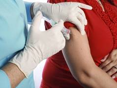
The U.S. Advisory Committee on Immunization Practices (ACIP) recommended the cocooning approach in 2005, but "programmatic challenges and institutional hurdles have prevented widespread implementation both in the United States and in other countries." In 2011, ACIP recommended vaccination during the second or third trimester as the preferred strategy. [Editor’s note: The new CDC immunization schedule, released in January 2013, recommends that a dose of the Tdap vaccine be administered to all women during each pregnancy, whether or not she has received the vaccine previously (MMWR 2013;62 [Suppl 1:9-19)].
Dr. Terranella and his colleagues compared the health benefits and costs of three approaches using a cohort model of the 4,131,019 infants born in the United States in 2009 and followed for 1 year.
They calculated the expected number of infant pertussis cases using information from a national database of notifiable diseases, assuming a conservative underreporting rate of 15%. Some studies have documented underreporting rates as high as 50%, they noted.
They calculated the probabilities of hospitalization, respiratory disease, encephalopathy, and death, given an annual rate of 62.6 cases of infant pertussis per 100,000 infants.
The model assumed 72% coverage of vaccination during pregnancy and the same rate of coverage for postpartum vaccination with and without and cocooning vaccination of the father in one grandparent. It also assumed vaccine effectiveness of 85%.
The model further assumed that transfer of maternal antibodies would be 100% and that the rate of infant protection would be 60% for the first 2 months of life. For postpartum vaccination, there would be a 2-week delay in protection of the infant, because it takes that long for sufficient antibodies to form in the mother after the inoculation.
The model showed that with no vaccination, an estimated 3,041 cases of infant pertussis would occur each year, causing 1,463 hospitalizations and 22 deaths. In comparison, postpartum vaccination alone could avert 596 infant cases annually, a 20% reduction from the base case. Postpartum vaccination with cocooning averted 987 cases annually, which represents a 32% reduction in incidence. Vaccination during pregnancy would avert even more – 1,012 cases each year, a 33% reduction in incidence, the investigators said (Pediatrics 2013 [doi:10.1542/peds.2012-3144]).
Vaccination during pregnancy prevented more infant deaths (a 49% reduction, compared with no vaccination) than did postpartum vaccination with cocooning (a 29% reduction). It also prevented more hospitalizations (a 38% reduction) than did postpartum vaccination with cocooning (a 32% reduction).
In addition, vaccination during pregnancy saved 396 quality-adjusted life years (QALYs) annually, compared with 253 QALYs saved with postpartum vaccination with cocooning.
The annual cost of vaccination during pregnancy was estimated to be $171 million, while that of postpartum vaccination with cocooning was estimated to be $513 million. The cost per QALY saved was calculated to be approximately $414,000 for vaccination during pregnancy, compared for both postpartum vaccination ($1.2 million) with cocooning ($2 million).
Several sensitivity analyses were performed to assess outcomes if a variety of underlying conditions were changed. In particular, changes in vaccine efficacy, vaccine coverage, maternal antibody effect, and potential infant "blunting" of vaccine efficacy (the interference of infant antibodies with the antibody response to the vaccine) were tested.
"Under nearly all scenarios, a pregnancy vaccination strategy would result in fewer overall cases and deaths at lower cost per case averted and per QALY saved," Dr. Terranella and his associates said.
In addition to these advantages, vaccination during pregnancy is more feasible than postpartum vaccination and cocooning because it can easily be administered during existing prenatal care visits. In contrast, reaching postpartum mothers and especially reaching close contacts such as fathers, grandparents, and siblings of the infant is more difficult, the researchers added.
No financial conflicts of interest were reported.
Vaccinating pregnant women to protect their newborns from pertussis appears to avert more cases of the disease, hospitalizations, and deaths than does vaccinating mothers immediately post partum, according to a report published online May 27 in Pediatrics.
In a decision-analysis modeling study, vaccination during pregnancy also was found to be more cost-effective than "postpartum cocooning" – a strategy of vaccinating close family contacts, ideally before the infant’s birth, and vaccinating the mother immediately post partum, said Dr. Andrew Terranella and his associates at the Centers for Disease Control and Prevention.
The U.S. Advisory Committee on Immunization Practices (ACIP) recommended the cocooning approach in 2005, but "programmatic challenges and institutional hurdles have prevented widespread implementation both in the United States and in other countries." In 2011, ACIP recommended vaccination during the second or third trimester as the preferred strategy. [Editor’s note: The new CDC immunization schedule, released in January 2013, recommends that a dose of the Tdap vaccine be administered to all women during each pregnancy, whether or not she has received the vaccine previously (MMWR 2013;62 [Suppl 1:9-19)].
Dr. Terranella and his colleagues compared the health benefits and costs of three approaches using a cohort model of the 4,131,019 infants born in the United States in 2009 and followed for 1 year.
They calculated the expected number of infant pertussis cases using information from a national database of notifiable diseases, assuming a conservative underreporting rate of 15%. Some studies have documented underreporting rates as high as 50%, they noted.
They calculated the probabilities of hospitalization, respiratory disease, encephalopathy, and death, given an annual rate of 62.6 cases of infant pertussis per 100,000 infants.
The model assumed 72% coverage of vaccination during pregnancy and the same rate of coverage for postpartum vaccination with and without and cocooning vaccination of the father in one grandparent. It also assumed vaccine effectiveness of 85%.
The model further assumed that transfer of maternal antibodies would be 100% and that the rate of infant protection would be 60% for the first 2 months of life. For postpartum vaccination, there would be a 2-week delay in protection of the infant, because it takes that long for sufficient antibodies to form in the mother after the inoculation.
The model showed that with no vaccination, an estimated 3,041 cases of infant pertussis would occur each year, causing 1,463 hospitalizations and 22 deaths. In comparison, postpartum vaccination alone could avert 596 infant cases annually, a 20% reduction from the base case. Postpartum vaccination with cocooning averted 987 cases annually, which represents a 32% reduction in incidence. Vaccination during pregnancy would avert even more – 1,012 cases each year, a 33% reduction in incidence, the investigators said (Pediatrics 2013 [doi:10.1542/peds.2012-3144]).
Vaccination during pregnancy prevented more infant deaths (a 49% reduction, compared with no vaccination) than did postpartum vaccination with cocooning (a 29% reduction). It also prevented more hospitalizations (a 38% reduction) than did postpartum vaccination with cocooning (a 32% reduction).
In addition, vaccination during pregnancy saved 396 quality-adjusted life years (QALYs) annually, compared with 253 QALYs saved with postpartum vaccination with cocooning.
The annual cost of vaccination during pregnancy was estimated to be $171 million, while that of postpartum vaccination with cocooning was estimated to be $513 million. The cost per QALY saved was calculated to be approximately $414,000 for vaccination during pregnancy, compared for both postpartum vaccination ($1.2 million) with cocooning ($2 million).
Several sensitivity analyses were performed to assess outcomes if a variety of underlying conditions were changed. In particular, changes in vaccine efficacy, vaccine coverage, maternal antibody effect, and potential infant "blunting" of vaccine efficacy (the interference of infant antibodies with the antibody response to the vaccine) were tested.
"Under nearly all scenarios, a pregnancy vaccination strategy would result in fewer overall cases and deaths at lower cost per case averted and per QALY saved," Dr. Terranella and his associates said.
In addition to these advantages, vaccination during pregnancy is more feasible than postpartum vaccination and cocooning because it can easily be administered during existing prenatal care visits. In contrast, reaching postpartum mothers and especially reaching close contacts such as fathers, grandparents, and siblings of the infant is more difficult, the researchers added.
No financial conflicts of interest were reported.
FROM PEDIATRICS
Major finding: Vaccination during pregnancy would reduce deaths from infant pertussis by 49%, reduce hospitalizations by 38%, and save 396 QALYs per year, while postpartum vaccination and cocooning would reduce deaths by 29%, reduce hospitalizations by 33%, and save 253 QALYs per year.
Data source: A decision-analysis modeling study using data from the cohort of over 4 million U.S. births in 2009.
Disclosures: No financial conflicts of interest were reported.

Real-world NLST results show CT scans find more early-stage lung cancers
For older Americans who are or used to be heavy smokers, annual CT screening for lung cancer appears to reduce mortality from the disease, according to a report published online in the New England Journal of Medicine.
The results from the initial round of screening in the National Lung Screening Trial show that over a 3-year period, annual low-dose helical CT detected significantly more early-stage lung cancers than conventional chest radiography did, said Dr. Timothy R. Church of the division of environmental health sciences, University of Minnesota School of Public Health, Minneapolis, and his associates in the NLST research group.
In the real-world clinical settings, "a reduction in mortality from lung cancer is achievable at U.S. screening centers that have staff experienced in chest CT," they added.
The findings were published online and simultaneously presented at the annual meeting of the American Thoracic Society.
For this first round of the study, 53,454 asymptomatic men and women aged 55-74 years participated. All had a history of at least 30 pack-years of smoking and were either current smokers or had quit within the preceding 15 years.
The study subjects were treated and followed at 33 sites across the country and were randomly assigned to undergo either low-dose helical CT (26,722 patients) or chest radiography (26,732 patients) to screen for lung cancer every year for 3 years. Eight of these subjects were found to have lung cancer before their first screening could be done and were excluded from the study.
The proportion of study subjects who had positive screening results was markedly higher with low-dose CT scanning (27.3%) than with radiography (9.2%). Lung cancer was diagnosed in 1.1% of the CT group, compared with 0.7% of the radiography group.
In the CT group, 92.5% of the patients who developed lung cancer had a positive screening result (a true-positive result) and 6.2% had a negative screening result (a false-negative result); the remaining subjects missed their scheduled screening visit.
In the radiography group, 71.6% of the patients who developed lung cancer had a true-positive screening result and 25.8% had a false-negative result; the remaining subjects missed their scheduled screening visit.
Thus, the sensitivity and specificity of lung CT were 93.8% and 73.4%, respectively, compared with 73.5% sensitivity and 91.3% specificity for radiography, Dr. Church and his colleagues said (N. Engl. J. Med. 2013 [doi:10.1056/NEJMoa1209120]).
"There were many more bronchioloalveolar carcinomas and adenocarcinomas in the low-dose CT group than in the radiography group (38 vs. 8 and 123 vs. 71, respectively), but the frequencies of other histologic features were similar in the two groups," the investigators said.
The difference between the two groups in the number of detected cancers was almost entirely accounted for by early, stage IA tumors, which were markedly more likely to be identified on CT screening. The rates of detection were fairly comparable between CT and radiography for all the more advanced malignancies.
The study was funded by the National Cancer Institute. No financial conflicts of interest were reported.
FROM THE NEW ENGLAND JOURNAL OF MEDICINE
Major Finding: The sensitivity and specificity of CT for lung cancer detection were 93.8% and 73.4%, respectively; for radiography, sensitivity was 73.5% and specificity was 91.3%.
Data Source: A descriptive study involving 53,454 asymptomatic current or past smokers randomly assigned to undergo annual lung cancer screening using either low-dose CT or chest radiography and followed for 3 years.
Disclosures: This study was funded by the National Cancer Institute. No financial conflicts of interest were reported.