User login
Warmer temperatures push malaria to higher elevations

Credit: Asnakew Yeshiwondim
Researchers say they have the first hard evidence that malaria creeps to higher elevations during warmer years and retreats to lower altitudes when temperatures cool.
The evidence comes from an analysis of highland regions in Ethiopia and Colombia.
It suggests that future climate warming will prompt a rise in malaria incidence in densely populated regions of Africa and
South America, unless efforts to monitor and control malaria are increased.
“We saw an upward expansion of malaria cases to higher altitudes in warmer years, which is a clear signal of a response by highland malaria to changes in climate,” said study author Mercedes Pascual, PhD, of the University of Michigan in Ann Arbor.
“This is indisputable evidence of a climate effect. The main implication is that, with warmer temperatures, we expect to see a higher number of people exposed to the risk of malaria in tropical highland areas like these.”
Dr Pascual and her colleagues reported these findings in Science.
It was more than 20 years ago that malaria was first identified as a disease that might be especially sensitive to climate change, because both the Plasmodium parasites that cause it and the Anopheles mosquitoes that spread it thrive as temperatures warm.
Some early studies concluded that climate change would lead to an increase in malaria cases as the disease expanded its range into higher elevations. But some of the assumptions behind those predictions were later criticized.
More recently, researchers have argued that improved socioeconomic conditions and more aggressive mosquito-control efforts will likely exert a far greater influence than climatic factors over the extent and intensity of malaria worldwide.
What’s been missing in this debate is an analysis of regional records with sufficient resolution to determine how the spatial distribution of malaria cases has changed in response to year-to-year temperature variations, especially in densely populated highlands that have historically provided havens from the disease.
So Dr Pascual and her colleagues looked for evidence of a changing spatial distribution of malaria with varying temperature in the highlands of Ethiopia and Colombia. They examined malaria case records from the Antioquia region of western Colombia from 1990 to 2005 and from the Debre Zeit area of central Ethiopia from 1993 to 2005.
By focusing solely on the altitudinal response to year-to-year temperature changes, the researchers were able to exclude other variables that can influence malaria case numbers, such as mosquito-control programs, resistance to antimalarial drugs, and fluctuations in rainfall amounts.
The team found that the median altitude of malaria cases shifted to higher elevations in warmer years and back to lower elevations in cooler years. This relatively simple analysis yielded a clear signal that can only be explained by temperature changes, the group said.
“Our latest research suggests that, with progressive global warming, malaria will creep up the mountains and spread to new high-altitude areas,” said study author Menno Bouma, MD, of the London School of Hygiene & Tropical Medicine in the UK.
“And because these populations lack protective immunity, they will be particularly vulnerable to severe morbidity and mortality.”
In addition, the study results suggest that climate change can explain malaria trends in both the highland regions in recent decades.
In the Debre Zeit region of Ethiopia, at an elevation range of between 5280 feet and 7920 feet, about 37 million people (roughly 43% of the country’s population) live in rural areas at risk of higher malaria exposure under a warming climate.
In a previous study, researchers estimated that a 1-degree temperature increase could result in an additional 3 million malaria cases annually in Ethiopia in the under-15 population, unless control efforts are strengthened.
“Our findings here underscore the size of the problem,” Dr Pascual said, “and emphasize the need for sustained intervention efforts in these regions, especially in Africa.” 
Credit: Asnakew Yeshiwondim
Researchers say they have the first hard evidence that malaria creeps to higher elevations during warmer years and retreats to lower altitudes when temperatures cool.
The evidence comes from an analysis of highland regions in Ethiopia and Colombia.
It suggests that future climate warming will prompt a rise in malaria incidence in densely populated regions of Africa and
South America, unless efforts to monitor and control malaria are increased.
“We saw an upward expansion of malaria cases to higher altitudes in warmer years, which is a clear signal of a response by highland malaria to changes in climate,” said study author Mercedes Pascual, PhD, of the University of Michigan in Ann Arbor.
“This is indisputable evidence of a climate effect. The main implication is that, with warmer temperatures, we expect to see a higher number of people exposed to the risk of malaria in tropical highland areas like these.”
Dr Pascual and her colleagues reported these findings in Science.
It was more than 20 years ago that malaria was first identified as a disease that might be especially sensitive to climate change, because both the Plasmodium parasites that cause it and the Anopheles mosquitoes that spread it thrive as temperatures warm.
Some early studies concluded that climate change would lead to an increase in malaria cases as the disease expanded its range into higher elevations. But some of the assumptions behind those predictions were later criticized.
More recently, researchers have argued that improved socioeconomic conditions and more aggressive mosquito-control efforts will likely exert a far greater influence than climatic factors over the extent and intensity of malaria worldwide.
What’s been missing in this debate is an analysis of regional records with sufficient resolution to determine how the spatial distribution of malaria cases has changed in response to year-to-year temperature variations, especially in densely populated highlands that have historically provided havens from the disease.
So Dr Pascual and her colleagues looked for evidence of a changing spatial distribution of malaria with varying temperature in the highlands of Ethiopia and Colombia. They examined malaria case records from the Antioquia region of western Colombia from 1990 to 2005 and from the Debre Zeit area of central Ethiopia from 1993 to 2005.
By focusing solely on the altitudinal response to year-to-year temperature changes, the researchers were able to exclude other variables that can influence malaria case numbers, such as mosquito-control programs, resistance to antimalarial drugs, and fluctuations in rainfall amounts.
The team found that the median altitude of malaria cases shifted to higher elevations in warmer years and back to lower elevations in cooler years. This relatively simple analysis yielded a clear signal that can only be explained by temperature changes, the group said.
“Our latest research suggests that, with progressive global warming, malaria will creep up the mountains and spread to new high-altitude areas,” said study author Menno Bouma, MD, of the London School of Hygiene & Tropical Medicine in the UK.
“And because these populations lack protective immunity, they will be particularly vulnerable to severe morbidity and mortality.”
In addition, the study results suggest that climate change can explain malaria trends in both the highland regions in recent decades.
In the Debre Zeit region of Ethiopia, at an elevation range of between 5280 feet and 7920 feet, about 37 million people (roughly 43% of the country’s population) live in rural areas at risk of higher malaria exposure under a warming climate.
In a previous study, researchers estimated that a 1-degree temperature increase could result in an additional 3 million malaria cases annually in Ethiopia in the under-15 population, unless control efforts are strengthened.
“Our findings here underscore the size of the problem,” Dr Pascual said, “and emphasize the need for sustained intervention efforts in these regions, especially in Africa.”
Credit: Asnakew Yeshiwondim
Researchers say they have the first hard evidence that malaria creeps to higher elevations during warmer years and retreats to lower altitudes when temperatures cool.
The evidence comes from an analysis of highland regions in Ethiopia and Colombia.
It suggests that future climate warming will prompt a rise in malaria incidence in densely populated regions of Africa and
South America, unless efforts to monitor and control malaria are increased.
“We saw an upward expansion of malaria cases to higher altitudes in warmer years, which is a clear signal of a response by highland malaria to changes in climate,” said study author Mercedes Pascual, PhD, of the University of Michigan in Ann Arbor.
“This is indisputable evidence of a climate effect. The main implication is that, with warmer temperatures, we expect to see a higher number of people exposed to the risk of malaria in tropical highland areas like these.”
Dr Pascual and her colleagues reported these findings in Science.
It was more than 20 years ago that malaria was first identified as a disease that might be especially sensitive to climate change, because both the Plasmodium parasites that cause it and the Anopheles mosquitoes that spread it thrive as temperatures warm.
Some early studies concluded that climate change would lead to an increase in malaria cases as the disease expanded its range into higher elevations. But some of the assumptions behind those predictions were later criticized.
More recently, researchers have argued that improved socioeconomic conditions and more aggressive mosquito-control efforts will likely exert a far greater influence than climatic factors over the extent and intensity of malaria worldwide.
What’s been missing in this debate is an analysis of regional records with sufficient resolution to determine how the spatial distribution of malaria cases has changed in response to year-to-year temperature variations, especially in densely populated highlands that have historically provided havens from the disease.
So Dr Pascual and her colleagues looked for evidence of a changing spatial distribution of malaria with varying temperature in the highlands of Ethiopia and Colombia. They examined malaria case records from the Antioquia region of western Colombia from 1990 to 2005 and from the Debre Zeit area of central Ethiopia from 1993 to 2005.
By focusing solely on the altitudinal response to year-to-year temperature changes, the researchers were able to exclude other variables that can influence malaria case numbers, such as mosquito-control programs, resistance to antimalarial drugs, and fluctuations in rainfall amounts.
The team found that the median altitude of malaria cases shifted to higher elevations in warmer years and back to lower elevations in cooler years. This relatively simple analysis yielded a clear signal that can only be explained by temperature changes, the group said.
“Our latest research suggests that, with progressive global warming, malaria will creep up the mountains and spread to new high-altitude areas,” said study author Menno Bouma, MD, of the London School of Hygiene & Tropical Medicine in the UK.
“And because these populations lack protective immunity, they will be particularly vulnerable to severe morbidity and mortality.”
In addition, the study results suggest that climate change can explain malaria trends in both the highland regions in recent decades.
In the Debre Zeit region of Ethiopia, at an elevation range of between 5280 feet and 7920 feet, about 37 million people (roughly 43% of the country’s population) live in rural areas at risk of higher malaria exposure under a warming climate.
In a previous study, researchers estimated that a 1-degree temperature increase could result in an additional 3 million malaria cases annually in Ethiopia in the under-15 population, unless control efforts are strengthened.
“Our findings here underscore the size of the problem,” Dr Pascual said, “and emphasize the need for sustained intervention efforts in these regions, especially in Africa.”
Pathway may drive treatment resistance in T-ALL

Experiments in zebrafish have revealed a mechanism that may drive relapse in human T-cell acute lymphoblastic leukemia (T-ALL).
Researchers identified a subset of T-ALL cells that spontaneously acquired activation of the Akt pathway.
This increased the frequency of leukemia-propagating cells (LPCs) and mediated resistance to treatment with dexamethasone. However, adding an Akt inhibitor to treatment overcame this resistance.
“The Akt pathway appears to be a major driver of treatment resistance,” said study author David Langenau, PhD, of the Harvard Stem Cell Institute in Boston.
“We also show that this same pathway increases overall growth of leukemic cells and increases the fraction of cells capable of driving relapse.”
Dr Langenau and his colleagues described these findings in Cancer Cell.
Previous research had shown that, if LPCs are retained following treatment, they will initiate disease relapse. And LPC frequency can increase over time. However, it was not clear if this was the result of continued clonal evolution or if a clone with high LPC frequency out-competed other cells.
So Dr Langenau and his colleagues used zebrafish models to study T-ALL clones. The team observed functional variation within individual clones and identified clones that enhanced growth rate and leukemia-propagating potential with time.
A subset of these clones acquired Akt pathway activation, which increased the number of LPCs by activating mTORC1. The cells also exhibited an elevated growth rate, which may have resulted from stabilizing the Myc protein.
Furthermore, the LPCs proved resistant to treatment with dexamethasone. But the researchers were able to reverse this resistance by combining dexamethasone with the Akt inhibitor MK2206. This approach proved effective both in zebrafish models and in human T-ALL cells.
“Our work will likely help in identifying patients that are prone to relapse and would benefit from co-treatment with inhibitors of the Akt pathway and typical front-line cancer therapy,” said Jessica Blackburn, PhD, a member of Dr Langenau’s lab.
She and her colleagues are now hoping to identify other mutations that lead to relapse, thereby pinpointing potential drug targets for patients with aggressive leukemia.
Experiments in zebrafish have revealed a mechanism that may drive relapse in human T-cell acute lymphoblastic leukemia (T-ALL).
Researchers identified a subset of T-ALL cells that spontaneously acquired activation of the Akt pathway.
This increased the frequency of leukemia-propagating cells (LPCs) and mediated resistance to treatment with dexamethasone. However, adding an Akt inhibitor to treatment overcame this resistance.
“The Akt pathway appears to be a major driver of treatment resistance,” said study author David Langenau, PhD, of the Harvard Stem Cell Institute in Boston.
“We also show that this same pathway increases overall growth of leukemic cells and increases the fraction of cells capable of driving relapse.”
Dr Langenau and his colleagues described these findings in Cancer Cell.
Previous research had shown that, if LPCs are retained following treatment, they will initiate disease relapse. And LPC frequency can increase over time. However, it was not clear if this was the result of continued clonal evolution or if a clone with high LPC frequency out-competed other cells.
So Dr Langenau and his colleagues used zebrafish models to study T-ALL clones. The team observed functional variation within individual clones and identified clones that enhanced growth rate and leukemia-propagating potential with time.
A subset of these clones acquired Akt pathway activation, which increased the number of LPCs by activating mTORC1. The cells also exhibited an elevated growth rate, which may have resulted from stabilizing the Myc protein.
Furthermore, the LPCs proved resistant to treatment with dexamethasone. But the researchers were able to reverse this resistance by combining dexamethasone with the Akt inhibitor MK2206. This approach proved effective both in zebrafish models and in human T-ALL cells.
“Our work will likely help in identifying patients that are prone to relapse and would benefit from co-treatment with inhibitors of the Akt pathway and typical front-line cancer therapy,” said Jessica Blackburn, PhD, a member of Dr Langenau’s lab.
She and her colleagues are now hoping to identify other mutations that lead to relapse, thereby pinpointing potential drug targets for patients with aggressive leukemia.
Experiments in zebrafish have revealed a mechanism that may drive relapse in human T-cell acute lymphoblastic leukemia (T-ALL).
Researchers identified a subset of T-ALL cells that spontaneously acquired activation of the Akt pathway.
This increased the frequency of leukemia-propagating cells (LPCs) and mediated resistance to treatment with dexamethasone. However, adding an Akt inhibitor to treatment overcame this resistance.
“The Akt pathway appears to be a major driver of treatment resistance,” said study author David Langenau, PhD, of the Harvard Stem Cell Institute in Boston.
“We also show that this same pathway increases overall growth of leukemic cells and increases the fraction of cells capable of driving relapse.”
Dr Langenau and his colleagues described these findings in Cancer Cell.
Previous research had shown that, if LPCs are retained following treatment, they will initiate disease relapse. And LPC frequency can increase over time. However, it was not clear if this was the result of continued clonal evolution or if a clone with high LPC frequency out-competed other cells.
So Dr Langenau and his colleagues used zebrafish models to study T-ALL clones. The team observed functional variation within individual clones and identified clones that enhanced growth rate and leukemia-propagating potential with time.
A subset of these clones acquired Akt pathway activation, which increased the number of LPCs by activating mTORC1. The cells also exhibited an elevated growth rate, which may have resulted from stabilizing the Myc protein.
Furthermore, the LPCs proved resistant to treatment with dexamethasone. But the researchers were able to reverse this resistance by combining dexamethasone with the Akt inhibitor MK2206. This approach proved effective both in zebrafish models and in human T-ALL cells.
“Our work will likely help in identifying patients that are prone to relapse and would benefit from co-treatment with inhibitors of the Akt pathway and typical front-line cancer therapy,” said Jessica Blackburn, PhD, a member of Dr Langenau’s lab.
She and her colleagues are now hoping to identify other mutations that lead to relapse, thereby pinpointing potential drug targets for patients with aggressive leukemia.
England’s Cancer Drugs Fund raises concerns

Credit: Rhoda Baer
Cancer patients in England are more likely to receive prescriptions for expensive drugs than patients in Wales, according to a study published in the British Journal of Cancer.
The research suggests this disparity is associated with the Cancer Drugs Fund (CDF), money set aside by the English government to pay for drugs that haven’t been approved by the National Institute for Health and Care Excellence (NICE) and aren’t available within the National Health Service (NHS).
The governments of Wales, Scotland, and Northern Ireland do not have access to the CDF or have similar programs of their own.
“There’s been much debate surrounding the Cancer Drugs Fund,” said study author Charlotte Chamberlain, MBBS, of the University of Bristol in the UK.
“The vast majority of Cancer Drugs Fund drugs do not cure the cancer but may extend life or improve symptoms in some people. The high cost of these drugs means that the NHS cannot afford other treatments, and, therefore, critics argue that public money is being spent inefficiently.”
To assess the impact of the CDF, Dr Chamberlain and her colleagues analyzed data from hospital pharmacies in England and Wales from August 2007 to December 2012. (The CDF was established in 2010, and the researchers wanted to capture data from before and after its introduction.)
The team evaluated 15 drugs that represent different categories of NICE approval—recommended, not recommended, and not yet appraised.
The results showed that, after the CDF was established, drugs recommended by NICE were not prescribed any more in England than in Wales.
However, drugs that were rejected by NICE because they were not cost-effective were prescribed up to 7 times more often in England than in Wales. For example, in the year before the CDF was introduced, prescription rates of imatinib (which is not recommended by NICE) were substantially higher in England than in Wales.
Immediately before the introduction of the CDF, following the first NICE rejection, imatinib prescribing declined in both countries. But it declined more slowly in England than in Wales, despite 2 additional NICE rejections. Regression analysis showed evidence of an association between the CDF and increased prescribing in England compared to Wales (P<0.001).
The research also revealed surprising information regarding the 3 most recently launched drugs—bendamustine, pazopanib, and abiraterone, which were awaiting NICE appraisal when the CDF was established but have since been approved.
These drugs were prescribed less often in England than in Wales. For instance, prescription rates of bendamustine were 25% lower in England.
This finding suggests that physicians in England have been slower to adopt newer drugs that are cost-effective, the researchers said.
“Our research has highlighted that the CDF has created an inequality between cancer sufferers in England and those in Wales,” Dr Chamberlain noted. “This raises ethical, moral, financial, and policy concerns.”
Credit: Rhoda Baer
Cancer patients in England are more likely to receive prescriptions for expensive drugs than patients in Wales, according to a study published in the British Journal of Cancer.
The research suggests this disparity is associated with the Cancer Drugs Fund (CDF), money set aside by the English government to pay for drugs that haven’t been approved by the National Institute for Health and Care Excellence (NICE) and aren’t available within the National Health Service (NHS).
The governments of Wales, Scotland, and Northern Ireland do not have access to the CDF or have similar programs of their own.
“There’s been much debate surrounding the Cancer Drugs Fund,” said study author Charlotte Chamberlain, MBBS, of the University of Bristol in the UK.
“The vast majority of Cancer Drugs Fund drugs do not cure the cancer but may extend life or improve symptoms in some people. The high cost of these drugs means that the NHS cannot afford other treatments, and, therefore, critics argue that public money is being spent inefficiently.”
To assess the impact of the CDF, Dr Chamberlain and her colleagues analyzed data from hospital pharmacies in England and Wales from August 2007 to December 2012. (The CDF was established in 2010, and the researchers wanted to capture data from before and after its introduction.)
The team evaluated 15 drugs that represent different categories of NICE approval—recommended, not recommended, and not yet appraised.
The results showed that, after the CDF was established, drugs recommended by NICE were not prescribed any more in England than in Wales.
However, drugs that were rejected by NICE because they were not cost-effective were prescribed up to 7 times more often in England than in Wales. For example, in the year before the CDF was introduced, prescription rates of imatinib (which is not recommended by NICE) were substantially higher in England than in Wales.
Immediately before the introduction of the CDF, following the first NICE rejection, imatinib prescribing declined in both countries. But it declined more slowly in England than in Wales, despite 2 additional NICE rejections. Regression analysis showed evidence of an association between the CDF and increased prescribing in England compared to Wales (P<0.001).
The research also revealed surprising information regarding the 3 most recently launched drugs—bendamustine, pazopanib, and abiraterone, which were awaiting NICE appraisal when the CDF was established but have since been approved.
These drugs were prescribed less often in England than in Wales. For instance, prescription rates of bendamustine were 25% lower in England.
This finding suggests that physicians in England have been slower to adopt newer drugs that are cost-effective, the researchers said.
“Our research has highlighted that the CDF has created an inequality between cancer sufferers in England and those in Wales,” Dr Chamberlain noted. “This raises ethical, moral, financial, and policy concerns.”
Credit: Rhoda Baer
Cancer patients in England are more likely to receive prescriptions for expensive drugs than patients in Wales, according to a study published in the British Journal of Cancer.
The research suggests this disparity is associated with the Cancer Drugs Fund (CDF), money set aside by the English government to pay for drugs that haven’t been approved by the National Institute for Health and Care Excellence (NICE) and aren’t available within the National Health Service (NHS).
The governments of Wales, Scotland, and Northern Ireland do not have access to the CDF or have similar programs of their own.
“There’s been much debate surrounding the Cancer Drugs Fund,” said study author Charlotte Chamberlain, MBBS, of the University of Bristol in the UK.
“The vast majority of Cancer Drugs Fund drugs do not cure the cancer but may extend life or improve symptoms in some people. The high cost of these drugs means that the NHS cannot afford other treatments, and, therefore, critics argue that public money is being spent inefficiently.”
To assess the impact of the CDF, Dr Chamberlain and her colleagues analyzed data from hospital pharmacies in England and Wales from August 2007 to December 2012. (The CDF was established in 2010, and the researchers wanted to capture data from before and after its introduction.)
The team evaluated 15 drugs that represent different categories of NICE approval—recommended, not recommended, and not yet appraised.
The results showed that, after the CDF was established, drugs recommended by NICE were not prescribed any more in England than in Wales.
However, drugs that were rejected by NICE because they were not cost-effective were prescribed up to 7 times more often in England than in Wales. For example, in the year before the CDF was introduced, prescription rates of imatinib (which is not recommended by NICE) were substantially higher in England than in Wales.
Immediately before the introduction of the CDF, following the first NICE rejection, imatinib prescribing declined in both countries. But it declined more slowly in England than in Wales, despite 2 additional NICE rejections. Regression analysis showed evidence of an association between the CDF and increased prescribing in England compared to Wales (P<0.001).
The research also revealed surprising information regarding the 3 most recently launched drugs—bendamustine, pazopanib, and abiraterone, which were awaiting NICE appraisal when the CDF was established but have since been approved.
These drugs were prescribed less often in England than in Wales. For instance, prescription rates of bendamustine were 25% lower in England.
This finding suggests that physicians in England have been slower to adopt newer drugs that are cost-effective, the researchers said.
“Our research has highlighted that the CDF has created an inequality between cancer sufferers in England and those in Wales,” Dr Chamberlain noted. “This raises ethical, moral, financial, and policy concerns.”
Nanoparticle therapy active in B-cell malignancies

An experimental nanoparticle therapy has shown preclinical activity against a range of B-cell malignancies, researchers have reported.
The therapy, SNS01-T, inhibited tumor growth and improved survival in mouse models of multiple myeloma, mantle cell lymphoma, and diffuse large B-cell lymphoma.
The treatment exhibited single-agent activity in these mice but also demonstrated synergy with lenalidomide and bortezomib.
Sarah Francis, PhD, of the University of Waterloo in Ontario, Canada, and her colleagues conducted this research and recounted the results in Molecular Therapy.
SNS01-T consists of 3 components: small interfering RNA targeting the native eukaryotic translation initiation factor 5A (eIF5A), plasmids expressing a pro-apoptotic mutant of elF5A under the control of a B-cell specific promoter, and a synthetic cationic polymer polyethylenimine, which serves as the delivery vehicle.
The small interfering RNA component of SNS01-T suppresses elF5A expression, thereby interfering with translation of eIF5A and reducing levels of hypusinated elF5A in cancer cells. This inhibits activation of NF-kB and induces apoptosis.
The B-cell specific plasmid component of SNS01-T expresses an arginine substituted form of eIF5A—eIF5AK50R—that cannot be hypusinated, which leads to selective induction of apoptosis in B cells.
Dr Francis and her colleagues found that SNS01-T is preferentially taken up by malignant B cells. In myeloma cells, uptake of SNS01-T was up to 5-fold higher than uptake by normal, naïve B cells.
Uptake into myeloma cells induced 45% cell death within 24 hours, but there was almost no measureable death of normal, naïve B cells.
SNS01-T also prompted dose-dependent inhibition of tumor growth in mouse models of multiple myeloma, mantle cell lymphoma, and diffuse large B-cell lymphoma, with up to 90% inhibition at the highest doses.
When administered at doses of 0.18 mg/kg or more, SNS01-T significantly extended the life span of treated mice. The researchers observed a reduction in the pro-survival form of the eIF5A protein in tumor tissue, which was consistent with drug activity.
SNS01-T also demonstrated synergy with bortezomib and lenalidomide. Mice that received SNS01-T and lenalidomide in combination had a 100% survival rate, compared to 60% for mice that received SNS01-T alone and 20% for mice that received lenalidomide alone.
A single 6-week cycle of SNS01-T and lenalidomide eradicated tumors in 67% of mice, and there was no regrowth after an additional 8 weeks without further treatment.
Similarly, combination SNS01-T and bortezomib inhibited tumor growth by 89%, compared to 59% for SNS01-T alone and 39% for bortezomib alone.
This research was supported by Senesco Technologies, Inc., the company developing SNS01-T.
An experimental nanoparticle therapy has shown preclinical activity against a range of B-cell malignancies, researchers have reported.
The therapy, SNS01-T, inhibited tumor growth and improved survival in mouse models of multiple myeloma, mantle cell lymphoma, and diffuse large B-cell lymphoma.
The treatment exhibited single-agent activity in these mice but also demonstrated synergy with lenalidomide and bortezomib.
Sarah Francis, PhD, of the University of Waterloo in Ontario, Canada, and her colleagues conducted this research and recounted the results in Molecular Therapy.
SNS01-T consists of 3 components: small interfering RNA targeting the native eukaryotic translation initiation factor 5A (eIF5A), plasmids expressing a pro-apoptotic mutant of elF5A under the control of a B-cell specific promoter, and a synthetic cationic polymer polyethylenimine, which serves as the delivery vehicle.
The small interfering RNA component of SNS01-T suppresses elF5A expression, thereby interfering with translation of eIF5A and reducing levels of hypusinated elF5A in cancer cells. This inhibits activation of NF-kB and induces apoptosis.
The B-cell specific plasmid component of SNS01-T expresses an arginine substituted form of eIF5A—eIF5AK50R—that cannot be hypusinated, which leads to selective induction of apoptosis in B cells.
Dr Francis and her colleagues found that SNS01-T is preferentially taken up by malignant B cells. In myeloma cells, uptake of SNS01-T was up to 5-fold higher than uptake by normal, naïve B cells.
Uptake into myeloma cells induced 45% cell death within 24 hours, but there was almost no measureable death of normal, naïve B cells.
SNS01-T also prompted dose-dependent inhibition of tumor growth in mouse models of multiple myeloma, mantle cell lymphoma, and diffuse large B-cell lymphoma, with up to 90% inhibition at the highest doses.
When administered at doses of 0.18 mg/kg or more, SNS01-T significantly extended the life span of treated mice. The researchers observed a reduction in the pro-survival form of the eIF5A protein in tumor tissue, which was consistent with drug activity.
SNS01-T also demonstrated synergy with bortezomib and lenalidomide. Mice that received SNS01-T and lenalidomide in combination had a 100% survival rate, compared to 60% for mice that received SNS01-T alone and 20% for mice that received lenalidomide alone.
A single 6-week cycle of SNS01-T and lenalidomide eradicated tumors in 67% of mice, and there was no regrowth after an additional 8 weeks without further treatment.
Similarly, combination SNS01-T and bortezomib inhibited tumor growth by 89%, compared to 59% for SNS01-T alone and 39% for bortezomib alone.
This research was supported by Senesco Technologies, Inc., the company developing SNS01-T.
An experimental nanoparticle therapy has shown preclinical activity against a range of B-cell malignancies, researchers have reported.
The therapy, SNS01-T, inhibited tumor growth and improved survival in mouse models of multiple myeloma, mantle cell lymphoma, and diffuse large B-cell lymphoma.
The treatment exhibited single-agent activity in these mice but also demonstrated synergy with lenalidomide and bortezomib.
Sarah Francis, PhD, of the University of Waterloo in Ontario, Canada, and her colleagues conducted this research and recounted the results in Molecular Therapy.
SNS01-T consists of 3 components: small interfering RNA targeting the native eukaryotic translation initiation factor 5A (eIF5A), plasmids expressing a pro-apoptotic mutant of elF5A under the control of a B-cell specific promoter, and a synthetic cationic polymer polyethylenimine, which serves as the delivery vehicle.
The small interfering RNA component of SNS01-T suppresses elF5A expression, thereby interfering with translation of eIF5A and reducing levels of hypusinated elF5A in cancer cells. This inhibits activation of NF-kB and induces apoptosis.
The B-cell specific plasmid component of SNS01-T expresses an arginine substituted form of eIF5A—eIF5AK50R—that cannot be hypusinated, which leads to selective induction of apoptosis in B cells.
Dr Francis and her colleagues found that SNS01-T is preferentially taken up by malignant B cells. In myeloma cells, uptake of SNS01-T was up to 5-fold higher than uptake by normal, naïve B cells.
Uptake into myeloma cells induced 45% cell death within 24 hours, but there was almost no measureable death of normal, naïve B cells.
SNS01-T also prompted dose-dependent inhibition of tumor growth in mouse models of multiple myeloma, mantle cell lymphoma, and diffuse large B-cell lymphoma, with up to 90% inhibition at the highest doses.
When administered at doses of 0.18 mg/kg or more, SNS01-T significantly extended the life span of treated mice. The researchers observed a reduction in the pro-survival form of the eIF5A protein in tumor tissue, which was consistent with drug activity.
SNS01-T also demonstrated synergy with bortezomib and lenalidomide. Mice that received SNS01-T and lenalidomide in combination had a 100% survival rate, compared to 60% for mice that received SNS01-T alone and 20% for mice that received lenalidomide alone.
A single 6-week cycle of SNS01-T and lenalidomide eradicated tumors in 67% of mice, and there was no regrowth after an additional 8 weeks without further treatment.
Similarly, combination SNS01-T and bortezomib inhibited tumor growth by 89%, compared to 59% for SNS01-T alone and 39% for bortezomib alone.
This research was supported by Senesco Technologies, Inc., the company developing SNS01-T.
Palliative chemo can have undesired outcomes

Credit: Rhoda Baer
Palliative chemotherapy can negatively impact the end of life for terminally ill cancer patients, according to a paper published in BMJ.
Investigators found that patients who received palliative chemotherapy in their last months of life had an increased risk of requiring intensive medical care, such as resuscitation, and dying in a place they did not choose, such as an intensive care unit.
The researchers therefore suggested that end-of-life discussions may be particularly important for patients who want to receive palliative chemotherapy.
“The results highlight the need for more effective communication by doctors of terminal prognoses and the likely outcomes of chemotherapy for these patients,” said study author Holly Prigerson, PhD, of Weill Cornell Medical College in New York.
“For patients to make informed choices about their care, they need to know if they are incurable and understand what their life expectancy is, that palliative chemotherapy is not intended to cure them, that it may not appreciably prolong their life, and that it may result in the receipt of very aggressive, life-prolonging care at the expense of their quality of life.”
Data have suggested that between 20% and 50% of patients with incurable cancers undergo palliative chemotherapy within 30 days of death. But it has not been clear whether the use of chemotherapy in a patient’s last months is associated with the need for intensive medical care in the last week of life or with the patient’s death.
So Dr Prigerson and her colleagues decided to study the use of palliative chemotherapy in patients with 6 or fewer months to live. The researchers used data from “Coping with Cancer,” a 6-year study of 386 terminally ill patients.
The patients were interviewed around the time of their decision regarding palliative chemotherapy. In the month after each patient died, caregivers were asked to rate their loved ones’ care, quality of life, and place of death as being where the patient would have wanted to die. The investigators then reviewed patients’ medical charts to determine the type of care they actually received in their last week.
Effects of palliative chemo
In all, 56% of patients opted to receive palliative chemotherapy. They were more likely to be younger, married, and better educated than patients not on the treatment.
Patients on chemotherapy also had better performance status, overall quality of life, physical functioning, and psychological well-being at study enrollment.
However, patients who received palliative chemotherapy had a greater risk of requiring cardiopulmonary resuscitation and/or mechanical ventilation (14% vs 2%), and they were more likely to need a feeding tube (11% vs 5%) in their last weeks of life.
Patients on chemotherapy had a greater risk of being admitted to an intensive care unit (14% vs 8%) and of having a late hospice referral (54% vs 37%).
They were also less likely to die where they wanted to (65% vs 80%). They had a greater risk of dying in an intensive care unit (11% vs 2%) and were less likely than their peers to die at home (47% vs 66%).
“It’s hard to see in these data much of a silver lining to palliative chemotherapy for patients in the terminal stage of their cancer,” Dr Prigerson said. “Until now, there hasn’t been evidence of harmful effects of palliative chemotherapy in the last few months of life.”
“This study is a first step in providing evidence that specifically demonstrates what negative outcomes may result. Additional studies are needed to confirm these troubling findings.”
Explaining the negative effects
Dr Prigerson said the harmful effects of palliative chemotherapy may be a result of misunderstanding, a lack of communication, and denial. Patients may not comprehend the purpose and likely consequences of palliative chemotherapy, and they may not fully acknowledge their own prognoses.
In the study, patients receiving palliative chemotherapy were less likely than their peers to talk to their oncologists about end-of-life care (37% vs 48%), to complete Do-Not-Resuscitate orders (36% vs 49%), or to acknowledge that they were terminally ill (35% vs 47%).
“Our finding that patients with terminal cancers were at higher risk of receiving intensive end-of-life care if they were treated with palliative chemotherapy months earlier underscores the importance of oncologists asking patients about their end-of-life wishes,” said Alexi Wright, MD, of the Dana-Farber Cancer Institute in Boston.
“We often wait until patients stop chemotherapy before asking them about where and how they want to die, but this study shows we need to ask patients about their preferences while they are receiving chemotherapy to ensure they receive the kind of care they want near death.”
Moving forward
The investigators stressed that the study results do not suggest patients should be denied palliative chemotherapy.
“The vast majority of patients in this study wanted palliative chemotherapy if it might increase their survival by as little as a week,” Dr Wright said. “This study is a step towards understanding some of the human costs and benefits of palliative chemotherapy.”
The researchers said additional studies should examine whether patients who are aware that chemotherapy is not intended to cure them still want to receive the treatment, confirm the negative outcomes of palliative chemotherapy, and determine if end-of-life discussions promote more informed decision-making and receipt of value-consistent care.
In a related editorial, Mike Rabow, MD, of the University of California, San Francisco, noted that although most patients with metastatic cancer choose to receive chemotherapy, evidence suggests most do not understand its intent.
He said Dr Prigerson’s study suggests the need to “better identify patients who are likely to benefit from chemotherapy near the end of life.” And he encouraged oncologists to discuss with patients the broader implications of chemotherapy when making decisions about treatment.
Credit: Rhoda Baer
Palliative chemotherapy can negatively impact the end of life for terminally ill cancer patients, according to a paper published in BMJ.
Investigators found that patients who received palliative chemotherapy in their last months of life had an increased risk of requiring intensive medical care, such as resuscitation, and dying in a place they did not choose, such as an intensive care unit.
The researchers therefore suggested that end-of-life discussions may be particularly important for patients who want to receive palliative chemotherapy.
“The results highlight the need for more effective communication by doctors of terminal prognoses and the likely outcomes of chemotherapy for these patients,” said study author Holly Prigerson, PhD, of Weill Cornell Medical College in New York.
“For patients to make informed choices about their care, they need to know if they are incurable and understand what their life expectancy is, that palliative chemotherapy is not intended to cure them, that it may not appreciably prolong their life, and that it may result in the receipt of very aggressive, life-prolonging care at the expense of their quality of life.”
Data have suggested that between 20% and 50% of patients with incurable cancers undergo palliative chemotherapy within 30 days of death. But it has not been clear whether the use of chemotherapy in a patient’s last months is associated with the need for intensive medical care in the last week of life or with the patient’s death.
So Dr Prigerson and her colleagues decided to study the use of palliative chemotherapy in patients with 6 or fewer months to live. The researchers used data from “Coping with Cancer,” a 6-year study of 386 terminally ill patients.
The patients were interviewed around the time of their decision regarding palliative chemotherapy. In the month after each patient died, caregivers were asked to rate their loved ones’ care, quality of life, and place of death as being where the patient would have wanted to die. The investigators then reviewed patients’ medical charts to determine the type of care they actually received in their last week.
Effects of palliative chemo
In all, 56% of patients opted to receive palliative chemotherapy. They were more likely to be younger, married, and better educated than patients not on the treatment.
Patients on chemotherapy also had better performance status, overall quality of life, physical functioning, and psychological well-being at study enrollment.
However, patients who received palliative chemotherapy had a greater risk of requiring cardiopulmonary resuscitation and/or mechanical ventilation (14% vs 2%), and they were more likely to need a feeding tube (11% vs 5%) in their last weeks of life.
Patients on chemotherapy had a greater risk of being admitted to an intensive care unit (14% vs 8%) and of having a late hospice referral (54% vs 37%).
They were also less likely to die where they wanted to (65% vs 80%). They had a greater risk of dying in an intensive care unit (11% vs 2%) and were less likely than their peers to die at home (47% vs 66%).
“It’s hard to see in these data much of a silver lining to palliative chemotherapy for patients in the terminal stage of their cancer,” Dr Prigerson said. “Until now, there hasn’t been evidence of harmful effects of palliative chemotherapy in the last few months of life.”
“This study is a first step in providing evidence that specifically demonstrates what negative outcomes may result. Additional studies are needed to confirm these troubling findings.”
Explaining the negative effects
Dr Prigerson said the harmful effects of palliative chemotherapy may be a result of misunderstanding, a lack of communication, and denial. Patients may not comprehend the purpose and likely consequences of palliative chemotherapy, and they may not fully acknowledge their own prognoses.
In the study, patients receiving palliative chemotherapy were less likely than their peers to talk to their oncologists about end-of-life care (37% vs 48%), to complete Do-Not-Resuscitate orders (36% vs 49%), or to acknowledge that they were terminally ill (35% vs 47%).
“Our finding that patients with terminal cancers were at higher risk of receiving intensive end-of-life care if they were treated with palliative chemotherapy months earlier underscores the importance of oncologists asking patients about their end-of-life wishes,” said Alexi Wright, MD, of the Dana-Farber Cancer Institute in Boston.
“We often wait until patients stop chemotherapy before asking them about where and how they want to die, but this study shows we need to ask patients about their preferences while they are receiving chemotherapy to ensure they receive the kind of care they want near death.”
Moving forward
The investigators stressed that the study results do not suggest patients should be denied palliative chemotherapy.
“The vast majority of patients in this study wanted palliative chemotherapy if it might increase their survival by as little as a week,” Dr Wright said. “This study is a step towards understanding some of the human costs and benefits of palliative chemotherapy.”
The researchers said additional studies should examine whether patients who are aware that chemotherapy is not intended to cure them still want to receive the treatment, confirm the negative outcomes of palliative chemotherapy, and determine if end-of-life discussions promote more informed decision-making and receipt of value-consistent care.
In a related editorial, Mike Rabow, MD, of the University of California, San Francisco, noted that although most patients with metastatic cancer choose to receive chemotherapy, evidence suggests most do not understand its intent.
He said Dr Prigerson’s study suggests the need to “better identify patients who are likely to benefit from chemotherapy near the end of life.” And he encouraged oncologists to discuss with patients the broader implications of chemotherapy when making decisions about treatment.
Credit: Rhoda Baer
Palliative chemotherapy can negatively impact the end of life for terminally ill cancer patients, according to a paper published in BMJ.
Investigators found that patients who received palliative chemotherapy in their last months of life had an increased risk of requiring intensive medical care, such as resuscitation, and dying in a place they did not choose, such as an intensive care unit.
The researchers therefore suggested that end-of-life discussions may be particularly important for patients who want to receive palliative chemotherapy.
“The results highlight the need for more effective communication by doctors of terminal prognoses and the likely outcomes of chemotherapy for these patients,” said study author Holly Prigerson, PhD, of Weill Cornell Medical College in New York.
“For patients to make informed choices about their care, they need to know if they are incurable and understand what their life expectancy is, that palliative chemotherapy is not intended to cure them, that it may not appreciably prolong their life, and that it may result in the receipt of very aggressive, life-prolonging care at the expense of their quality of life.”
Data have suggested that between 20% and 50% of patients with incurable cancers undergo palliative chemotherapy within 30 days of death. But it has not been clear whether the use of chemotherapy in a patient’s last months is associated with the need for intensive medical care in the last week of life or with the patient’s death.
So Dr Prigerson and her colleagues decided to study the use of palliative chemotherapy in patients with 6 or fewer months to live. The researchers used data from “Coping with Cancer,” a 6-year study of 386 terminally ill patients.
The patients were interviewed around the time of their decision regarding palliative chemotherapy. In the month after each patient died, caregivers were asked to rate their loved ones’ care, quality of life, and place of death as being where the patient would have wanted to die. The investigators then reviewed patients’ medical charts to determine the type of care they actually received in their last week.
Effects of palliative chemo
In all, 56% of patients opted to receive palliative chemotherapy. They were more likely to be younger, married, and better educated than patients not on the treatment.
Patients on chemotherapy also had better performance status, overall quality of life, physical functioning, and psychological well-being at study enrollment.
However, patients who received palliative chemotherapy had a greater risk of requiring cardiopulmonary resuscitation and/or mechanical ventilation (14% vs 2%), and they were more likely to need a feeding tube (11% vs 5%) in their last weeks of life.
Patients on chemotherapy had a greater risk of being admitted to an intensive care unit (14% vs 8%) and of having a late hospice referral (54% vs 37%).
They were also less likely to die where they wanted to (65% vs 80%). They had a greater risk of dying in an intensive care unit (11% vs 2%) and were less likely than their peers to die at home (47% vs 66%).
“It’s hard to see in these data much of a silver lining to palliative chemotherapy for patients in the terminal stage of their cancer,” Dr Prigerson said. “Until now, there hasn’t been evidence of harmful effects of palliative chemotherapy in the last few months of life.”
“This study is a first step in providing evidence that specifically demonstrates what negative outcomes may result. Additional studies are needed to confirm these troubling findings.”
Explaining the negative effects
Dr Prigerson said the harmful effects of palliative chemotherapy may be a result of misunderstanding, a lack of communication, and denial. Patients may not comprehend the purpose and likely consequences of palliative chemotherapy, and they may not fully acknowledge their own prognoses.
In the study, patients receiving palliative chemotherapy were less likely than their peers to talk to their oncologists about end-of-life care (37% vs 48%), to complete Do-Not-Resuscitate orders (36% vs 49%), or to acknowledge that they were terminally ill (35% vs 47%).
“Our finding that patients with terminal cancers were at higher risk of receiving intensive end-of-life care if they were treated with palliative chemotherapy months earlier underscores the importance of oncologists asking patients about their end-of-life wishes,” said Alexi Wright, MD, of the Dana-Farber Cancer Institute in Boston.
“We often wait until patients stop chemotherapy before asking them about where and how they want to die, but this study shows we need to ask patients about their preferences while they are receiving chemotherapy to ensure they receive the kind of care they want near death.”
Moving forward
The investigators stressed that the study results do not suggest patients should be denied palliative chemotherapy.
“The vast majority of patients in this study wanted palliative chemotherapy if it might increase their survival by as little as a week,” Dr Wright said. “This study is a step towards understanding some of the human costs and benefits of palliative chemotherapy.”
The researchers said additional studies should examine whether patients who are aware that chemotherapy is not intended to cure them still want to receive the treatment, confirm the negative outcomes of palliative chemotherapy, and determine if end-of-life discussions promote more informed decision-making and receipt of value-consistent care.
In a related editorial, Mike Rabow, MD, of the University of California, San Francisco, noted that although most patients with metastatic cancer choose to receive chemotherapy, evidence suggests most do not understand its intent.
He said Dr Prigerson’s study suggests the need to “better identify patients who are likely to benefit from chemotherapy near the end of life.” And he encouraged oncologists to discuss with patients the broader implications of chemotherapy when making decisions about treatment.
Histones’ role in gene regulation

Credit: Eric Smith
Researchers say they’ve discovered how histones control PARP1’s ability to activate genes and repair DNA damage.
Their findings, published in Molecular Cell, appear to have implications for cancer treatment.
Specifically, the investigators found that chemical modification of the histone H2Av leads to substantial changes in nucleosome shape.
As a consequence, a previously hidden portion of the nucleosome becomes exposed and activates PARP1.
Upon activation, PARP1 assembles long branching molecules of Poly(ADP-ribose), which appear to open the DNA packaging around the site of PARP1 activation, thereby exposing specific genes for activation.
“[T]he nucleosome is often portrayed as a stable, inert structure, or a tiny ball,” said study author Alexei V. Tulin, PhD, of Fox Chase Cancer Center in Philadelphia.
“We found that the nucleosome is actually a quite dynamic structure. When we modified one histone, we changed the whole nucleosome.”
In addition to revealing new information about how histones control gene activation, Dr Tulin’s research elucidated a new mechanism of PARP1 regulation.
“This mechanism of PARP1 regulation by histones is still very new,” Dr Tulin said. “People believe that PARP1 is mainly activated through interactions with DNA, but we have found that the main pathway of PARP1 activation is through interactions with the nucleosome.”
Previous research suggested that combining standard anticancer agents with drugs that inhibit PARP1 can more effectively kill cancer cells. But clinical trials testing PARP1 inhibitors in cancer patients have produced disappointing results.
“I believe that, to a large extent, the previous setbacks were caused by a general misconception of the role of PARP1 in living cells and the mechanisms of PARP1 regulation,” Dr Tulin said. “Now that we know this mechanism of PARP1 regulation, we can design approaches to inhibit this protein in an effective way to better treat cancer.”
Dr Tulin and his colleagues are now developing the next generation of PARP1 inhibitors. Designed to block the newly identified mechanism of PARP1 activation, these inhibitors will specifically target PARP1, in contrast to the PARP1 inhibitors currently being tested in clinical trials.
Credit: Eric Smith
Researchers say they’ve discovered how histones control PARP1’s ability to activate genes and repair DNA damage.
Their findings, published in Molecular Cell, appear to have implications for cancer treatment.
Specifically, the investigators found that chemical modification of the histone H2Av leads to substantial changes in nucleosome shape.
As a consequence, a previously hidden portion of the nucleosome becomes exposed and activates PARP1.
Upon activation, PARP1 assembles long branching molecules of Poly(ADP-ribose), which appear to open the DNA packaging around the site of PARP1 activation, thereby exposing specific genes for activation.
“[T]he nucleosome is often portrayed as a stable, inert structure, or a tiny ball,” said study author Alexei V. Tulin, PhD, of Fox Chase Cancer Center in Philadelphia.
“We found that the nucleosome is actually a quite dynamic structure. When we modified one histone, we changed the whole nucleosome.”
In addition to revealing new information about how histones control gene activation, Dr Tulin’s research elucidated a new mechanism of PARP1 regulation.
“This mechanism of PARP1 regulation by histones is still very new,” Dr Tulin said. “People believe that PARP1 is mainly activated through interactions with DNA, but we have found that the main pathway of PARP1 activation is through interactions with the nucleosome.”
Previous research suggested that combining standard anticancer agents with drugs that inhibit PARP1 can more effectively kill cancer cells. But clinical trials testing PARP1 inhibitors in cancer patients have produced disappointing results.
“I believe that, to a large extent, the previous setbacks were caused by a general misconception of the role of PARP1 in living cells and the mechanisms of PARP1 regulation,” Dr Tulin said. “Now that we know this mechanism of PARP1 regulation, we can design approaches to inhibit this protein in an effective way to better treat cancer.”
Dr Tulin and his colleagues are now developing the next generation of PARP1 inhibitors. Designed to block the newly identified mechanism of PARP1 activation, these inhibitors will specifically target PARP1, in contrast to the PARP1 inhibitors currently being tested in clinical trials.
Credit: Eric Smith
Researchers say they’ve discovered how histones control PARP1’s ability to activate genes and repair DNA damage.
Their findings, published in Molecular Cell, appear to have implications for cancer treatment.
Specifically, the investigators found that chemical modification of the histone H2Av leads to substantial changes in nucleosome shape.
As a consequence, a previously hidden portion of the nucleosome becomes exposed and activates PARP1.
Upon activation, PARP1 assembles long branching molecules of Poly(ADP-ribose), which appear to open the DNA packaging around the site of PARP1 activation, thereby exposing specific genes for activation.
“[T]he nucleosome is often portrayed as a stable, inert structure, or a tiny ball,” said study author Alexei V. Tulin, PhD, of Fox Chase Cancer Center in Philadelphia.
“We found that the nucleosome is actually a quite dynamic structure. When we modified one histone, we changed the whole nucleosome.”
In addition to revealing new information about how histones control gene activation, Dr Tulin’s research elucidated a new mechanism of PARP1 regulation.
“This mechanism of PARP1 regulation by histones is still very new,” Dr Tulin said. “People believe that PARP1 is mainly activated through interactions with DNA, but we have found that the main pathway of PARP1 activation is through interactions with the nucleosome.”
Previous research suggested that combining standard anticancer agents with drugs that inhibit PARP1 can more effectively kill cancer cells. But clinical trials testing PARP1 inhibitors in cancer patients have produced disappointing results.
“I believe that, to a large extent, the previous setbacks were caused by a general misconception of the role of PARP1 in living cells and the mechanisms of PARP1 regulation,” Dr Tulin said. “Now that we know this mechanism of PARP1 regulation, we can design approaches to inhibit this protein in an effective way to better treat cancer.”
Dr Tulin and his colleagues are now developing the next generation of PARP1 inhibitors. Designed to block the newly identified mechanism of PARP1 activation, these inhibitors will specifically target PARP1, in contrast to the PARP1 inhibitors currently being tested in clinical trials.
Drugs get orphan designation for AML, MM

Credit: FDA
The US Food and Drug Administration (FDA) has granted orphan designation for the histone deacetylase (HDAC) inhibitor pracinostat to treat acute myeloid leukemia (AML) and the proteasome inhibitor marizomib to treat multiple myeloma (MM).
Orphan designation is available for drugs that treat or prevent rare diseases affecting fewer than 200,000 people in the US.
The designation qualifies the sponsor of a drug for development incentives, including tax credits for qualified clinical testing, prescription drug user fee exemptions, and 7-year marketing exclusivity upon FDA approval.
About pracinostat
The oral HDAC inhibitor pracinostat has been tested in phase 1 and 2 trials of adult and pediatric patients with advanced hematologic disorders and solid tumors.
The drug has been generally well tolerated in more than 200 patients to date, according to the drug’s maker, MEI Pharma.
In a dose-escalation phase 1 trial, pracinostat demonstrated single-agent activity in elderly AML patients. Two of 14 patients (14%) achieved a complete remission, with responses persisting more than 206 days and 362 days.
Researchers are currently conducting a phase 2 trial of pracinostat in combination with azacitidine in elderly patients with newly diagnosed AML. Preliminary data from this trial are expected to be available by December 2014.
About marizomib
The proteasome inhibitor marizomib is under development for the treatment of MM and other malignancies.
Intravenous marizomib has been evaluated in more than 230 patients across 4 phase 1/2 studies, as a single agent or in combination with dexamethasone or an HDAC inhibitor.
Researchers are currently evaluating marizomib in combination with dexamethasone in an ongoing phase 2 trial of highly refractory MM patients, including those who are refractory to carfilzomib.
Marizomib is also being tested in combination with pomalidomide and dexamethasone in a phase 1/2 study of patients with relapsed and refractory MM.
The drug’s maker, Triphase Accelerator Corporation, is currently developing an oral formulation of marizomib.
Credit: FDA
The US Food and Drug Administration (FDA) has granted orphan designation for the histone deacetylase (HDAC) inhibitor pracinostat to treat acute myeloid leukemia (AML) and the proteasome inhibitor marizomib to treat multiple myeloma (MM).
Orphan designation is available for drugs that treat or prevent rare diseases affecting fewer than 200,000 people in the US.
The designation qualifies the sponsor of a drug for development incentives, including tax credits for qualified clinical testing, prescription drug user fee exemptions, and 7-year marketing exclusivity upon FDA approval.
About pracinostat
The oral HDAC inhibitor pracinostat has been tested in phase 1 and 2 trials of adult and pediatric patients with advanced hematologic disorders and solid tumors.
The drug has been generally well tolerated in more than 200 patients to date, according to the drug’s maker, MEI Pharma.
In a dose-escalation phase 1 trial, pracinostat demonstrated single-agent activity in elderly AML patients. Two of 14 patients (14%) achieved a complete remission, with responses persisting more than 206 days and 362 days.
Researchers are currently conducting a phase 2 trial of pracinostat in combination with azacitidine in elderly patients with newly diagnosed AML. Preliminary data from this trial are expected to be available by December 2014.
About marizomib
The proteasome inhibitor marizomib is under development for the treatment of MM and other malignancies.
Intravenous marizomib has been evaluated in more than 230 patients across 4 phase 1/2 studies, as a single agent or in combination with dexamethasone or an HDAC inhibitor.
Researchers are currently evaluating marizomib in combination with dexamethasone in an ongoing phase 2 trial of highly refractory MM patients, including those who are refractory to carfilzomib.
Marizomib is also being tested in combination with pomalidomide and dexamethasone in a phase 1/2 study of patients with relapsed and refractory MM.
The drug’s maker, Triphase Accelerator Corporation, is currently developing an oral formulation of marizomib.
Credit: FDA
The US Food and Drug Administration (FDA) has granted orphan designation for the histone deacetylase (HDAC) inhibitor pracinostat to treat acute myeloid leukemia (AML) and the proteasome inhibitor marizomib to treat multiple myeloma (MM).
Orphan designation is available for drugs that treat or prevent rare diseases affecting fewer than 200,000 people in the US.
The designation qualifies the sponsor of a drug for development incentives, including tax credits for qualified clinical testing, prescription drug user fee exemptions, and 7-year marketing exclusivity upon FDA approval.
About pracinostat
The oral HDAC inhibitor pracinostat has been tested in phase 1 and 2 trials of adult and pediatric patients with advanced hematologic disorders and solid tumors.
The drug has been generally well tolerated in more than 200 patients to date, according to the drug’s maker, MEI Pharma.
In a dose-escalation phase 1 trial, pracinostat demonstrated single-agent activity in elderly AML patients. Two of 14 patients (14%) achieved a complete remission, with responses persisting more than 206 days and 362 days.
Researchers are currently conducting a phase 2 trial of pracinostat in combination with azacitidine in elderly patients with newly diagnosed AML. Preliminary data from this trial are expected to be available by December 2014.
About marizomib
The proteasome inhibitor marizomib is under development for the treatment of MM and other malignancies.
Intravenous marizomib has been evaluated in more than 230 patients across 4 phase 1/2 studies, as a single agent or in combination with dexamethasone or an HDAC inhibitor.
Researchers are currently evaluating marizomib in combination with dexamethasone in an ongoing phase 2 trial of highly refractory MM patients, including those who are refractory to carfilzomib.
Marizomib is also being tested in combination with pomalidomide and dexamethasone in a phase 1/2 study of patients with relapsed and refractory MM.
The drug’s maker, Triphase Accelerator Corporation, is currently developing an oral formulation of marizomib.
Neutropenia prophylaxis, incidence increase with age

Credit: Volker Brinkmann
New research suggests that older non-Hodgkin lymphoma patients are more likely than their younger counterparts to receive prophylaxis for neutropenia, but the older patients still have a higher incidence of severe neutropenia.
Most of the patients studied had received granulocyte colony-stimulating factor (G-CSF) as neutropenia prophylaxis, but more than half of patients in each age group developed grade 3/4 neutropenia.
And although patients aged 65 and older were more likely to receive G-CSF, they had a higher incidence of grade 3/4 neutropenia than patients who were younger than 65.
Lee S. Schwartzberg, MD, of The West Clinic in Memphis, Tennessee, and his colleagues reported these findings in Supportive Care in Cancer.
The researchers conducted a review of 1579 patients with non-Hodgkin lymphoma. Nearly 46% of patients were 65 years of age or older, and 54.1% were younger than 65.
Most patients had received treatment with R-CHOP every 3 weeks. And the dose levels were about the same in both groups of patients. The mean relative dose intensity was 80.4% among younger patients and 73.9% among the older patients.
The incidence of treatment delays was similar between the 2 groups—24.6% among the older patients and 26.5% among the younger patients.
But older patients were more likely to experience dose reductions—24.9% compared to 9.6% of younger patients.
A majority of all patients—86.9%—received G-CSF, but older patients were more likely to receive it upfront.
Among the older patients, 80.1% received G-CSF as primary prophylaxis, 11.6% received it as secondary prophylaxis, and 8.3% received it as treatment.
Among the younger patients, 71.9% received G-CSF as primary prophylaxis, 17.4% received it as secondary prophylaxis, and 10.7% received it as treatment.
The incidence of grade 3/4 neutropenia was 52.3% for the younger patients and 63.2% for the older patients.
Credit: Volker Brinkmann
New research suggests that older non-Hodgkin lymphoma patients are more likely than their younger counterparts to receive prophylaxis for neutropenia, but the older patients still have a higher incidence of severe neutropenia.
Most of the patients studied had received granulocyte colony-stimulating factor (G-CSF) as neutropenia prophylaxis, but more than half of patients in each age group developed grade 3/4 neutropenia.
And although patients aged 65 and older were more likely to receive G-CSF, they had a higher incidence of grade 3/4 neutropenia than patients who were younger than 65.
Lee S. Schwartzberg, MD, of The West Clinic in Memphis, Tennessee, and his colleagues reported these findings in Supportive Care in Cancer.
The researchers conducted a review of 1579 patients with non-Hodgkin lymphoma. Nearly 46% of patients were 65 years of age or older, and 54.1% were younger than 65.
Most patients had received treatment with R-CHOP every 3 weeks. And the dose levels were about the same in both groups of patients. The mean relative dose intensity was 80.4% among younger patients and 73.9% among the older patients.
The incidence of treatment delays was similar between the 2 groups—24.6% among the older patients and 26.5% among the younger patients.
But older patients were more likely to experience dose reductions—24.9% compared to 9.6% of younger patients.
A majority of all patients—86.9%—received G-CSF, but older patients were more likely to receive it upfront.
Among the older patients, 80.1% received G-CSF as primary prophylaxis, 11.6% received it as secondary prophylaxis, and 8.3% received it as treatment.
Among the younger patients, 71.9% received G-CSF as primary prophylaxis, 17.4% received it as secondary prophylaxis, and 10.7% received it as treatment.
The incidence of grade 3/4 neutropenia was 52.3% for the younger patients and 63.2% for the older patients.
Credit: Volker Brinkmann
New research suggests that older non-Hodgkin lymphoma patients are more likely than their younger counterparts to receive prophylaxis for neutropenia, but the older patients still have a higher incidence of severe neutropenia.
Most of the patients studied had received granulocyte colony-stimulating factor (G-CSF) as neutropenia prophylaxis, but more than half of patients in each age group developed grade 3/4 neutropenia.
And although patients aged 65 and older were more likely to receive G-CSF, they had a higher incidence of grade 3/4 neutropenia than patients who were younger than 65.
Lee S. Schwartzberg, MD, of The West Clinic in Memphis, Tennessee, and his colleagues reported these findings in Supportive Care in Cancer.
The researchers conducted a review of 1579 patients with non-Hodgkin lymphoma. Nearly 46% of patients were 65 years of age or older, and 54.1% were younger than 65.
Most patients had received treatment with R-CHOP every 3 weeks. And the dose levels were about the same in both groups of patients. The mean relative dose intensity was 80.4% among younger patients and 73.9% among the older patients.
The incidence of treatment delays was similar between the 2 groups—24.6% among the older patients and 26.5% among the younger patients.
But older patients were more likely to experience dose reductions—24.9% compared to 9.6% of younger patients.
A majority of all patients—86.9%—received G-CSF, but older patients were more likely to receive it upfront.
Among the older patients, 80.1% received G-CSF as primary prophylaxis, 11.6% received it as secondary prophylaxis, and 8.3% received it as treatment.
Among the younger patients, 71.9% received G-CSF as primary prophylaxis, 17.4% received it as secondary prophylaxis, and 10.7% received it as treatment.
The incidence of grade 3/4 neutropenia was 52.3% for the younger patients and 63.2% for the older patients.
New approach for treating PNH
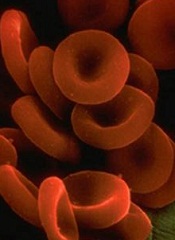
Investigators have identified a novel strategy for treating paroxysmal nocturnal hemoglobinuria (PNH), according to a paper published in Blood.
In patients with PNH, defective expression of regulatory proteins on the surface of red blood cells leaves the cells vulnerable to attack by the complement immune system.
This can lead to hemolysis, which results in severe anemia and contributes to a high risk of thrombosis.
Eculizumab is the only approved therapeutic for PNH. The drug reduces hemolysis and can provide patients with relief from blood transfusions.
However, eculizumab is costly (currently more than $400,000 per year per patient), and one third of PNH patients who receive eculizumab continue to require blood transfusions to manage their anemia.
Investigators previously discovered that this non-response is due to fragments of complement C3 proteins on the surface of red blood cells, which are eventually attacked by immune cells.
Therefore, John Lambris, PhD, of the University of Pennsylvania, and his colleagues hypothesized that using small molecules to inhibit the complement cascade at the level of C3 proteins might be an effective strategy for treating PNH.
The team thought this method would prevent both hemolysis and immune cell recognition, and it might be more cost-effective than the current antibody-based treatment.
So they investigated the effect of a C3 inhibitor called Cp40 and its long-acting form, PEG-Cp40, on self-attack and resulting hemolysis using human PNH cells. Both compounds effectively inhibited hemolysis and efficiently prevented deposition of C3 fragments on PNH red blood cells.
In non-human primates, a single injection of PEG-Cp40 had an elimination half-life of more than 5 days. However, the investigators found evidence to suggest the drug may affect plasma levels of C3.
“We think these 2 compounds are excellent and potentially cost-effective candidates for further clinical investigation,” Dr Lambris said.
He hopes the compounds will be tested in clinical trials by 2015. Dr Lambris and his colleague, Daniel Ricklin, PhD, are the inventors of patents and patent applications owned by the University of Pennsylvania that describe the use of complement inhibitors for therapeutic purposes.
And Dr Lambris is a founder and equity holder of Amyndas Pharmaceuticals, which has exclusively licensed the Cp40 and PEG-Cp40 technologies from the university and is developing complement inhibitors for clinical applications.
Investigators have identified a novel strategy for treating paroxysmal nocturnal hemoglobinuria (PNH), according to a paper published in Blood.
In patients with PNH, defective expression of regulatory proteins on the surface of red blood cells leaves the cells vulnerable to attack by the complement immune system.
This can lead to hemolysis, which results in severe anemia and contributes to a high risk of thrombosis.
Eculizumab is the only approved therapeutic for PNH. The drug reduces hemolysis and can provide patients with relief from blood transfusions.
However, eculizumab is costly (currently more than $400,000 per year per patient), and one third of PNH patients who receive eculizumab continue to require blood transfusions to manage their anemia.
Investigators previously discovered that this non-response is due to fragments of complement C3 proteins on the surface of red blood cells, which are eventually attacked by immune cells.
Therefore, John Lambris, PhD, of the University of Pennsylvania, and his colleagues hypothesized that using small molecules to inhibit the complement cascade at the level of C3 proteins might be an effective strategy for treating PNH.
The team thought this method would prevent both hemolysis and immune cell recognition, and it might be more cost-effective than the current antibody-based treatment.
So they investigated the effect of a C3 inhibitor called Cp40 and its long-acting form, PEG-Cp40, on self-attack and resulting hemolysis using human PNH cells. Both compounds effectively inhibited hemolysis and efficiently prevented deposition of C3 fragments on PNH red blood cells.
In non-human primates, a single injection of PEG-Cp40 had an elimination half-life of more than 5 days. However, the investigators found evidence to suggest the drug may affect plasma levels of C3.
“We think these 2 compounds are excellent and potentially cost-effective candidates for further clinical investigation,” Dr Lambris said.
He hopes the compounds will be tested in clinical trials by 2015. Dr Lambris and his colleague, Daniel Ricklin, PhD, are the inventors of patents and patent applications owned by the University of Pennsylvania that describe the use of complement inhibitors for therapeutic purposes.
And Dr Lambris is a founder and equity holder of Amyndas Pharmaceuticals, which has exclusively licensed the Cp40 and PEG-Cp40 technologies from the university and is developing complement inhibitors for clinical applications.
Investigators have identified a novel strategy for treating paroxysmal nocturnal hemoglobinuria (PNH), according to a paper published in Blood.
In patients with PNH, defective expression of regulatory proteins on the surface of red blood cells leaves the cells vulnerable to attack by the complement immune system.
This can lead to hemolysis, which results in severe anemia and contributes to a high risk of thrombosis.
Eculizumab is the only approved therapeutic for PNH. The drug reduces hemolysis and can provide patients with relief from blood transfusions.
However, eculizumab is costly (currently more than $400,000 per year per patient), and one third of PNH patients who receive eculizumab continue to require blood transfusions to manage their anemia.
Investigators previously discovered that this non-response is due to fragments of complement C3 proteins on the surface of red blood cells, which are eventually attacked by immune cells.
Therefore, John Lambris, PhD, of the University of Pennsylvania, and his colleagues hypothesized that using small molecules to inhibit the complement cascade at the level of C3 proteins might be an effective strategy for treating PNH.
The team thought this method would prevent both hemolysis and immune cell recognition, and it might be more cost-effective than the current antibody-based treatment.
So they investigated the effect of a C3 inhibitor called Cp40 and its long-acting form, PEG-Cp40, on self-attack and resulting hemolysis using human PNH cells. Both compounds effectively inhibited hemolysis and efficiently prevented deposition of C3 fragments on PNH red blood cells.
In non-human primates, a single injection of PEG-Cp40 had an elimination half-life of more than 5 days. However, the investigators found evidence to suggest the drug may affect plasma levels of C3.
“We think these 2 compounds are excellent and potentially cost-effective candidates for further clinical investigation,” Dr Lambris said.
He hopes the compounds will be tested in clinical trials by 2015. Dr Lambris and his colleague, Daniel Ricklin, PhD, are the inventors of patents and patent applications owned by the University of Pennsylvania that describe the use of complement inhibitors for therapeutic purposes.
And Dr Lambris is a founder and equity holder of Amyndas Pharmaceuticals, which has exclusively licensed the Cp40 and PEG-Cp40 technologies from the university and is developing complement inhibitors for clinical applications.
Researchers create reversible LMWH
Scientists say they’ve created a synthetic form of low-molecular-weight heparin (LMWH) that is both reversible and safe for patients with poor kidney function.
In the event of uncontrolled bleeding, this synthetic heparin can be reversed by an existing drug.
And the LMWH is cleared by the liver rather than the kidneys.
The team described their creation of the drug in Nature Chemical Biology.
“When doctors talk to me about the kind of heparin they want to use during and after surgery, they want it reversible, and they want it to not go through the kidneys,” said study author Jian Liu, PhD, of the University of North Carolina, Chapel Hill.
Dr Liu noted that up to 5% of patients receiving heparin experience some form of uncontrolled bleeding. Patients receiving unfractionated heparin are in less danger because there is an existing FDA-approved antidote available, protamine.
But protamine is not as effective in reversing LMWH. So Dr Liu and his colleagues tweaked the drug’s molecular structure so that protamine is able to deactivate LMWH.
The team used a chemo-enzymatic process to synthesize the LMWH, an approach they developed in research on a simpler anticoagulant published in Science in 2011. Synthesizing the LMWH allowed them to make improvements on the animal-derived form of the drug.
That form of LMWH is cleared from the body by the kidneys, which can make it unsuitable for patients with a weakened renal system. So the researchers made changes that allowed their LMWH to bind to receptors that clear it through the liver.
“If a person’s kidneys aren’t effectively clearing heparin from the blood, the drug stays active in the body for longer than expected,” said study author Nigel Key, MB ChB, also of the University of North Carolina.
“That can represent a potentially dangerous situation for the physician, pharmacist, and patient.”
LMWH did prove dangerous in 2008, when more than 80 people died and hundreds of others suffered adverse reactions to the drug. Authorities linked the problems to a contaminant in raw natural heparin from China.
“Whenever you mix the food chain and the drug chain together, you end up with potential for disaster,” said study author Robert Linhardt, PhD, of the Rensselaer Polytechnic Institute in Troy, New York.
“Whether it comes from contamination, adulteration, impurities like viruses or prions—any of those possibilities are much more likely when you make something in an uncontrolled environment. This is a drug that millions of people rely upon, and it’s important to develop a safe, synthetic alternative to the current supply chain.”
LMWH makes up more than half the US market for heparin. The researchers said the new version they created is a safe, economically viable alternative to the existing animal-derived supply.
“The pig stuff has served us well for 50 years and is very inexpensive, but if we cannot control the supply chain, we cannot ensure the safety of the drug,” Dr Liu said. “I am working for the day when synthetic heparin can be brewed in large laboratories at a low cost.”
Scientists say they’ve created a synthetic form of low-molecular-weight heparin (LMWH) that is both reversible and safe for patients with poor kidney function.
In the event of uncontrolled bleeding, this synthetic heparin can be reversed by an existing drug.
And the LMWH is cleared by the liver rather than the kidneys.
The team described their creation of the drug in Nature Chemical Biology.
“When doctors talk to me about the kind of heparin they want to use during and after surgery, they want it reversible, and they want it to not go through the kidneys,” said study author Jian Liu, PhD, of the University of North Carolina, Chapel Hill.
Dr Liu noted that up to 5% of patients receiving heparin experience some form of uncontrolled bleeding. Patients receiving unfractionated heparin are in less danger because there is an existing FDA-approved antidote available, protamine.
But protamine is not as effective in reversing LMWH. So Dr Liu and his colleagues tweaked the drug’s molecular structure so that protamine is able to deactivate LMWH.
The team used a chemo-enzymatic process to synthesize the LMWH, an approach they developed in research on a simpler anticoagulant published in Science in 2011. Synthesizing the LMWH allowed them to make improvements on the animal-derived form of the drug.
That form of LMWH is cleared from the body by the kidneys, which can make it unsuitable for patients with a weakened renal system. So the researchers made changes that allowed their LMWH to bind to receptors that clear it through the liver.
“If a person’s kidneys aren’t effectively clearing heparin from the blood, the drug stays active in the body for longer than expected,” said study author Nigel Key, MB ChB, also of the University of North Carolina.
“That can represent a potentially dangerous situation for the physician, pharmacist, and patient.”
LMWH did prove dangerous in 2008, when more than 80 people died and hundreds of others suffered adverse reactions to the drug. Authorities linked the problems to a contaminant in raw natural heparin from China.
“Whenever you mix the food chain and the drug chain together, you end up with potential for disaster,” said study author Robert Linhardt, PhD, of the Rensselaer Polytechnic Institute in Troy, New York.
“Whether it comes from contamination, adulteration, impurities like viruses or prions—any of those possibilities are much more likely when you make something in an uncontrolled environment. This is a drug that millions of people rely upon, and it’s important to develop a safe, synthetic alternative to the current supply chain.”
LMWH makes up more than half the US market for heparin. The researchers said the new version they created is a safe, economically viable alternative to the existing animal-derived supply.
“The pig stuff has served us well for 50 years and is very inexpensive, but if we cannot control the supply chain, we cannot ensure the safety of the drug,” Dr Liu said. “I am working for the day when synthetic heparin can be brewed in large laboratories at a low cost.”
Scientists say they’ve created a synthetic form of low-molecular-weight heparin (LMWH) that is both reversible and safe for patients with poor kidney function.
In the event of uncontrolled bleeding, this synthetic heparin can be reversed by an existing drug.
And the LMWH is cleared by the liver rather than the kidneys.
The team described their creation of the drug in Nature Chemical Biology.
“When doctors talk to me about the kind of heparin they want to use during and after surgery, they want it reversible, and they want it to not go through the kidneys,” said study author Jian Liu, PhD, of the University of North Carolina, Chapel Hill.
Dr Liu noted that up to 5% of patients receiving heparin experience some form of uncontrolled bleeding. Patients receiving unfractionated heparin are in less danger because there is an existing FDA-approved antidote available, protamine.
But protamine is not as effective in reversing LMWH. So Dr Liu and his colleagues tweaked the drug’s molecular structure so that protamine is able to deactivate LMWH.
The team used a chemo-enzymatic process to synthesize the LMWH, an approach they developed in research on a simpler anticoagulant published in Science in 2011. Synthesizing the LMWH allowed them to make improvements on the animal-derived form of the drug.
That form of LMWH is cleared from the body by the kidneys, which can make it unsuitable for patients with a weakened renal system. So the researchers made changes that allowed their LMWH to bind to receptors that clear it through the liver.
“If a person’s kidneys aren’t effectively clearing heparin from the blood, the drug stays active in the body for longer than expected,” said study author Nigel Key, MB ChB, also of the University of North Carolina.
“That can represent a potentially dangerous situation for the physician, pharmacist, and patient.”
LMWH did prove dangerous in 2008, when more than 80 people died and hundreds of others suffered adverse reactions to the drug. Authorities linked the problems to a contaminant in raw natural heparin from China.
“Whenever you mix the food chain and the drug chain together, you end up with potential for disaster,” said study author Robert Linhardt, PhD, of the Rensselaer Polytechnic Institute in Troy, New York.
“Whether it comes from contamination, adulteration, impurities like viruses or prions—any of those possibilities are much more likely when you make something in an uncontrolled environment. This is a drug that millions of people rely upon, and it’s important to develop a safe, synthetic alternative to the current supply chain.”
LMWH makes up more than half the US market for heparin. The researchers said the new version they created is a safe, economically viable alternative to the existing animal-derived supply.
“The pig stuff has served us well for 50 years and is very inexpensive, but if we cannot control the supply chain, we cannot ensure the safety of the drug,” Dr Liu said. “I am working for the day when synthetic heparin can be brewed in large laboratories at a low cost.”