User login
CDC reports update data on HAIs

Staphylococcus infection
Credit: Bill Branson
About 1 in 25 US patients will contract at least 1 healthcare-associated infection (HAI) during the course of hospital care, according to data from the Centers for Disease Control and Prevention (CDC).
The agency has released 2 new reports on the topic.
The first, published in NEJM, is a survey of nearly 200 hospitals, which researchers used to estimate the national burden of HAIs in 2011.
The second is a 2012 annual report on the progress made toward US Health and Human Services HAI
prevention goals.
Together, the reports suggest that US hospitals have made progress in their effort to eliminate HAIs, but more work is needed to improve patient safety.
“Although there has been some progress, today and every day, more than 200 Americans with healthcare-associated infections will die during their hospital stay,” said CDC Director Tom Frieden, MD, MPH.
“The most advanced medical care won’t work if clinicians don’t prevent infections through basic things such as regular hand hygiene. Healthcare workers want the best for their patients. Following standard infection control practices every time will help ensure their patients’ safety.”
Estimating HAI incidence
The CDC Multistate Point-Prevalence Survey of Health Care-Associated Infections, published in NEJM, used 2011 data from 183 US hospitals to estimate the burden of a wide range of infections in hospital patients.
That year, about 721,800 infections occurred in 648,000 hospital patients. About 75,000 patients with HAIs died during their hospitalizations.
The most common infections were pneumonia (22%), surgical-site infections (22%), gastrointestinal infections (17%), urinary tract infections (13%), and bloodstream infections (10%).
The most common causes of HAIs were Clostridium difficile (12%), Staphylococcus aureus (including MRSA; 11%), Klebsiella (10%), Escherichia coli (9%), Enterococcus (9%), and Pseudomonas (7%).
Tracking national progress
The second report, CDC’s National and State Healthcare-associated Infection Progress Report, includes a subset of infection types that are often required to be reported to CDC.
The report revealed a 44% decrease in central line-associated bloodstream infections between 2008 and 2012, as well as a 20% decrease in infections related to 10 surgical procedures between 2008 and 2012.
There was a 4% decrease in hospital-onset MRSA between 2011 and 2012 and a 2% decrease in hospital-onset C difficile infections between 2011 and 2012.
On the other hand, there was a 3% increase in catheter-associated urinary tract infections between 2009 and 2012.
To access both reports and see the updated HAI data, visit the CDC website. 
Staphylococcus infection
Credit: Bill Branson
About 1 in 25 US patients will contract at least 1 healthcare-associated infection (HAI) during the course of hospital care, according to data from the Centers for Disease Control and Prevention (CDC).
The agency has released 2 new reports on the topic.
The first, published in NEJM, is a survey of nearly 200 hospitals, which researchers used to estimate the national burden of HAIs in 2011.
The second is a 2012 annual report on the progress made toward US Health and Human Services HAI
prevention goals.
Together, the reports suggest that US hospitals have made progress in their effort to eliminate HAIs, but more work is needed to improve patient safety.
“Although there has been some progress, today and every day, more than 200 Americans with healthcare-associated infections will die during their hospital stay,” said CDC Director Tom Frieden, MD, MPH.
“The most advanced medical care won’t work if clinicians don’t prevent infections through basic things such as regular hand hygiene. Healthcare workers want the best for their patients. Following standard infection control practices every time will help ensure their patients’ safety.”
Estimating HAI incidence
The CDC Multistate Point-Prevalence Survey of Health Care-Associated Infections, published in NEJM, used 2011 data from 183 US hospitals to estimate the burden of a wide range of infections in hospital patients.
That year, about 721,800 infections occurred in 648,000 hospital patients. About 75,000 patients with HAIs died during their hospitalizations.
The most common infections were pneumonia (22%), surgical-site infections (22%), gastrointestinal infections (17%), urinary tract infections (13%), and bloodstream infections (10%).
The most common causes of HAIs were Clostridium difficile (12%), Staphylococcus aureus (including MRSA; 11%), Klebsiella (10%), Escherichia coli (9%), Enterococcus (9%), and Pseudomonas (7%).
Tracking national progress
The second report, CDC’s National and State Healthcare-associated Infection Progress Report, includes a subset of infection types that are often required to be reported to CDC.
The report revealed a 44% decrease in central line-associated bloodstream infections between 2008 and 2012, as well as a 20% decrease in infections related to 10 surgical procedures between 2008 and 2012.
There was a 4% decrease in hospital-onset MRSA between 2011 and 2012 and a 2% decrease in hospital-onset C difficile infections between 2011 and 2012.
On the other hand, there was a 3% increase in catheter-associated urinary tract infections between 2009 and 2012.
To access both reports and see the updated HAI data, visit the CDC website.
Staphylococcus infection
Credit: Bill Branson
About 1 in 25 US patients will contract at least 1 healthcare-associated infection (HAI) during the course of hospital care, according to data from the Centers for Disease Control and Prevention (CDC).
The agency has released 2 new reports on the topic.
The first, published in NEJM, is a survey of nearly 200 hospitals, which researchers used to estimate the national burden of HAIs in 2011.
The second is a 2012 annual report on the progress made toward US Health and Human Services HAI
prevention goals.
Together, the reports suggest that US hospitals have made progress in their effort to eliminate HAIs, but more work is needed to improve patient safety.
“Although there has been some progress, today and every day, more than 200 Americans with healthcare-associated infections will die during their hospital stay,” said CDC Director Tom Frieden, MD, MPH.
“The most advanced medical care won’t work if clinicians don’t prevent infections through basic things such as regular hand hygiene. Healthcare workers want the best for their patients. Following standard infection control practices every time will help ensure their patients’ safety.”
Estimating HAI incidence
The CDC Multistate Point-Prevalence Survey of Health Care-Associated Infections, published in NEJM, used 2011 data from 183 US hospitals to estimate the burden of a wide range of infections in hospital patients.
That year, about 721,800 infections occurred in 648,000 hospital patients. About 75,000 patients with HAIs died during their hospitalizations.
The most common infections were pneumonia (22%), surgical-site infections (22%), gastrointestinal infections (17%), urinary tract infections (13%), and bloodstream infections (10%).
The most common causes of HAIs were Clostridium difficile (12%), Staphylococcus aureus (including MRSA; 11%), Klebsiella (10%), Escherichia coli (9%), Enterococcus (9%), and Pseudomonas (7%).
Tracking national progress
The second report, CDC’s National and State Healthcare-associated Infection Progress Report, includes a subset of infection types that are often required to be reported to CDC.
The report revealed a 44% decrease in central line-associated bloodstream infections between 2008 and 2012, as well as a 20% decrease in infections related to 10 surgical procedures between 2008 and 2012.
There was a 4% decrease in hospital-onset MRSA between 2011 and 2012 and a 2% decrease in hospital-onset C difficile infections between 2011 and 2012.
On the other hand, there was a 3% increase in catheter-associated urinary tract infections between 2009 and 2012.
To access both reports and see the updated HAI data, visit the CDC website.
Group aims to make African blood supply safer

Credit: UAB Hospital
Three organizations have joined together to adapt a pathogen inactivation system so that it works in whole blood and can be used in sub-Saharan Africa.
The INTERCEPT Blood System is currently used to inactivate bacteria, viruses, parasites, and leukocytes in donated platelets and plasma.
However, as the common practice in many African countries is to transfuse whole blood, the organizations want to adapt the system so it can be used with whole blood.
They also want to ensure the system can function in regions that may not have the infrastructure to support complex devices or have access to controlled temperature storage. Ideally, the system will not require electricity to inactivate pathogens or leukocytes.
For this endeavor, the company that makes the INTERCEPT system, Cerus Corporation, has partnered with SRTS Geneva and Swiss Transfusion SRC.
The Humanitarian Foundation Swiss Red Cross has granted funds to Swiss Transfusion SRC for the project. The initial funding of 1.5 million Swiss Francs will support the feasibility phase of the project and the completion of in vitro studies to support clinical trials.
“We believe pathogen inactivation for whole blood has the potential to improve the safety of transfusions in sub-Saharan Africa, where diminished blood availability due to severe anemia from malaria, HIV, and obstetric bleeding is common,” said Rudolf Schwabe, chief executive officer of the Swiss Red Cross.
“Based on our experience over the past 3 years with the INTERCEPT system, we have seen first-hand the substantial impact that pathogen inactivation has had in reducing transfusion-transmitted infectious risk in platelets and plasma.”
“This technology should be made available to developing countries such as those in sub-Saharan Africa, where the risk of bacterial contamination is about 2500 times greater than in Switzerland, and 10% to 15% of HIV infections are caused by contaminated transfusions.”
Cerus currently sells the INTERCEPT Blood System for both platelets and plasma in Europe, the Commonwealth of Independent States, the Middle East, and select countries in other regions around the world.
In the US, Cerus is seeking regulatory approval of the INTERCEPT Blood System for plasma and platelets.
The INTERCEPT red blood cell system is in clinical development. For more information, see the Cerus website.
Credit: UAB Hospital
Three organizations have joined together to adapt a pathogen inactivation system so that it works in whole blood and can be used in sub-Saharan Africa.
The INTERCEPT Blood System is currently used to inactivate bacteria, viruses, parasites, and leukocytes in donated platelets and plasma.
However, as the common practice in many African countries is to transfuse whole blood, the organizations want to adapt the system so it can be used with whole blood.
They also want to ensure the system can function in regions that may not have the infrastructure to support complex devices or have access to controlled temperature storage. Ideally, the system will not require electricity to inactivate pathogens or leukocytes.
For this endeavor, the company that makes the INTERCEPT system, Cerus Corporation, has partnered with SRTS Geneva and Swiss Transfusion SRC.
The Humanitarian Foundation Swiss Red Cross has granted funds to Swiss Transfusion SRC for the project. The initial funding of 1.5 million Swiss Francs will support the feasibility phase of the project and the completion of in vitro studies to support clinical trials.
“We believe pathogen inactivation for whole blood has the potential to improve the safety of transfusions in sub-Saharan Africa, where diminished blood availability due to severe anemia from malaria, HIV, and obstetric bleeding is common,” said Rudolf Schwabe, chief executive officer of the Swiss Red Cross.
“Based on our experience over the past 3 years with the INTERCEPT system, we have seen first-hand the substantial impact that pathogen inactivation has had in reducing transfusion-transmitted infectious risk in platelets and plasma.”
“This technology should be made available to developing countries such as those in sub-Saharan Africa, where the risk of bacterial contamination is about 2500 times greater than in Switzerland, and 10% to 15% of HIV infections are caused by contaminated transfusions.”
Cerus currently sells the INTERCEPT Blood System for both platelets and plasma in Europe, the Commonwealth of Independent States, the Middle East, and select countries in other regions around the world.
In the US, Cerus is seeking regulatory approval of the INTERCEPT Blood System for plasma and platelets.
The INTERCEPT red blood cell system is in clinical development. For more information, see the Cerus website.
Credit: UAB Hospital
Three organizations have joined together to adapt a pathogen inactivation system so that it works in whole blood and can be used in sub-Saharan Africa.
The INTERCEPT Blood System is currently used to inactivate bacteria, viruses, parasites, and leukocytes in donated platelets and plasma.
However, as the common practice in many African countries is to transfuse whole blood, the organizations want to adapt the system so it can be used with whole blood.
They also want to ensure the system can function in regions that may not have the infrastructure to support complex devices or have access to controlled temperature storage. Ideally, the system will not require electricity to inactivate pathogens or leukocytes.
For this endeavor, the company that makes the INTERCEPT system, Cerus Corporation, has partnered with SRTS Geneva and Swiss Transfusion SRC.
The Humanitarian Foundation Swiss Red Cross has granted funds to Swiss Transfusion SRC for the project. The initial funding of 1.5 million Swiss Francs will support the feasibility phase of the project and the completion of in vitro studies to support clinical trials.
“We believe pathogen inactivation for whole blood has the potential to improve the safety of transfusions in sub-Saharan Africa, where diminished blood availability due to severe anemia from malaria, HIV, and obstetric bleeding is common,” said Rudolf Schwabe, chief executive officer of the Swiss Red Cross.
“Based on our experience over the past 3 years with the INTERCEPT system, we have seen first-hand the substantial impact that pathogen inactivation has had in reducing transfusion-transmitted infectious risk in platelets and plasma.”
“This technology should be made available to developing countries such as those in sub-Saharan Africa, where the risk of bacterial contamination is about 2500 times greater than in Switzerland, and 10% to 15% of HIV infections are caused by contaminated transfusions.”
Cerus currently sells the INTERCEPT Blood System for both platelets and plasma in Europe, the Commonwealth of Independent States, the Middle East, and select countries in other regions around the world.
In the US, Cerus is seeking regulatory approval of the INTERCEPT Blood System for plasma and platelets.
The INTERCEPT red blood cell system is in clinical development. For more information, see the Cerus website.
FANTOM investigators map blood cell landscape
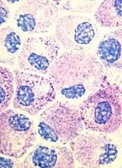
Research by an international consortium has revealed new information on hematopoiesis and shed new light on the etiology
of blood diseases.
The consortium, Functional Annotation of the Mammalian Genome (FANTOM), reported the results of this research in Blood.*
The investigators identified epigenetic regulators of hematopoiesis and uncovered the epigenetic and transcriptional changes that occur during granulopoiesis.
They “redefined” the mast cell transcriptome and mapped the enhancer and promoter landscapes of monocytes, conventional T cells, and regulatory T cells.
By pinpointing the locations of enhancers and promoters, the group was able to correlate them with activity in specific genes.
“Until this point, researchers could only recognize the unique signatures of enhancers and promoters,” said FANTOM investigator Alistair R.R. Forrest, PhD, of the RIKEN Centre for Life Science Technology in Yokohama, Japan.
“However, their exact location, as well as the association of specific enhancers to specific blood cells, remained unclear.”
Knowing the location of enhancers and promoters will improve experiments designed to determine how genes become activated, according to the investigators. And this could potentially lead to strategies for preventing or treating malignancies.
“The specific genetic alterations that are responsible for a normal cell turning into a cancer cell show up in the levels of messenger RNA in the cell, and these differences are often very subtle,” Dr Forrest said.
“Now that we have these incredibly detailed pictures of each of these cell types, we can now work backwards to compare cancer cells to the cells they came from originally to better understand what may have triggered the cells to malfunction, so we will be better equipped to develop new and more effective therapies.”
*The studies include:
High-throughput transcription profiling identifies putative epigenetic regulators of hematopoiesis
Redefinition of the human mast cell transcriptome by deep-CAGE sequencing
Transcription and enhancer profiling in human monocyte subsets
The enhancer and promoter landscape of human regulatory and conventional T-cell subpopulations.
Research by an international consortium has revealed new information on hematopoiesis and shed new light on the etiology
of blood diseases.
The consortium, Functional Annotation of the Mammalian Genome (FANTOM), reported the results of this research in Blood.*
The investigators identified epigenetic regulators of hematopoiesis and uncovered the epigenetic and transcriptional changes that occur during granulopoiesis.
They “redefined” the mast cell transcriptome and mapped the enhancer and promoter landscapes of monocytes, conventional T cells, and regulatory T cells.
By pinpointing the locations of enhancers and promoters, the group was able to correlate them with activity in specific genes.
“Until this point, researchers could only recognize the unique signatures of enhancers and promoters,” said FANTOM investigator Alistair R.R. Forrest, PhD, of the RIKEN Centre for Life Science Technology in Yokohama, Japan.
“However, their exact location, as well as the association of specific enhancers to specific blood cells, remained unclear.”
Knowing the location of enhancers and promoters will improve experiments designed to determine how genes become activated, according to the investigators. And this could potentially lead to strategies for preventing or treating malignancies.
“The specific genetic alterations that are responsible for a normal cell turning into a cancer cell show up in the levels of messenger RNA in the cell, and these differences are often very subtle,” Dr Forrest said.
“Now that we have these incredibly detailed pictures of each of these cell types, we can now work backwards to compare cancer cells to the cells they came from originally to better understand what may have triggered the cells to malfunction, so we will be better equipped to develop new and more effective therapies.”
*The studies include:
High-throughput transcription profiling identifies putative epigenetic regulators of hematopoiesis
Redefinition of the human mast cell transcriptome by deep-CAGE sequencing
Transcription and enhancer profiling in human monocyte subsets
The enhancer and promoter landscape of human regulatory and conventional T-cell subpopulations.
Research by an international consortium has revealed new information on hematopoiesis and shed new light on the etiology
of blood diseases.
The consortium, Functional Annotation of the Mammalian Genome (FANTOM), reported the results of this research in Blood.*
The investigators identified epigenetic regulators of hematopoiesis and uncovered the epigenetic and transcriptional changes that occur during granulopoiesis.
They “redefined” the mast cell transcriptome and mapped the enhancer and promoter landscapes of monocytes, conventional T cells, and regulatory T cells.
By pinpointing the locations of enhancers and promoters, the group was able to correlate them with activity in specific genes.
“Until this point, researchers could only recognize the unique signatures of enhancers and promoters,” said FANTOM investigator Alistair R.R. Forrest, PhD, of the RIKEN Centre for Life Science Technology in Yokohama, Japan.
“However, their exact location, as well as the association of specific enhancers to specific blood cells, remained unclear.”
Knowing the location of enhancers and promoters will improve experiments designed to determine how genes become activated, according to the investigators. And this could potentially lead to strategies for preventing or treating malignancies.
“The specific genetic alterations that are responsible for a normal cell turning into a cancer cell show up in the levels of messenger RNA in the cell, and these differences are often very subtle,” Dr Forrest said.
“Now that we have these incredibly detailed pictures of each of these cell types, we can now work backwards to compare cancer cells to the cells they came from originally to better understand what may have triggered the cells to malfunction, so we will be better equipped to develop new and more effective therapies.”
*The studies include:
High-throughput transcription profiling identifies putative epigenetic regulators of hematopoiesis
Redefinition of the human mast cell transcriptome by deep-CAGE sequencing
Transcription and enhancer profiling in human monocyte subsets
The enhancer and promoter landscape of human regulatory and conventional T-cell subpopulations.
NK cell findings may have treatment implications

Credit: St Jude Children’s
Research Hospital
Researchers say they’ve gained new insight into the production of natural killer (NK) cells.
And their findings may help them generate greater numbers of the cells in culture, which could have implications for the treatment of leukemia and other malignancies.
A previous study conducted by the same team revealed that the gene E4bp4 must be switched on to allow the immune system to produce NK cells.
Their new work suggests that E4bp4 expression is required for progenitor cells to commit to the NK lineage. And the gene promotes NK-cell development by regulating expression of the transcription factors Eomes and Id2.
The researchers described these discoveries in the Journal of Experimental Medicine.
“We are excited to find that E4bp4 has such a crucial role in determining the decisive point where blood progenitor cells become NK cells,” said study author Hugh Brady, of Imperial College London in the UK.
“We are now starting to apply this to human blood stem cells to work out how switching on E4bp4 can allow us to make lots of robust human NK cells in culture. We are hoping to make human NK cells that will have improved survival and be very toxic to cancer cells when transfused into patients. Hopefully, this will allow a big reduction in the number of NK cells needed to treat an individual patient.”
To gain insight into NK-cell production, Dr Brady and his colleagues evaluated 2 types of mice with NK-cell deficiencies. The Il15ra knockout mouse model cannot mediate IL-15 signaling, which is critical for NK-cell production. And the T-bet (Tbx21) knockout model lacks a transcription factor that’s crucial for NK-cell development.
Analysis of the Il15ra model revealed that the absence of E4bp4 perturbs NK-cell development earlier than the absence of IL-15 signaling. This suggests E4bp4 acts before IL-15, which was previously considered the definitive factor required for NK-cell production.
The researchers also found that E4bp4 is required for the production of NK progenitors, but T-bet is not. And this suggests E4bp4 acts before T-bet in NK-cell development.
To investigate these findings further, the team took cells at various stages of NK-cell differentiation from wild-type bone marrow and measured their expression of transcription factor mRNAs.
They detected E4bp4 transcript in both lymphoid-primed multipotent progenitors (LMPPs) and common lymphoid progenitors (CLPs), and E4bp4 expression increased at later stages of NK-cell development.
Based on these results, the researchers speculated that E4bp4 might be a lineage commitment factor controlling the development of NK progenitors from CLPs. To test that theory, they restored E4bp4 expression in purified E4bp4-/- CLPs to see if this could re-establish NK-cell development.
The team sorted CLPs from E4bp4-/- bone marrow, cultured them in lymphocyte-inducing conditions, transduced them with E4bp4 or empty vector, and moved on to NK-cell-inducing conditions. But neither cell type produced NK cells.
So the researchers decided to initiate the culture at an earlier developmental stage, using LMPPs. They cultured LMPPs, which exhibited a CLP phenotype at the time of transduction. And CLPs transduced with E4bp4 gave rise to NK cells, but CLPs transduced with empty vector did not.
As these results suggest that E4bp4 acts at the earliest possible point in NK-cell development, the team wanted to characterize E4bp4’s relationship with transcription factors that are likely to act downstream.
They tested several transcription factors known to play a part in NK-cell production and function. But only Eomes and Id2 proved essential for E4bp4 to direct the production of fully functional, mature NK cells.
Credit: St Jude Children’s
Research Hospital
Researchers say they’ve gained new insight into the production of natural killer (NK) cells.
And their findings may help them generate greater numbers of the cells in culture, which could have implications for the treatment of leukemia and other malignancies.
A previous study conducted by the same team revealed that the gene E4bp4 must be switched on to allow the immune system to produce NK cells.
Their new work suggests that E4bp4 expression is required for progenitor cells to commit to the NK lineage. And the gene promotes NK-cell development by regulating expression of the transcription factors Eomes and Id2.
The researchers described these discoveries in the Journal of Experimental Medicine.
“We are excited to find that E4bp4 has such a crucial role in determining the decisive point where blood progenitor cells become NK cells,” said study author Hugh Brady, of Imperial College London in the UK.
“We are now starting to apply this to human blood stem cells to work out how switching on E4bp4 can allow us to make lots of robust human NK cells in culture. We are hoping to make human NK cells that will have improved survival and be very toxic to cancer cells when transfused into patients. Hopefully, this will allow a big reduction in the number of NK cells needed to treat an individual patient.”
To gain insight into NK-cell production, Dr Brady and his colleagues evaluated 2 types of mice with NK-cell deficiencies. The Il15ra knockout mouse model cannot mediate IL-15 signaling, which is critical for NK-cell production. And the T-bet (Tbx21) knockout model lacks a transcription factor that’s crucial for NK-cell development.
Analysis of the Il15ra model revealed that the absence of E4bp4 perturbs NK-cell development earlier than the absence of IL-15 signaling. This suggests E4bp4 acts before IL-15, which was previously considered the definitive factor required for NK-cell production.
The researchers also found that E4bp4 is required for the production of NK progenitors, but T-bet is not. And this suggests E4bp4 acts before T-bet in NK-cell development.
To investigate these findings further, the team took cells at various stages of NK-cell differentiation from wild-type bone marrow and measured their expression of transcription factor mRNAs.
They detected E4bp4 transcript in both lymphoid-primed multipotent progenitors (LMPPs) and common lymphoid progenitors (CLPs), and E4bp4 expression increased at later stages of NK-cell development.
Based on these results, the researchers speculated that E4bp4 might be a lineage commitment factor controlling the development of NK progenitors from CLPs. To test that theory, they restored E4bp4 expression in purified E4bp4-/- CLPs to see if this could re-establish NK-cell development.
The team sorted CLPs from E4bp4-/- bone marrow, cultured them in lymphocyte-inducing conditions, transduced them with E4bp4 or empty vector, and moved on to NK-cell-inducing conditions. But neither cell type produced NK cells.
So the researchers decided to initiate the culture at an earlier developmental stage, using LMPPs. They cultured LMPPs, which exhibited a CLP phenotype at the time of transduction. And CLPs transduced with E4bp4 gave rise to NK cells, but CLPs transduced with empty vector did not.
As these results suggest that E4bp4 acts at the earliest possible point in NK-cell development, the team wanted to characterize E4bp4’s relationship with transcription factors that are likely to act downstream.
They tested several transcription factors known to play a part in NK-cell production and function. But only Eomes and Id2 proved essential for E4bp4 to direct the production of fully functional, mature NK cells.
Credit: St Jude Children’s
Research Hospital
Researchers say they’ve gained new insight into the production of natural killer (NK) cells.
And their findings may help them generate greater numbers of the cells in culture, which could have implications for the treatment of leukemia and other malignancies.
A previous study conducted by the same team revealed that the gene E4bp4 must be switched on to allow the immune system to produce NK cells.
Their new work suggests that E4bp4 expression is required for progenitor cells to commit to the NK lineage. And the gene promotes NK-cell development by regulating expression of the transcription factors Eomes and Id2.
The researchers described these discoveries in the Journal of Experimental Medicine.
“We are excited to find that E4bp4 has such a crucial role in determining the decisive point where blood progenitor cells become NK cells,” said study author Hugh Brady, of Imperial College London in the UK.
“We are now starting to apply this to human blood stem cells to work out how switching on E4bp4 can allow us to make lots of robust human NK cells in culture. We are hoping to make human NK cells that will have improved survival and be very toxic to cancer cells when transfused into patients. Hopefully, this will allow a big reduction in the number of NK cells needed to treat an individual patient.”
To gain insight into NK-cell production, Dr Brady and his colleagues evaluated 2 types of mice with NK-cell deficiencies. The Il15ra knockout mouse model cannot mediate IL-15 signaling, which is critical for NK-cell production. And the T-bet (Tbx21) knockout model lacks a transcription factor that’s crucial for NK-cell development.
Analysis of the Il15ra model revealed that the absence of E4bp4 perturbs NK-cell development earlier than the absence of IL-15 signaling. This suggests E4bp4 acts before IL-15, which was previously considered the definitive factor required for NK-cell production.
The researchers also found that E4bp4 is required for the production of NK progenitors, but T-bet is not. And this suggests E4bp4 acts before T-bet in NK-cell development.
To investigate these findings further, the team took cells at various stages of NK-cell differentiation from wild-type bone marrow and measured their expression of transcription factor mRNAs.
They detected E4bp4 transcript in both lymphoid-primed multipotent progenitors (LMPPs) and common lymphoid progenitors (CLPs), and E4bp4 expression increased at later stages of NK-cell development.
Based on these results, the researchers speculated that E4bp4 might be a lineage commitment factor controlling the development of NK progenitors from CLPs. To test that theory, they restored E4bp4 expression in purified E4bp4-/- CLPs to see if this could re-establish NK-cell development.
The team sorted CLPs from E4bp4-/- bone marrow, cultured them in lymphocyte-inducing conditions, transduced them with E4bp4 or empty vector, and moved on to NK-cell-inducing conditions. But neither cell type produced NK cells.
So the researchers decided to initiate the culture at an earlier developmental stage, using LMPPs. They cultured LMPPs, which exhibited a CLP phenotype at the time of transduction. And CLPs transduced with E4bp4 gave rise to NK cells, but CLPs transduced with empty vector did not.
As these results suggest that E4bp4 acts at the earliest possible point in NK-cell development, the team wanted to characterize E4bp4’s relationship with transcription factors that are likely to act downstream.
They tested several transcription factors known to play a part in NK-cell production and function. But only Eomes and Id2 proved essential for E4bp4 to direct the production of fully functional, mature NK cells.
Video glasses can curb patient anxiety

Credit: CDC
SAN DIEGO—Watching videos through special glasses can calm patients undergoing a biopsy or other minimally invasive treatment, according to research presented at the Society of Interventional Radiology’s 39th Annual Scientific Meeting.
Researchers have explored strategies other than medication to reduce anxiety in these patients, including having patients listen to music or undergo hypnosis. But these methods have had modest benefits.
“Our study—the first of its kind for interventional radiology treatments—puts a spin on using modern technology to provide a safe, potentially cost-effective strategy of reducing anxiety, which can help and improve patient care,” said David L. Waldman, MD, PhD, of the University of Rochester Medical Center in New York.
“Whether they were watching a children’s movie or a nature show, patients wearing video glasses were successful at tuning out their surroundings. It’s an effective distraction technique that helps focus the individual’s attention away from the treatment.”
The study involved 49 patients (33 men and 16 women, ages 18-87) who were undergoing an outpatient interventional radiology treatment, such as a biopsy or placement of a catheter in the arm or chest to receive medication for treating cancer or infection.
Twenty-five of the patients donned video glasses prior to undergoing the treatment, and 24 did not. The video viewers chose from 20 videos, none of which were violent.
All of the patients filled out a standard 20-question test called the State-Trait Anxiety Inventory Form Y before and after the procedure to assess their level of anxiety.
Patients who wore the video glasses were 18.1% less anxious after the treatment than they were before, while those who didn’t wear video glasses were 7.5% less anxious afterward.
And the presence of the video glasses did not bother either the patient or the doctor, Dr Waldman said.
The glasses had no significant effect on blood pressure, heart rate, respiratory rate, pain, procedure time, or the amount of sedation or pain medication used.
“Patients told us the video glasses really helped calm them down and took their mind off the treatment, and we now offer video glasses to help distract patients from medical treatment going on mere inches away,” Dr Waldman said. “It is really comforting for patients, especially the ones who tend to be more nervous.
Dr Waldman and his colleagues presented these results at the meeting as abstract 126.
Credit: CDC
SAN DIEGO—Watching videos through special glasses can calm patients undergoing a biopsy or other minimally invasive treatment, according to research presented at the Society of Interventional Radiology’s 39th Annual Scientific Meeting.
Researchers have explored strategies other than medication to reduce anxiety in these patients, including having patients listen to music or undergo hypnosis. But these methods have had modest benefits.
“Our study—the first of its kind for interventional radiology treatments—puts a spin on using modern technology to provide a safe, potentially cost-effective strategy of reducing anxiety, which can help and improve patient care,” said David L. Waldman, MD, PhD, of the University of Rochester Medical Center in New York.
“Whether they were watching a children’s movie or a nature show, patients wearing video glasses were successful at tuning out their surroundings. It’s an effective distraction technique that helps focus the individual’s attention away from the treatment.”
The study involved 49 patients (33 men and 16 women, ages 18-87) who were undergoing an outpatient interventional radiology treatment, such as a biopsy or placement of a catheter in the arm or chest to receive medication for treating cancer or infection.
Twenty-five of the patients donned video glasses prior to undergoing the treatment, and 24 did not. The video viewers chose from 20 videos, none of which were violent.
All of the patients filled out a standard 20-question test called the State-Trait Anxiety Inventory Form Y before and after the procedure to assess their level of anxiety.
Patients who wore the video glasses were 18.1% less anxious after the treatment than they were before, while those who didn’t wear video glasses were 7.5% less anxious afterward.
And the presence of the video glasses did not bother either the patient or the doctor, Dr Waldman said.
The glasses had no significant effect on blood pressure, heart rate, respiratory rate, pain, procedure time, or the amount of sedation or pain medication used.
“Patients told us the video glasses really helped calm them down and took their mind off the treatment, and we now offer video glasses to help distract patients from medical treatment going on mere inches away,” Dr Waldman said. “It is really comforting for patients, especially the ones who tend to be more nervous.
Dr Waldman and his colleagues presented these results at the meeting as abstract 126.
Credit: CDC
SAN DIEGO—Watching videos through special glasses can calm patients undergoing a biopsy or other minimally invasive treatment, according to research presented at the Society of Interventional Radiology’s 39th Annual Scientific Meeting.
Researchers have explored strategies other than medication to reduce anxiety in these patients, including having patients listen to music or undergo hypnosis. But these methods have had modest benefits.
“Our study—the first of its kind for interventional radiology treatments—puts a spin on using modern technology to provide a safe, potentially cost-effective strategy of reducing anxiety, which can help and improve patient care,” said David L. Waldman, MD, PhD, of the University of Rochester Medical Center in New York.
“Whether they were watching a children’s movie or a nature show, patients wearing video glasses were successful at tuning out their surroundings. It’s an effective distraction technique that helps focus the individual’s attention away from the treatment.”
The study involved 49 patients (33 men and 16 women, ages 18-87) who were undergoing an outpatient interventional radiology treatment, such as a biopsy or placement of a catheter in the arm or chest to receive medication for treating cancer or infection.
Twenty-five of the patients donned video glasses prior to undergoing the treatment, and 24 did not. The video viewers chose from 20 videos, none of which were violent.
All of the patients filled out a standard 20-question test called the State-Trait Anxiety Inventory Form Y before and after the procedure to assess their level of anxiety.
Patients who wore the video glasses were 18.1% less anxious after the treatment than they were before, while those who didn’t wear video glasses were 7.5% less anxious afterward.
And the presence of the video glasses did not bother either the patient or the doctor, Dr Waldman said.
The glasses had no significant effect on blood pressure, heart rate, respiratory rate, pain, procedure time, or the amount of sedation or pain medication used.
“Patients told us the video glasses really helped calm them down and took their mind off the treatment, and we now offer video glasses to help distract patients from medical treatment going on mere inches away,” Dr Waldman said. “It is really comforting for patients, especially the ones who tend to be more nervous.
Dr Waldman and his colleagues presented these results at the meeting as abstract 126.
Vorinostat demonstrates antitumor activity in FL
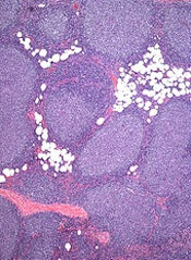
Single-agent treatment with the HDAC inhibitor vorinostat can be effective in certain patients with indolent non-Hodgkin lymphoma (NHL), according to research published in the British Journal of Haematology.
In the phase 2 study, vorinostat prompted a 49% overall response rate among 39 patients with relapsed or refractory follicular lymphoma (FL).
However, none of the 4 patients with previously treated mantle cell lymphoma (MCL) achieved a response.
Michinori Ogura, MD, PhD, of Nagoya Daini Red Cross Hospital in Japan, and his colleagues set out to analyze the effects of vorinostat in 56 patients with indolent NHL. Six patients were excluded from the final analysis, as their diseases could not be classified.
Thirty-nine patients had FL, 4 had extranodal marginal zone lymphoma (MZL) of MALT type, 4 had MCL, 2 had small B-cell lymphoma not otherwise specified (NOS), and 1 had small lymphocytic lymphoma.
The median age was 60 years (range, 33-75), and the median number of prior therapies was 2 (range, 1-4). These therapies included rituximab (n=40), alkylating agents (n=7), purine analogs (n=5), and radioimmunotherapy (n=3).
The patients received vorinostat for a median of 8 months. The planned dosage was 200 mg twice daily for 14 consecutive days in a 21-day cycle.
At the first data cutoff point (1 year from the last patient’s enrollment), 18 patients remained on treatment. Thirty-eight had discontinued due to disease progression (n=25), drug-related adverse events (n=9), or withdrawn consent (n=4).
The overall response rate was 49% among FL patients. Ten percent (n=4) achieved a complete response, 8% (n=3) achieved an unconfirmed complete response, and 31% (n=12) achieved a partial response.
None of the patients with MCL responded, but 3 of the 7 (43%) patients with other indolent NHLs achieved a response.
That included 2 patients with small B-cell lymphoma NOS and 1 with extranodal MZL of MALT type. One of the patients with small B-cell lymphoma NOS achieved a complete response.
Approximately 81% of all 56 patients remained alive at 2 years after the last patients had enrolled (the second data cutoff point).
At that point, the median overall survival had not been reached. And the median progression-free survival was 26 months among the FL patients who responded.
There were no treatment-related deaths. The most common drug-related events (in all 56 patients) were thrombocytopenia (93%), diarrhea (68%), neutropenia (68%), decreased appetite (63%), nausea (61%), leukopenia (55%), and fatigue (52%).
Eighty percent of patients (n=45) experienced grade 3/4 adverse events, the most common of which were thrombocytopenia (23% grade 3; 25% grade 4) and neutropenia (36% grade 3; 5% grade 4).
However, all of the patients with thrombocytopenia or neutropenia recovered after they received adequate supportive care and their vorinostat dose was reduced or treatment was interrupted or discontinued.
Taking these results together, the researchers concluded that vorinostat offers sustained antitumor activity and has an acceptable safety profile for patients with relapsed or refractory FL. The team noted, however, that because this was a single-arm study with limited data, a comparative study is needed.
Single-agent treatment with the HDAC inhibitor vorinostat can be effective in certain patients with indolent non-Hodgkin lymphoma (NHL), according to research published in the British Journal of Haematology.
In the phase 2 study, vorinostat prompted a 49% overall response rate among 39 patients with relapsed or refractory follicular lymphoma (FL).
However, none of the 4 patients with previously treated mantle cell lymphoma (MCL) achieved a response.
Michinori Ogura, MD, PhD, of Nagoya Daini Red Cross Hospital in Japan, and his colleagues set out to analyze the effects of vorinostat in 56 patients with indolent NHL. Six patients were excluded from the final analysis, as their diseases could not be classified.
Thirty-nine patients had FL, 4 had extranodal marginal zone lymphoma (MZL) of MALT type, 4 had MCL, 2 had small B-cell lymphoma not otherwise specified (NOS), and 1 had small lymphocytic lymphoma.
The median age was 60 years (range, 33-75), and the median number of prior therapies was 2 (range, 1-4). These therapies included rituximab (n=40), alkylating agents (n=7), purine analogs (n=5), and radioimmunotherapy (n=3).
The patients received vorinostat for a median of 8 months. The planned dosage was 200 mg twice daily for 14 consecutive days in a 21-day cycle.
At the first data cutoff point (1 year from the last patient’s enrollment), 18 patients remained on treatment. Thirty-eight had discontinued due to disease progression (n=25), drug-related adverse events (n=9), or withdrawn consent (n=4).
The overall response rate was 49% among FL patients. Ten percent (n=4) achieved a complete response, 8% (n=3) achieved an unconfirmed complete response, and 31% (n=12) achieved a partial response.
None of the patients with MCL responded, but 3 of the 7 (43%) patients with other indolent NHLs achieved a response.
That included 2 patients with small B-cell lymphoma NOS and 1 with extranodal MZL of MALT type. One of the patients with small B-cell lymphoma NOS achieved a complete response.
Approximately 81% of all 56 patients remained alive at 2 years after the last patients had enrolled (the second data cutoff point).
At that point, the median overall survival had not been reached. And the median progression-free survival was 26 months among the FL patients who responded.
There were no treatment-related deaths. The most common drug-related events (in all 56 patients) were thrombocytopenia (93%), diarrhea (68%), neutropenia (68%), decreased appetite (63%), nausea (61%), leukopenia (55%), and fatigue (52%).
Eighty percent of patients (n=45) experienced grade 3/4 adverse events, the most common of which were thrombocytopenia (23% grade 3; 25% grade 4) and neutropenia (36% grade 3; 5% grade 4).
However, all of the patients with thrombocytopenia or neutropenia recovered after they received adequate supportive care and their vorinostat dose was reduced or treatment was interrupted or discontinued.
Taking these results together, the researchers concluded that vorinostat offers sustained antitumor activity and has an acceptable safety profile for patients with relapsed or refractory FL. The team noted, however, that because this was a single-arm study with limited data, a comparative study is needed.
Single-agent treatment with the HDAC inhibitor vorinostat can be effective in certain patients with indolent non-Hodgkin lymphoma (NHL), according to research published in the British Journal of Haematology.
In the phase 2 study, vorinostat prompted a 49% overall response rate among 39 patients with relapsed or refractory follicular lymphoma (FL).
However, none of the 4 patients with previously treated mantle cell lymphoma (MCL) achieved a response.
Michinori Ogura, MD, PhD, of Nagoya Daini Red Cross Hospital in Japan, and his colleagues set out to analyze the effects of vorinostat in 56 patients with indolent NHL. Six patients were excluded from the final analysis, as their diseases could not be classified.
Thirty-nine patients had FL, 4 had extranodal marginal zone lymphoma (MZL) of MALT type, 4 had MCL, 2 had small B-cell lymphoma not otherwise specified (NOS), and 1 had small lymphocytic lymphoma.
The median age was 60 years (range, 33-75), and the median number of prior therapies was 2 (range, 1-4). These therapies included rituximab (n=40), alkylating agents (n=7), purine analogs (n=5), and radioimmunotherapy (n=3).
The patients received vorinostat for a median of 8 months. The planned dosage was 200 mg twice daily for 14 consecutive days in a 21-day cycle.
At the first data cutoff point (1 year from the last patient’s enrollment), 18 patients remained on treatment. Thirty-eight had discontinued due to disease progression (n=25), drug-related adverse events (n=9), or withdrawn consent (n=4).
The overall response rate was 49% among FL patients. Ten percent (n=4) achieved a complete response, 8% (n=3) achieved an unconfirmed complete response, and 31% (n=12) achieved a partial response.
None of the patients with MCL responded, but 3 of the 7 (43%) patients with other indolent NHLs achieved a response.
That included 2 patients with small B-cell lymphoma NOS and 1 with extranodal MZL of MALT type. One of the patients with small B-cell lymphoma NOS achieved a complete response.
Approximately 81% of all 56 patients remained alive at 2 years after the last patients had enrolled (the second data cutoff point).
At that point, the median overall survival had not been reached. And the median progression-free survival was 26 months among the FL patients who responded.
There were no treatment-related deaths. The most common drug-related events (in all 56 patients) were thrombocytopenia (93%), diarrhea (68%), neutropenia (68%), decreased appetite (63%), nausea (61%), leukopenia (55%), and fatigue (52%).
Eighty percent of patients (n=45) experienced grade 3/4 adverse events, the most common of which were thrombocytopenia (23% grade 3; 25% grade 4) and neutropenia (36% grade 3; 5% grade 4).
However, all of the patients with thrombocytopenia or neutropenia recovered after they received adequate supportive care and their vorinostat dose was reduced or treatment was interrupted or discontinued.
Taking these results together, the researchers concluded that vorinostat offers sustained antitumor activity and has an acceptable safety profile for patients with relapsed or refractory FL. The team noted, however, that because this was a single-arm study with limited data, a comparative study is needed.
Immunotherapy shows promise in CBCL

diffuse large B-cell lymphoma
Credit: Leszek Woźniak
& Krzysztof W. Zieliński
An immunotherapeutic agent can confer clinical benefit in patients with relapsed cutaneous B-cell lymphoma (CBCL), results of a small phase 2 study suggest.
The therapy, TG1042, is human adenovirus type 5 engineered to express interferon-gamma.
Repeat injections of TG1042 elicited responses in 11 of 12 evaluable patients, with complete responses in 7.
All 13 of the patients enrolled on the study experienced an adverse event that may have been related to TG1042, but most were grade 1 or 2 in severity.
“Intralesional TG1042 therapy is well-tolerated and results in lasting tumor regressions,” said study author Reinhard Dummer, MD, of the University of Zurich in Switzerland.
He and his colleagues reported these results in PLOS ONE. The study was sponsored by Transgene SA, makers of TG1042.
The trial consisted of 13 patients with primary CBCL, including primary cutaneous marginal zone B-cell lymphoma, primary cutaneous follicle center B-cell lymphoma, primary cutaneous diffuse large B-cell lymphoma other than leg type, and T-cell/histiocyte-rich B-cell lymphoma.
Patients were required to have either relapsed or active disease after at least 1 first-line treatment.
The patients received intralesional injections of TG1042 at 5 x 10¹⁰ viral particles per lesion. They could receive injections in up to 6 lesions, which were treated simultaneously on days 1, 8, and 15 of a 4-week cycle.
Patients did not receive treatment during the fourth week. At the end of the cycle, the researchers evaluated tumors for response.
If patients’ disease did not progress, they could receive an additional cycle, up to a maximum of 4. If patients responded to treatment and their lesions disappeared, they were eligible to receive a second series of injections in untreated lesions.
Of the 13 patients treated, 12 were evaluable for response. Eleven of the patients (85%) achieved an objective response—7 complete responses and 4 partial responses. One patient had stable disease.
All reviewed skin biopsies showed that lesions improved after treatment, with a decrease of the lymphoid infiltrate.
The median time to first objective response was 3.2 months (rage, 1-17.5 months). Among complete responders, the median time to response was 4.3 months (range, 1.4-17.5 months).
The median time to progression was 23.5 months (range, 6.5-26.4+ months).
All 13 patients were included in the safety evaluation, and all experienced 1 or more adverse events that were considered possibly or probably related to the treatment.
One patient discontinued treatment due to influenza-like illness, pyrexia, headache, and skin blisters that were possibly related to TG1042.
Another patient had grade 3 increased lipase, but this was thought to be unrelated to TG1042. And it resolved without treatment.
All other adverse events were grade 1 or 2 in nature. They included fatigue, headache, pyrexia, chills, influenza-like illness, injection site irritation, injection site erythema, and injection site pain.
All of these reactions resolved after treatment discontinuation.
diffuse large B-cell lymphoma
Credit: Leszek Woźniak
& Krzysztof W. Zieliński
An immunotherapeutic agent can confer clinical benefit in patients with relapsed cutaneous B-cell lymphoma (CBCL), results of a small phase 2 study suggest.
The therapy, TG1042, is human adenovirus type 5 engineered to express interferon-gamma.
Repeat injections of TG1042 elicited responses in 11 of 12 evaluable patients, with complete responses in 7.
All 13 of the patients enrolled on the study experienced an adverse event that may have been related to TG1042, but most were grade 1 or 2 in severity.
“Intralesional TG1042 therapy is well-tolerated and results in lasting tumor regressions,” said study author Reinhard Dummer, MD, of the University of Zurich in Switzerland.
He and his colleagues reported these results in PLOS ONE. The study was sponsored by Transgene SA, makers of TG1042.
The trial consisted of 13 patients with primary CBCL, including primary cutaneous marginal zone B-cell lymphoma, primary cutaneous follicle center B-cell lymphoma, primary cutaneous diffuse large B-cell lymphoma other than leg type, and T-cell/histiocyte-rich B-cell lymphoma.
Patients were required to have either relapsed or active disease after at least 1 first-line treatment.
The patients received intralesional injections of TG1042 at 5 x 10¹⁰ viral particles per lesion. They could receive injections in up to 6 lesions, which were treated simultaneously on days 1, 8, and 15 of a 4-week cycle.
Patients did not receive treatment during the fourth week. At the end of the cycle, the researchers evaluated tumors for response.
If patients’ disease did not progress, they could receive an additional cycle, up to a maximum of 4. If patients responded to treatment and their lesions disappeared, they were eligible to receive a second series of injections in untreated lesions.
Of the 13 patients treated, 12 were evaluable for response. Eleven of the patients (85%) achieved an objective response—7 complete responses and 4 partial responses. One patient had stable disease.
All reviewed skin biopsies showed that lesions improved after treatment, with a decrease of the lymphoid infiltrate.
The median time to first objective response was 3.2 months (rage, 1-17.5 months). Among complete responders, the median time to response was 4.3 months (range, 1.4-17.5 months).
The median time to progression was 23.5 months (range, 6.5-26.4+ months).
All 13 patients were included in the safety evaluation, and all experienced 1 or more adverse events that were considered possibly or probably related to the treatment.
One patient discontinued treatment due to influenza-like illness, pyrexia, headache, and skin blisters that were possibly related to TG1042.
Another patient had grade 3 increased lipase, but this was thought to be unrelated to TG1042. And it resolved without treatment.
All other adverse events were grade 1 or 2 in nature. They included fatigue, headache, pyrexia, chills, influenza-like illness, injection site irritation, injection site erythema, and injection site pain.
All of these reactions resolved after treatment discontinuation.
diffuse large B-cell lymphoma
Credit: Leszek Woźniak
& Krzysztof W. Zieliński
An immunotherapeutic agent can confer clinical benefit in patients with relapsed cutaneous B-cell lymphoma (CBCL), results of a small phase 2 study suggest.
The therapy, TG1042, is human adenovirus type 5 engineered to express interferon-gamma.
Repeat injections of TG1042 elicited responses in 11 of 12 evaluable patients, with complete responses in 7.
All 13 of the patients enrolled on the study experienced an adverse event that may have been related to TG1042, but most were grade 1 or 2 in severity.
“Intralesional TG1042 therapy is well-tolerated and results in lasting tumor regressions,” said study author Reinhard Dummer, MD, of the University of Zurich in Switzerland.
He and his colleagues reported these results in PLOS ONE. The study was sponsored by Transgene SA, makers of TG1042.
The trial consisted of 13 patients with primary CBCL, including primary cutaneous marginal zone B-cell lymphoma, primary cutaneous follicle center B-cell lymphoma, primary cutaneous diffuse large B-cell lymphoma other than leg type, and T-cell/histiocyte-rich B-cell lymphoma.
Patients were required to have either relapsed or active disease after at least 1 first-line treatment.
The patients received intralesional injections of TG1042 at 5 x 10¹⁰ viral particles per lesion. They could receive injections in up to 6 lesions, which were treated simultaneously on days 1, 8, and 15 of a 4-week cycle.
Patients did not receive treatment during the fourth week. At the end of the cycle, the researchers evaluated tumors for response.
If patients’ disease did not progress, they could receive an additional cycle, up to a maximum of 4. If patients responded to treatment and their lesions disappeared, they were eligible to receive a second series of injections in untreated lesions.
Of the 13 patients treated, 12 were evaluable for response. Eleven of the patients (85%) achieved an objective response—7 complete responses and 4 partial responses. One patient had stable disease.
All reviewed skin biopsies showed that lesions improved after treatment, with a decrease of the lymphoid infiltrate.
The median time to first objective response was 3.2 months (rage, 1-17.5 months). Among complete responders, the median time to response was 4.3 months (range, 1.4-17.5 months).
The median time to progression was 23.5 months (range, 6.5-26.4+ months).
All 13 patients were included in the safety evaluation, and all experienced 1 or more adverse events that were considered possibly or probably related to the treatment.
One patient discontinued treatment due to influenza-like illness, pyrexia, headache, and skin blisters that were possibly related to TG1042.
Another patient had grade 3 increased lipase, but this was thought to be unrelated to TG1042. And it resolved without treatment.
All other adverse events were grade 1 or 2 in nature. They included fatigue, headache, pyrexia, chills, influenza-like illness, injection site irritation, injection site erythema, and injection site pain.
All of these reactions resolved after treatment discontinuation.
Device measures drugs’ antithrombotic activity

on a microfluidic chip
Credit: Rob Felt
New research suggests a microfluidic device could help physicians choose the appropriate antiplatelet therapy for patients in need of thromboprophylaxis.
Study investigators ran patient blood samples through the microfluidic device to compare the effects of 3 antiplatelet agents—aspirin, eptifibatide, and heparin—on occlusion times and thrombus detachment.
They found that shear rates had a significant impact on treatment effects.
Although aspirin was effective at low shear rates, it proved largely ineffective at high shear rates, even with increasing doses. And the same was true of heparin.
Eptifibatide was affected by shear rates but still lengthened occlusion times and decreased the risk of thrombus detachment. At higher doses, the drug could prevent occlusion regardless of shear rate.
Melissa Li, PhD, of the University of Washington in Seattle, and her colleagues reported these results in PLOS ONE.
“Doctors have many drug options, and it is difficult for them to determine how well each of those options is going to work for a patient,” Dr Li said. “This study is the first time that a prototype benchtop diagnostic device has tried to address this problem using varying shear rates and patient dosing and tried to make it more personalized.”
The researchers used the microfluidic device to measure occlusion times and thrombus detachment for a range of shear rates (500, 1500, 4000, and 10000 s-1) and drug concentrations (0-2.4 μM for eptifibatide, 0-2 mM for aspirin, and 3.5-40 units/L for heparin).
The team took blood samples from healthy patients, added the drugs to the samples, and ran them through the device. The device has 4 channels that mimic the coronary arteries, which allowed the investigators to study clotting under a variety of conditions.
Results showed that eptifibatide lengthened occlusion time, but the time decreased as shear rates increased. Increasing doses of the drug reduced the risk of occlusion, however. At doses greater than 1.2 μM, eptifibatide prevented occlusion at all shear rates.
Aspirin was unable to fully prevent occlusive thrombosis at high shear rates (4000 and 10000 s-1), even at doses of 2 mM, which is 20 times the recommended daily oral dose.
However, the drug did prevent nearly all occlusive thromboses at low shear rates (500 and 1500 s-1), even at the lowest doses tested. The researchers said they observed identical trends in aspirin and heparin.
When they controlled for shear rate, dosage, and inter-subject variability, the investigators found that occlusion rates were significantly different with the 3 therapies.
Compared to samples treated with eptifibatide, those treated with aspirin had a 4-fold greater risk of occlusion, and those treated with heparin had a 9-fold greater risk. An untreated sample had 14 times the risk of occlusion as an eptifibatide-treated sample.
The researchers also assessed the risk of thrombus detachment and found that eptifibatide lowered the risk, while aspirin raised it. Compared to no therapy, the odds ratios were 0.72 for eptifibatide and 4.48 for aspirin.
Dr Li said this evidence suggests that aspirin can prevent thrombosis in some patients but may not be effective in others, particularly those with atherosclerosis. The team’s device could one day help physicians identify which patients can benefit from aspirin and which cannot, although more testing is needed.
“This finding is something that’s been echoed in the literature by physicians who would find that a number of patients who would take aspirin were not receiving any clinical benefit,” Dr Li said. “What we showed is a good explanation for the conditions under which aspirin resistance occurs and one that matches up with what other people have found.”
On the other hand, as eptifibatide could prevent thrombosis across all shear rates, this research suggests the drug (and other GPIIb/IIIa inhibitors) could be effective for patients whether or not they had atherosclerosis. Dr Li noted that clinical evidence supports this finding.
on a microfluidic chip
Credit: Rob Felt
New research suggests a microfluidic device could help physicians choose the appropriate antiplatelet therapy for patients in need of thromboprophylaxis.
Study investigators ran patient blood samples through the microfluidic device to compare the effects of 3 antiplatelet agents—aspirin, eptifibatide, and heparin—on occlusion times and thrombus detachment.
They found that shear rates had a significant impact on treatment effects.
Although aspirin was effective at low shear rates, it proved largely ineffective at high shear rates, even with increasing doses. And the same was true of heparin.
Eptifibatide was affected by shear rates but still lengthened occlusion times and decreased the risk of thrombus detachment. At higher doses, the drug could prevent occlusion regardless of shear rate.
Melissa Li, PhD, of the University of Washington in Seattle, and her colleagues reported these results in PLOS ONE.
“Doctors have many drug options, and it is difficult for them to determine how well each of those options is going to work for a patient,” Dr Li said. “This study is the first time that a prototype benchtop diagnostic device has tried to address this problem using varying shear rates and patient dosing and tried to make it more personalized.”
The researchers used the microfluidic device to measure occlusion times and thrombus detachment for a range of shear rates (500, 1500, 4000, and 10000 s-1) and drug concentrations (0-2.4 μM for eptifibatide, 0-2 mM for aspirin, and 3.5-40 units/L for heparin).
The team took blood samples from healthy patients, added the drugs to the samples, and ran them through the device. The device has 4 channels that mimic the coronary arteries, which allowed the investigators to study clotting under a variety of conditions.
Results showed that eptifibatide lengthened occlusion time, but the time decreased as shear rates increased. Increasing doses of the drug reduced the risk of occlusion, however. At doses greater than 1.2 μM, eptifibatide prevented occlusion at all shear rates.
Aspirin was unable to fully prevent occlusive thrombosis at high shear rates (4000 and 10000 s-1), even at doses of 2 mM, which is 20 times the recommended daily oral dose.
However, the drug did prevent nearly all occlusive thromboses at low shear rates (500 and 1500 s-1), even at the lowest doses tested. The researchers said they observed identical trends in aspirin and heparin.
When they controlled for shear rate, dosage, and inter-subject variability, the investigators found that occlusion rates were significantly different with the 3 therapies.
Compared to samples treated with eptifibatide, those treated with aspirin had a 4-fold greater risk of occlusion, and those treated with heparin had a 9-fold greater risk. An untreated sample had 14 times the risk of occlusion as an eptifibatide-treated sample.
The researchers also assessed the risk of thrombus detachment and found that eptifibatide lowered the risk, while aspirin raised it. Compared to no therapy, the odds ratios were 0.72 for eptifibatide and 4.48 for aspirin.
Dr Li said this evidence suggests that aspirin can prevent thrombosis in some patients but may not be effective in others, particularly those with atherosclerosis. The team’s device could one day help physicians identify which patients can benefit from aspirin and which cannot, although more testing is needed.
“This finding is something that’s been echoed in the literature by physicians who would find that a number of patients who would take aspirin were not receiving any clinical benefit,” Dr Li said. “What we showed is a good explanation for the conditions under which aspirin resistance occurs and one that matches up with what other people have found.”
On the other hand, as eptifibatide could prevent thrombosis across all shear rates, this research suggests the drug (and other GPIIb/IIIa inhibitors) could be effective for patients whether or not they had atherosclerosis. Dr Li noted that clinical evidence supports this finding.
on a microfluidic chip
Credit: Rob Felt
New research suggests a microfluidic device could help physicians choose the appropriate antiplatelet therapy for patients in need of thromboprophylaxis.
Study investigators ran patient blood samples through the microfluidic device to compare the effects of 3 antiplatelet agents—aspirin, eptifibatide, and heparin—on occlusion times and thrombus detachment.
They found that shear rates had a significant impact on treatment effects.
Although aspirin was effective at low shear rates, it proved largely ineffective at high shear rates, even with increasing doses. And the same was true of heparin.
Eptifibatide was affected by shear rates but still lengthened occlusion times and decreased the risk of thrombus detachment. At higher doses, the drug could prevent occlusion regardless of shear rate.
Melissa Li, PhD, of the University of Washington in Seattle, and her colleagues reported these results in PLOS ONE.
“Doctors have many drug options, and it is difficult for them to determine how well each of those options is going to work for a patient,” Dr Li said. “This study is the first time that a prototype benchtop diagnostic device has tried to address this problem using varying shear rates and patient dosing and tried to make it more personalized.”
The researchers used the microfluidic device to measure occlusion times and thrombus detachment for a range of shear rates (500, 1500, 4000, and 10000 s-1) and drug concentrations (0-2.4 μM for eptifibatide, 0-2 mM for aspirin, and 3.5-40 units/L for heparin).
The team took blood samples from healthy patients, added the drugs to the samples, and ran them through the device. The device has 4 channels that mimic the coronary arteries, which allowed the investigators to study clotting under a variety of conditions.
Results showed that eptifibatide lengthened occlusion time, but the time decreased as shear rates increased. Increasing doses of the drug reduced the risk of occlusion, however. At doses greater than 1.2 μM, eptifibatide prevented occlusion at all shear rates.
Aspirin was unable to fully prevent occlusive thrombosis at high shear rates (4000 and 10000 s-1), even at doses of 2 mM, which is 20 times the recommended daily oral dose.
However, the drug did prevent nearly all occlusive thromboses at low shear rates (500 and 1500 s-1), even at the lowest doses tested. The researchers said they observed identical trends in aspirin and heparin.
When they controlled for shear rate, dosage, and inter-subject variability, the investigators found that occlusion rates were significantly different with the 3 therapies.
Compared to samples treated with eptifibatide, those treated with aspirin had a 4-fold greater risk of occlusion, and those treated with heparin had a 9-fold greater risk. An untreated sample had 14 times the risk of occlusion as an eptifibatide-treated sample.
The researchers also assessed the risk of thrombus detachment and found that eptifibatide lowered the risk, while aspirin raised it. Compared to no therapy, the odds ratios were 0.72 for eptifibatide and 4.48 for aspirin.
Dr Li said this evidence suggests that aspirin can prevent thrombosis in some patients but may not be effective in others, particularly those with atherosclerosis. The team’s device could one day help physicians identify which patients can benefit from aspirin and which cannot, although more testing is needed.
“This finding is something that’s been echoed in the literature by physicians who would find that a number of patients who would take aspirin were not receiving any clinical benefit,” Dr Li said. “What we showed is a good explanation for the conditions under which aspirin resistance occurs and one that matches up with what other people have found.”
On the other hand, as eptifibatide could prevent thrombosis across all shear rates, this research suggests the drug (and other GPIIb/IIIa inhibitors) could be effective for patients whether or not they had atherosclerosis. Dr Li noted that clinical evidence supports this finding.
Some docs are wary of multiplex genomic testing

General Medical Sciences
Results of a survey suggest that some physicians aren’t ready to embrace multiplex genomic testing in cancer patients, while others plan to offer the testing to most of their patients.
The variation in attitudes is, in part, associated with physician confidence.
Physicians who reported having a lot of confidence in their ability to use and explain genomic findings were more likely to want to prescribe tests and consider using test results when making treatment recommendations.
But physicians with lower levels of confidence were more reluctant to offer such testing.
Researchers were surprised by these findings because the survey was carried out at the Dana-Farber/Brigham and Women’s Cancer Center, which has a research program that allows all consenting patients to undergo tests to reveal mutations and other DNA changes that drive their cancer.
The team was perplexed by another finding as well. Forty-two percent of responding oncologists approved of telling patients about test results even when their significance for the patient’s outlook and treatment is uncertain.
This issue comes with the growing use of multiplex testing, which can hunt for tens or hundreds of mutant genes simultaneously and often detects rare DNA variants that may or may not be relevant to an individual’s cancer.
“Some oncologists said we shouldn’t return these results to the patient, and others say, ‘Of course, we should give them to the patient,’” said study author Stacy W. Gray, MD, of the Dana-Farber Cancer Institute in Boston.
“The fact that we found so much variation in physicians’ confidence about their ability to use genetic data at a tertiary care National Cancer Institute-designated Comprehensive Cancer Center makes us pause and wonder about how confident physicians in the community are about dealing with this. It begs the question at a national level: how are we going to make sure that this technology for cancer care is adequately delivered?”
The survey was conducted in 2011 and early 2012 as a baseline assessment of physicians’ attitudes prior to the rollout of a tumor profiling project called Profile. The Profile technology platform includes complete DNA sequencing of more than 300 genomic regions to detect known and unknown cancer-related mutations.
The technology can also examine those regions for gains and losses of DNA sequences and rearrangements of DNA on chromosomes. The results are entered into a database for research purposes, but, if patients agree, the clinically important findings can also be returned to their doctor for use in the clinic.
A total of 160 cancer physicians—including medical oncologists, surgeons, and radiation oncologists—participated in the survey.
They were asked about their current use of somatic testing, their attitudes about multiplex testing, and their confidence in their ability to understand and use genomic data. The survey did not include a direct test of the physicians’ knowledge.
Respondents said they ordered tumor genomic testing on an average of 24% of patients. Twenty-two percent of the doctors reported low confidence in their knowledge about genomics.
Fourteen percent lacked knowledge in explaining these concepts to patients, and 26% doubted their ability to make treatment recommendations based on genomic data. Perhaps for these reasons, 18% of the physicians said they planned to use multiplex tumor testing infrequently.
Dr Gray and her colleagues concluded that there is “little consensus” on how physicians plan to use this technology for personalized cancer care and suggested the need for evidence-based guidelines to help doctors determine when testing is indicated.
The researchers reported their findings in the Journal of Clinical Oncology. A related editorial is available in the journal as well.
General Medical Sciences
Results of a survey suggest that some physicians aren’t ready to embrace multiplex genomic testing in cancer patients, while others plan to offer the testing to most of their patients.
The variation in attitudes is, in part, associated with physician confidence.
Physicians who reported having a lot of confidence in their ability to use and explain genomic findings were more likely to want to prescribe tests and consider using test results when making treatment recommendations.
But physicians with lower levels of confidence were more reluctant to offer such testing.
Researchers were surprised by these findings because the survey was carried out at the Dana-Farber/Brigham and Women’s Cancer Center, which has a research program that allows all consenting patients to undergo tests to reveal mutations and other DNA changes that drive their cancer.
The team was perplexed by another finding as well. Forty-two percent of responding oncologists approved of telling patients about test results even when their significance for the patient’s outlook and treatment is uncertain.
This issue comes with the growing use of multiplex testing, which can hunt for tens or hundreds of mutant genes simultaneously and often detects rare DNA variants that may or may not be relevant to an individual’s cancer.
“Some oncologists said we shouldn’t return these results to the patient, and others say, ‘Of course, we should give them to the patient,’” said study author Stacy W. Gray, MD, of the Dana-Farber Cancer Institute in Boston.
“The fact that we found so much variation in physicians’ confidence about their ability to use genetic data at a tertiary care National Cancer Institute-designated Comprehensive Cancer Center makes us pause and wonder about how confident physicians in the community are about dealing with this. It begs the question at a national level: how are we going to make sure that this technology for cancer care is adequately delivered?”
The survey was conducted in 2011 and early 2012 as a baseline assessment of physicians’ attitudes prior to the rollout of a tumor profiling project called Profile. The Profile technology platform includes complete DNA sequencing of more than 300 genomic regions to detect known and unknown cancer-related mutations.
The technology can also examine those regions for gains and losses of DNA sequences and rearrangements of DNA on chromosomes. The results are entered into a database for research purposes, but, if patients agree, the clinically important findings can also be returned to their doctor for use in the clinic.
A total of 160 cancer physicians—including medical oncologists, surgeons, and radiation oncologists—participated in the survey.
They were asked about their current use of somatic testing, their attitudes about multiplex testing, and their confidence in their ability to understand and use genomic data. The survey did not include a direct test of the physicians’ knowledge.
Respondents said they ordered tumor genomic testing on an average of 24% of patients. Twenty-two percent of the doctors reported low confidence in their knowledge about genomics.
Fourteen percent lacked knowledge in explaining these concepts to patients, and 26% doubted their ability to make treatment recommendations based on genomic data. Perhaps for these reasons, 18% of the physicians said they planned to use multiplex tumor testing infrequently.
Dr Gray and her colleagues concluded that there is “little consensus” on how physicians plan to use this technology for personalized cancer care and suggested the need for evidence-based guidelines to help doctors determine when testing is indicated.
The researchers reported their findings in the Journal of Clinical Oncology. A related editorial is available in the journal as well.
General Medical Sciences
Results of a survey suggest that some physicians aren’t ready to embrace multiplex genomic testing in cancer patients, while others plan to offer the testing to most of their patients.
The variation in attitudes is, in part, associated with physician confidence.
Physicians who reported having a lot of confidence in their ability to use and explain genomic findings were more likely to want to prescribe tests and consider using test results when making treatment recommendations.
But physicians with lower levels of confidence were more reluctant to offer such testing.
Researchers were surprised by these findings because the survey was carried out at the Dana-Farber/Brigham and Women’s Cancer Center, which has a research program that allows all consenting patients to undergo tests to reveal mutations and other DNA changes that drive their cancer.
The team was perplexed by another finding as well. Forty-two percent of responding oncologists approved of telling patients about test results even when their significance for the patient’s outlook and treatment is uncertain.
This issue comes with the growing use of multiplex testing, which can hunt for tens or hundreds of mutant genes simultaneously and often detects rare DNA variants that may or may not be relevant to an individual’s cancer.
“Some oncologists said we shouldn’t return these results to the patient, and others say, ‘Of course, we should give them to the patient,’” said study author Stacy W. Gray, MD, of the Dana-Farber Cancer Institute in Boston.
“The fact that we found so much variation in physicians’ confidence about their ability to use genetic data at a tertiary care National Cancer Institute-designated Comprehensive Cancer Center makes us pause and wonder about how confident physicians in the community are about dealing with this. It begs the question at a national level: how are we going to make sure that this technology for cancer care is adequately delivered?”
The survey was conducted in 2011 and early 2012 as a baseline assessment of physicians’ attitudes prior to the rollout of a tumor profiling project called Profile. The Profile technology platform includes complete DNA sequencing of more than 300 genomic regions to detect known and unknown cancer-related mutations.
The technology can also examine those regions for gains and losses of DNA sequences and rearrangements of DNA on chromosomes. The results are entered into a database for research purposes, but, if patients agree, the clinically important findings can also be returned to their doctor for use in the clinic.
A total of 160 cancer physicians—including medical oncologists, surgeons, and radiation oncologists—participated in the survey.
They were asked about their current use of somatic testing, their attitudes about multiplex testing, and their confidence in their ability to understand and use genomic data. The survey did not include a direct test of the physicians’ knowledge.
Respondents said they ordered tumor genomic testing on an average of 24% of patients. Twenty-two percent of the doctors reported low confidence in their knowledge about genomics.
Fourteen percent lacked knowledge in explaining these concepts to patients, and 26% doubted their ability to make treatment recommendations based on genomic data. Perhaps for these reasons, 18% of the physicians said they planned to use multiplex tumor testing infrequently.
Dr Gray and her colleagues concluded that there is “little consensus” on how physicians plan to use this technology for personalized cancer care and suggested the need for evidence-based guidelines to help doctors determine when testing is indicated.
The researchers reported their findings in the Journal of Clinical Oncology. A related editorial is available in the journal as well.
Endothelial cells seem to support lymphoma growth
Credit: NIH
Researchers have found evidence to suggest that endothelial cells produce proteins that nurture lymphoma, thereby turning a slow-growing malignancy into an aggressive, treatment-resistant disease.
Their findings, published in Cancer Cell, challenge previous theories about cancer growth and development.
The research suggests it is not simply the number of genetic mutations in cancer cells that determines the aggressiveness of the disease.
Rather, lethality occurs when the cancer hijacks the reparative function of blood vessels, a step that ensures tumor cells’ ability to spread and resist treatment.
The researchers also found the crucial nurturing molecules that cancer co-opts from tumor blood vessels to promote invasiveness and resistance to chemotherapy. Experiments in mice showed that shutting down these previously unrecognized biological signals makes lymphoma less aggressive and improves survival.
“The endothelial cells that line the vessels orchestrate a wide variety of biological processes, good and bad,” said study author Shahin Rafii, MD, of Weill Cornell Medical College in New York.
“The understanding and control of blood vessel function and how this changes the malignant behaviors of cancer cells is a transformative concept and will pave the way for designing innovative treatments that disrupt signals from the local environment housing the tumor cells—a strategy that has been unappreciated.”
Dr Rafii and his colleagues studied human B-cell lymphoma cells in vitro and in mice. The team found that although the lymphoma cells harbor the same mutations, it is their interaction with and support from endothelial cells that dictates the fate and features of the disease.
Specifically, when slow-growing tumor cells come into contact with endothelial cells expressing the protein Jagged1 (Jag1), they become more aggressive and resistant to chemotherapy. However, when Jag1 is not available from surrounding blood vessels, the lethal features of the tumor cells are absent.
The researchers also found that when Jag1 binds to and activates the receptor Notch2 on tumor cells, the lymphoma becomes more tolerant of chemotherapy.
“We think signals from these abnormally stimulated tumor endothelial cells modulate the malignant features of lymphoma cells,” said Joseph Scandura, MD, PhD, of Weill Cornell. “This is a reversible process dictated by the location of the tumor cells rather than their genetics.”
“This is a critical finding because it suggests that targeting the endothelial cells with agents that disrupt their specific pro-tumorigenic signals can transform aggressive cancers into slow-growing cancers that are more sensitive to chemotherapy.”
The researchers found, for example, that blocking the Notch2 receptor in lymphoma cells or Jag1 on blood vessels made the lymphoma cells significantly more vulnerable to chemotherapy.
“This new approach to treatment would interfere with the nurturing proteins produced by tumor blood vessels,” said Bi-Sen Ding, PhD, of Weill Cornell. “It is different from traditional anti-angiogenic therapy that aims to eradicate all blood vessels in the tumor and prevent them from bringing oxygen and nutrients to the cancer.”
Dr Ding noted that conventional anti-angiogenic therapy can sometimes increase tumor cell aggressiveness by enhancing the expansion of tumor blood vessels.
But blocking specific proteins produced by the tumor blood vessels, such as Jag1, without altering oxygen and nutrient delivery, can circumvent this problem. And this approach could be translated to the clinical setting.
“[W]e can target tumor blood vessels by delivering biological cruise missiles loaded with inhibitory agents for specific cancer-promoting proteins,” Dr Ding said. This could halt tumor growth and increase sensitivity to chemotherapy.
The researchers also believe this study suggests that screening for anticancer drugs may be more effective if tumor cells are assayed in the context of signals derived from the subverted blood vessels.
Credit: NIH
Researchers have found evidence to suggest that endothelial cells produce proteins that nurture lymphoma, thereby turning a slow-growing malignancy into an aggressive, treatment-resistant disease.
Their findings, published in Cancer Cell, challenge previous theories about cancer growth and development.
The research suggests it is not simply the number of genetic mutations in cancer cells that determines the aggressiveness of the disease.
Rather, lethality occurs when the cancer hijacks the reparative function of blood vessels, a step that ensures tumor cells’ ability to spread and resist treatment.
The researchers also found the crucial nurturing molecules that cancer co-opts from tumor blood vessels to promote invasiveness and resistance to chemotherapy. Experiments in mice showed that shutting down these previously unrecognized biological signals makes lymphoma less aggressive and improves survival.
“The endothelial cells that line the vessels orchestrate a wide variety of biological processes, good and bad,” said study author Shahin Rafii, MD, of Weill Cornell Medical College in New York.
“The understanding and control of blood vessel function and how this changes the malignant behaviors of cancer cells is a transformative concept and will pave the way for designing innovative treatments that disrupt signals from the local environment housing the tumor cells—a strategy that has been unappreciated.”
Dr Rafii and his colleagues studied human B-cell lymphoma cells in vitro and in mice. The team found that although the lymphoma cells harbor the same mutations, it is their interaction with and support from endothelial cells that dictates the fate and features of the disease.
Specifically, when slow-growing tumor cells come into contact with endothelial cells expressing the protein Jagged1 (Jag1), they become more aggressive and resistant to chemotherapy. However, when Jag1 is not available from surrounding blood vessels, the lethal features of the tumor cells are absent.
The researchers also found that when Jag1 binds to and activates the receptor Notch2 on tumor cells, the lymphoma becomes more tolerant of chemotherapy.
“We think signals from these abnormally stimulated tumor endothelial cells modulate the malignant features of lymphoma cells,” said Joseph Scandura, MD, PhD, of Weill Cornell. “This is a reversible process dictated by the location of the tumor cells rather than their genetics.”
“This is a critical finding because it suggests that targeting the endothelial cells with agents that disrupt their specific pro-tumorigenic signals can transform aggressive cancers into slow-growing cancers that are more sensitive to chemotherapy.”
The researchers found, for example, that blocking the Notch2 receptor in lymphoma cells or Jag1 on blood vessels made the lymphoma cells significantly more vulnerable to chemotherapy.
“This new approach to treatment would interfere with the nurturing proteins produced by tumor blood vessels,” said Bi-Sen Ding, PhD, of Weill Cornell. “It is different from traditional anti-angiogenic therapy that aims to eradicate all blood vessels in the tumor and prevent them from bringing oxygen and nutrients to the cancer.”
Dr Ding noted that conventional anti-angiogenic therapy can sometimes increase tumor cell aggressiveness by enhancing the expansion of tumor blood vessels.
But blocking specific proteins produced by the tumor blood vessels, such as Jag1, without altering oxygen and nutrient delivery, can circumvent this problem. And this approach could be translated to the clinical setting.
“[W]e can target tumor blood vessels by delivering biological cruise missiles loaded with inhibitory agents for specific cancer-promoting proteins,” Dr Ding said. This could halt tumor growth and increase sensitivity to chemotherapy.
The researchers also believe this study suggests that screening for anticancer drugs may be more effective if tumor cells are assayed in the context of signals derived from the subverted blood vessels.
Credit: NIH
Researchers have found evidence to suggest that endothelial cells produce proteins that nurture lymphoma, thereby turning a slow-growing malignancy into an aggressive, treatment-resistant disease.
Their findings, published in Cancer Cell, challenge previous theories about cancer growth and development.
The research suggests it is not simply the number of genetic mutations in cancer cells that determines the aggressiveness of the disease.
Rather, lethality occurs when the cancer hijacks the reparative function of blood vessels, a step that ensures tumor cells’ ability to spread and resist treatment.
The researchers also found the crucial nurturing molecules that cancer co-opts from tumor blood vessels to promote invasiveness and resistance to chemotherapy. Experiments in mice showed that shutting down these previously unrecognized biological signals makes lymphoma less aggressive and improves survival.
“The endothelial cells that line the vessels orchestrate a wide variety of biological processes, good and bad,” said study author Shahin Rafii, MD, of Weill Cornell Medical College in New York.
“The understanding and control of blood vessel function and how this changes the malignant behaviors of cancer cells is a transformative concept and will pave the way for designing innovative treatments that disrupt signals from the local environment housing the tumor cells—a strategy that has been unappreciated.”
Dr Rafii and his colleagues studied human B-cell lymphoma cells in vitro and in mice. The team found that although the lymphoma cells harbor the same mutations, it is their interaction with and support from endothelial cells that dictates the fate and features of the disease.
Specifically, when slow-growing tumor cells come into contact with endothelial cells expressing the protein Jagged1 (Jag1), they become more aggressive and resistant to chemotherapy. However, when Jag1 is not available from surrounding blood vessels, the lethal features of the tumor cells are absent.
The researchers also found that when Jag1 binds to and activates the receptor Notch2 on tumor cells, the lymphoma becomes more tolerant of chemotherapy.
“We think signals from these abnormally stimulated tumor endothelial cells modulate the malignant features of lymphoma cells,” said Joseph Scandura, MD, PhD, of Weill Cornell. “This is a reversible process dictated by the location of the tumor cells rather than their genetics.”
“This is a critical finding because it suggests that targeting the endothelial cells with agents that disrupt their specific pro-tumorigenic signals can transform aggressive cancers into slow-growing cancers that are more sensitive to chemotherapy.”
The researchers found, for example, that blocking the Notch2 receptor in lymphoma cells or Jag1 on blood vessels made the lymphoma cells significantly more vulnerable to chemotherapy.
“This new approach to treatment would interfere with the nurturing proteins produced by tumor blood vessels,” said Bi-Sen Ding, PhD, of Weill Cornell. “It is different from traditional anti-angiogenic therapy that aims to eradicate all blood vessels in the tumor and prevent them from bringing oxygen and nutrients to the cancer.”
Dr Ding noted that conventional anti-angiogenic therapy can sometimes increase tumor cell aggressiveness by enhancing the expansion of tumor blood vessels.
But blocking specific proteins produced by the tumor blood vessels, such as Jag1, without altering oxygen and nutrient delivery, can circumvent this problem. And this approach could be translated to the clinical setting.
“[W]e can target tumor blood vessels by delivering biological cruise missiles loaded with inhibitory agents for specific cancer-promoting proteins,” Dr Ding said. This could halt tumor growth and increase sensitivity to chemotherapy.
The researchers also believe this study suggests that screening for anticancer drugs may be more effective if tumor cells are assayed in the context of signals derived from the subverted blood vessels.