User login
How MRP-14 triggers thrombosis

artery; platelets loaded with
MRP-14 shown in yellow.
Journal of Clinical Investigation
Investigators say they’ve discovered how myeloid related protein-14 (MRP-14) generates thrombi that can trigger myocardial infarction (MI) or stroke.
Previous research showed that MRP-14 is elevated in platelets from patients who present with acute MI.
In the current study, researchers found that platelet-derived MRP-14 directly regulates thrombosis, and CD36 is required for this process.
The team therefore believes we could target this platelet-dependent pathway to treat atherothrombotic disorders.
“This is exciting because we have now closed the loop of our original finding that MRP-14 is a heart attack gene,” said investigator Daniel I. Simon, MD, of the University Hospitals Harrington Heart & Vascular Institute in Cleveland, Ohio.
“We now describe a whole new pathway that shows clotting platelets have MRP-14 inside them, that platelets secrete MRP-14, and that MRP-14 binds to a platelet receptor called CD36 to activate platelets.”
Dr Simon and his colleagues recounted these findings in The Journal of Clinical Investigation.
The research alternated between the cardiac catheterization lab (where researchers were investigating MI patients) to the basic research lab (where the investigators were probing mechanisms of disease).
The clinical portion of this research yielded thrombi—extracted from an occluded heart artery—that were loaded with platelets containing MRP-14.
“It is remarkable that this abundant platelet protein promoting thrombosis could have gone undetected until now,” Dr Simon said.
In experiments on MRP-14-deficient mice, he and his colleagues observed MRP-14 in action. One key finding was that, while MRP-14 is required for pathologic thrombosis, it does not appear to be involved in the natural, primary hemostasis response to prevent bleeding.
“The practical significance of this research is that it may provide a new target to develop more effective and safer antithrombotic agents,” Dr Simon said.
“If we could develop an agent that affects pathologic clotting and not hemostasis, that would be a home run. You would have a safer medication to treat pathologic clotting in heart attack and stroke.” 
artery; platelets loaded with
MRP-14 shown in yellow.
Journal of Clinical Investigation
Investigators say they’ve discovered how myeloid related protein-14 (MRP-14) generates thrombi that can trigger myocardial infarction (MI) or stroke.
Previous research showed that MRP-14 is elevated in platelets from patients who present with acute MI.
In the current study, researchers found that platelet-derived MRP-14 directly regulates thrombosis, and CD36 is required for this process.
The team therefore believes we could target this platelet-dependent pathway to treat atherothrombotic disorders.
“This is exciting because we have now closed the loop of our original finding that MRP-14 is a heart attack gene,” said investigator Daniel I. Simon, MD, of the University Hospitals Harrington Heart & Vascular Institute in Cleveland, Ohio.
“We now describe a whole new pathway that shows clotting platelets have MRP-14 inside them, that platelets secrete MRP-14, and that MRP-14 binds to a platelet receptor called CD36 to activate platelets.”
Dr Simon and his colleagues recounted these findings in The Journal of Clinical Investigation.
The research alternated between the cardiac catheterization lab (where researchers were investigating MI patients) to the basic research lab (where the investigators were probing mechanisms of disease).
The clinical portion of this research yielded thrombi—extracted from an occluded heart artery—that were loaded with platelets containing MRP-14.
“It is remarkable that this abundant platelet protein promoting thrombosis could have gone undetected until now,” Dr Simon said.
In experiments on MRP-14-deficient mice, he and his colleagues observed MRP-14 in action. One key finding was that, while MRP-14 is required for pathologic thrombosis, it does not appear to be involved in the natural, primary hemostasis response to prevent bleeding.
“The practical significance of this research is that it may provide a new target to develop more effective and safer antithrombotic agents,” Dr Simon said.
“If we could develop an agent that affects pathologic clotting and not hemostasis, that would be a home run. You would have a safer medication to treat pathologic clotting in heart attack and stroke.”
artery; platelets loaded with
MRP-14 shown in yellow.
Journal of Clinical Investigation
Investigators say they’ve discovered how myeloid related protein-14 (MRP-14) generates thrombi that can trigger myocardial infarction (MI) or stroke.
Previous research showed that MRP-14 is elevated in platelets from patients who present with acute MI.
In the current study, researchers found that platelet-derived MRP-14 directly regulates thrombosis, and CD36 is required for this process.
The team therefore believes we could target this platelet-dependent pathway to treat atherothrombotic disorders.
“This is exciting because we have now closed the loop of our original finding that MRP-14 is a heart attack gene,” said investigator Daniel I. Simon, MD, of the University Hospitals Harrington Heart & Vascular Institute in Cleveland, Ohio.
“We now describe a whole new pathway that shows clotting platelets have MRP-14 inside them, that platelets secrete MRP-14, and that MRP-14 binds to a platelet receptor called CD36 to activate platelets.”
Dr Simon and his colleagues recounted these findings in The Journal of Clinical Investigation.
The research alternated between the cardiac catheterization lab (where researchers were investigating MI patients) to the basic research lab (where the investigators were probing mechanisms of disease).
The clinical portion of this research yielded thrombi—extracted from an occluded heart artery—that were loaded with platelets containing MRP-14.
“It is remarkable that this abundant platelet protein promoting thrombosis could have gone undetected until now,” Dr Simon said.
In experiments on MRP-14-deficient mice, he and his colleagues observed MRP-14 in action. One key finding was that, while MRP-14 is required for pathologic thrombosis, it does not appear to be involved in the natural, primary hemostasis response to prevent bleeding.
“The practical significance of this research is that it may provide a new target to develop more effective and safer antithrombotic agents,” Dr Simon said.
“If we could develop an agent that affects pathologic clotting and not hemostasis, that would be a home run. You would have a safer medication to treat pathologic clotting in heart attack and stroke.”
Helping SCD patients transition to adult care

A questionnaire may help aid the transition from pediatric to adult care for patients with sickle cell disease (SCD), according to a paper published in the Journal of Pediatric Hematology/Oncology.
Researchers showed that the questionnaire could pinpoint areas in which young SCD patients may need help to transition to an adult clinic.
The questionnaire measured 5 knowledge skill sets—medical, educational/vocational, health benefits, social, and independent living—as well as 3 psychological assessments—feelings, stress, and self-efficacy.
To test how effective the questionnaire can be, Amy Sobota, MD, of Boston Medical Center, and her colleagues looked at the answers provided by 33 patients between the ages of 18 and 22.
Most respondents had good medical knowledge of SCD. Ninety-seven percent said they could explain SCD to another person and understood “how they got” the disease.
Ninety-four percent of patients also understood that SCD might be passed on to their children, and 71% of women said they knew how SCD could affect their pregnancy. However, only 30% of patients reported knowing what their baseline hemoglobin level is.
Likewise, the questionnaire suggested some knowledge gaps with regard to health benefits. Sixty-four percent of patients said they understood the various types of health insurance available to them, and 61% knew how their age could affect their health benefits.
Patients’ educational/vocational knowledge and capabilities were promising overall. Ninety-one percent of patients said they had a specific plan for the future, and 94% said they knew the education or employment required for their job choice. Seventy-six percent said they could identify the type of work that could cause problems related to SCD.
As for independent living, 91% of patients said they could fill their prescriptions on their own, 85% could make doctor’s appointments on their own, and 79% reported going to doctor’s appointments on their own.
With regard to social support, 97% of patients said they had a good social support system. But fewer (70%) had friends they could talk to about SCD, and only 48% knew about community-based SCD programs.
Most patients said they were worried that SCD would hinder them in some ways. Seventy-six percent worried about SCD getting in the way of school or work, and 51% worried it might prevent them from doing things they enjoy.
However, most patients felt sure they could function well. Eighty-eight percent said they could keep doing most of the things they do day-to-day, and 54% said they had ways of managing their pain without medication.
A minority of patients were worried or anxious about transitioning to adult care. Twenty-five percent were “quite a bit” or “extremely” worried, and 9% were similarly anxious about the transition. Sixteen percent said they felt “not at all” or “a little bit” all right to transition to an adult health care setting.
“Our study indicates that this assessment tool—the only one of its kind—provides important information to physicians of patients with sickle cell disease who are transitioning from pediatric to adult care,” Dr Sobota said. “Caregivers can use this information from patients in order to effectively tailor and guide their treatment and education through this transition.”
A questionnaire may help aid the transition from pediatric to adult care for patients with sickle cell disease (SCD), according to a paper published in the Journal of Pediatric Hematology/Oncology.
Researchers showed that the questionnaire could pinpoint areas in which young SCD patients may need help to transition to an adult clinic.
The questionnaire measured 5 knowledge skill sets—medical, educational/vocational, health benefits, social, and independent living—as well as 3 psychological assessments—feelings, stress, and self-efficacy.
To test how effective the questionnaire can be, Amy Sobota, MD, of Boston Medical Center, and her colleagues looked at the answers provided by 33 patients between the ages of 18 and 22.
Most respondents had good medical knowledge of SCD. Ninety-seven percent said they could explain SCD to another person and understood “how they got” the disease.
Ninety-four percent of patients also understood that SCD might be passed on to their children, and 71% of women said they knew how SCD could affect their pregnancy. However, only 30% of patients reported knowing what their baseline hemoglobin level is.
Likewise, the questionnaire suggested some knowledge gaps with regard to health benefits. Sixty-four percent of patients said they understood the various types of health insurance available to them, and 61% knew how their age could affect their health benefits.
Patients’ educational/vocational knowledge and capabilities were promising overall. Ninety-one percent of patients said they had a specific plan for the future, and 94% said they knew the education or employment required for their job choice. Seventy-six percent said they could identify the type of work that could cause problems related to SCD.
As for independent living, 91% of patients said they could fill their prescriptions on their own, 85% could make doctor’s appointments on their own, and 79% reported going to doctor’s appointments on their own.
With regard to social support, 97% of patients said they had a good social support system. But fewer (70%) had friends they could talk to about SCD, and only 48% knew about community-based SCD programs.
Most patients said they were worried that SCD would hinder them in some ways. Seventy-six percent worried about SCD getting in the way of school or work, and 51% worried it might prevent them from doing things they enjoy.
However, most patients felt sure they could function well. Eighty-eight percent said they could keep doing most of the things they do day-to-day, and 54% said they had ways of managing their pain without medication.
A minority of patients were worried or anxious about transitioning to adult care. Twenty-five percent were “quite a bit” or “extremely” worried, and 9% were similarly anxious about the transition. Sixteen percent said they felt “not at all” or “a little bit” all right to transition to an adult health care setting.
“Our study indicates that this assessment tool—the only one of its kind—provides important information to physicians of patients with sickle cell disease who are transitioning from pediatric to adult care,” Dr Sobota said. “Caregivers can use this information from patients in order to effectively tailor and guide their treatment and education through this transition.”
A questionnaire may help aid the transition from pediatric to adult care for patients with sickle cell disease (SCD), according to a paper published in the Journal of Pediatric Hematology/Oncology.
Researchers showed that the questionnaire could pinpoint areas in which young SCD patients may need help to transition to an adult clinic.
The questionnaire measured 5 knowledge skill sets—medical, educational/vocational, health benefits, social, and independent living—as well as 3 psychological assessments—feelings, stress, and self-efficacy.
To test how effective the questionnaire can be, Amy Sobota, MD, of Boston Medical Center, and her colleagues looked at the answers provided by 33 patients between the ages of 18 and 22.
Most respondents had good medical knowledge of SCD. Ninety-seven percent said they could explain SCD to another person and understood “how they got” the disease.
Ninety-four percent of patients also understood that SCD might be passed on to their children, and 71% of women said they knew how SCD could affect their pregnancy. However, only 30% of patients reported knowing what their baseline hemoglobin level is.
Likewise, the questionnaire suggested some knowledge gaps with regard to health benefits. Sixty-four percent of patients said they understood the various types of health insurance available to them, and 61% knew how their age could affect their health benefits.
Patients’ educational/vocational knowledge and capabilities were promising overall. Ninety-one percent of patients said they had a specific plan for the future, and 94% said they knew the education or employment required for their job choice. Seventy-six percent said they could identify the type of work that could cause problems related to SCD.
As for independent living, 91% of patients said they could fill their prescriptions on their own, 85% could make doctor’s appointments on their own, and 79% reported going to doctor’s appointments on their own.
With regard to social support, 97% of patients said they had a good social support system. But fewer (70%) had friends they could talk to about SCD, and only 48% knew about community-based SCD programs.
Most patients said they were worried that SCD would hinder them in some ways. Seventy-six percent worried about SCD getting in the way of school or work, and 51% worried it might prevent them from doing things they enjoy.
However, most patients felt sure they could function well. Eighty-eight percent said they could keep doing most of the things they do day-to-day, and 54% said they had ways of managing their pain without medication.
A minority of patients were worried or anxious about transitioning to adult care. Twenty-five percent were “quite a bit” or “extremely” worried, and 9% were similarly anxious about the transition. Sixteen percent said they felt “not at all” or “a little bit” all right to transition to an adult health care setting.
“Our study indicates that this assessment tool—the only one of its kind—provides important information to physicians of patients with sickle cell disease who are transitioning from pediatric to adult care,” Dr Sobota said. “Caregivers can use this information from patients in order to effectively tailor and guide their treatment and education through this transition.”
Method may extend survival in patients with PE

pulmonary embolism
Medical College of Georgia
WASHINGTON, DC—Results of the SEATTLE II trial indicate that ultrasound-facilitated, catheter-directed, low-dose fibrinolysis can improve outcomes in patients with acute, massive or submassive pulmonary embolism (PE).
Overall, the treatment appeared to improve right ventricle function, minimize the risk of intracranial hemorrhage, and decrease the risk of death in this cohort of 150 patients.
However, some patients experienced major bleeding following treatment. There were 17 major bleeding events, including 1 severe event.
Gregory Piazza, MD, of Brigham and Women’s Hospital in Boston, presented these results at the American College of Cardiology’s 63rd Annual Scientific Session & Expo (presentation 407-04).
SEATTLE II is a prospective, single-arm, multicenter trial designed to evaluate the safety and efficacy of ultrasound-facilitated, catheter-directed, low-dose thrombolysis using the EkoSonic Endovascular System. The study was sponsored by the system’s makers, EKOS Corporation.
Researchers enrolled 150 patients with acute massive (N=31) or submassive (N=119) PE. Chest CT had to demonstrate proximal PE and a dilated right ventricle (RV/LV ratio ≥ 0.9) for patients to be eligible to participate.
Patients received 24 mg of tissue plasminogen activator (tPA), administered either as 1 mg/hour for 24 hours with a unilateral catheter or 1 mg/hour/catheter for 12 hours with bilateral catheters.
The treatment appeared to confer an improvement in right ventricle function. Overall, the mean RV/LV ratio decreased from 1.55 pre-procedure to 1.13 at 48 hours post-procedure, a difference of 0.42 (P<0.0001).
Previous research has suggested that massive PE has a mortality rate of about 52% at 90 days. In this study, there were 31 patients presenting with massive PE manifested by syncope and hypotension.
None of these patients died within the 30 day follow-up period. Of the 150 patients in the overall study, 1 death was directly attributed to PE.
There were no intracranial hemorrhages and no fatal bleeding events. Major bleeds occurred in 17 patients, including 1 severe bleed and 16 moderate bleeds.
Six of the major bleeds occurred in patients with comorbidities known to be associated with an increased risk of bleeding during thrombolytic therapy.
“This trial represents a breakthrough in demonstrating the safety and efficacy of thrombolytic therapy for acute PE,” said Samuel Z. Goldhaber, MD, a professor at Harvard Medical School and principal investigator for SEATTLE II.
“The reduction of the RV/LV ratio by 0.42 is substantial and clinically significant, without any intracranial hemorrhage and using a much-reduced lytic dose. These findings establish a new rationale for considering thrombolysis in both massive and submassive PE.”
About the EkoSonic Endovascular System
The EkoSonic Endovascular device is approved by the US Food and Drug Administration for controlled and selective infusion of physician-specified fluids, including thrombolytics, into the peripheral vasculature. The EkoSonic System is cleared for the infusion of solutions into the pulmonary arteries, but it is not designed for peripheral vasculature dilation purposes.
EkoSonic and MicroSonic products have earned the CE mark for the following indications. The EkoSonic Endovascular Device, consisting of the Intelligent Drug Delivery Catheter and the MicroSonic Device, is intended for controlled and selective infusion of physician-specified fluids into the peripheral vasculature.
The EkoSonic Endovascular System is intended for the treatment of PE patients with a 50% or greater clot burden in one or both main pulmonary arteries or lobar pulmonary arteries, and evidence of right heart dysfunction based on right heart pressures (mean pulmonary artery pressure ≥ 25mmHg) or echocardiographic evaluation.
pulmonary embolism
Medical College of Georgia
WASHINGTON, DC—Results of the SEATTLE II trial indicate that ultrasound-facilitated, catheter-directed, low-dose fibrinolysis can improve outcomes in patients with acute, massive or submassive pulmonary embolism (PE).
Overall, the treatment appeared to improve right ventricle function, minimize the risk of intracranial hemorrhage, and decrease the risk of death in this cohort of 150 patients.
However, some patients experienced major bleeding following treatment. There were 17 major bleeding events, including 1 severe event.
Gregory Piazza, MD, of Brigham and Women’s Hospital in Boston, presented these results at the American College of Cardiology’s 63rd Annual Scientific Session & Expo (presentation 407-04).
SEATTLE II is a prospective, single-arm, multicenter trial designed to evaluate the safety and efficacy of ultrasound-facilitated, catheter-directed, low-dose thrombolysis using the EkoSonic Endovascular System. The study was sponsored by the system’s makers, EKOS Corporation.
Researchers enrolled 150 patients with acute massive (N=31) or submassive (N=119) PE. Chest CT had to demonstrate proximal PE and a dilated right ventricle (RV/LV ratio ≥ 0.9) for patients to be eligible to participate.
Patients received 24 mg of tissue plasminogen activator (tPA), administered either as 1 mg/hour for 24 hours with a unilateral catheter or 1 mg/hour/catheter for 12 hours with bilateral catheters.
The treatment appeared to confer an improvement in right ventricle function. Overall, the mean RV/LV ratio decreased from 1.55 pre-procedure to 1.13 at 48 hours post-procedure, a difference of 0.42 (P<0.0001).
Previous research has suggested that massive PE has a mortality rate of about 52% at 90 days. In this study, there were 31 patients presenting with massive PE manifested by syncope and hypotension.
None of these patients died within the 30 day follow-up period. Of the 150 patients in the overall study, 1 death was directly attributed to PE.
There were no intracranial hemorrhages and no fatal bleeding events. Major bleeds occurred in 17 patients, including 1 severe bleed and 16 moderate bleeds.
Six of the major bleeds occurred in patients with comorbidities known to be associated with an increased risk of bleeding during thrombolytic therapy.
“This trial represents a breakthrough in demonstrating the safety and efficacy of thrombolytic therapy for acute PE,” said Samuel Z. Goldhaber, MD, a professor at Harvard Medical School and principal investigator for SEATTLE II.
“The reduction of the RV/LV ratio by 0.42 is substantial and clinically significant, without any intracranial hemorrhage and using a much-reduced lytic dose. These findings establish a new rationale for considering thrombolysis in both massive and submassive PE.”
About the EkoSonic Endovascular System
The EkoSonic Endovascular device is approved by the US Food and Drug Administration for controlled and selective infusion of physician-specified fluids, including thrombolytics, into the peripheral vasculature. The EkoSonic System is cleared for the infusion of solutions into the pulmonary arteries, but it is not designed for peripheral vasculature dilation purposes.
EkoSonic and MicroSonic products have earned the CE mark for the following indications. The EkoSonic Endovascular Device, consisting of the Intelligent Drug Delivery Catheter and the MicroSonic Device, is intended for controlled and selective infusion of physician-specified fluids into the peripheral vasculature.
The EkoSonic Endovascular System is intended for the treatment of PE patients with a 50% or greater clot burden in one or both main pulmonary arteries or lobar pulmonary arteries, and evidence of right heart dysfunction based on right heart pressures (mean pulmonary artery pressure ≥ 25mmHg) or echocardiographic evaluation.
pulmonary embolism
Medical College of Georgia
WASHINGTON, DC—Results of the SEATTLE II trial indicate that ultrasound-facilitated, catheter-directed, low-dose fibrinolysis can improve outcomes in patients with acute, massive or submassive pulmonary embolism (PE).
Overall, the treatment appeared to improve right ventricle function, minimize the risk of intracranial hemorrhage, and decrease the risk of death in this cohort of 150 patients.
However, some patients experienced major bleeding following treatment. There were 17 major bleeding events, including 1 severe event.
Gregory Piazza, MD, of Brigham and Women’s Hospital in Boston, presented these results at the American College of Cardiology’s 63rd Annual Scientific Session & Expo (presentation 407-04).
SEATTLE II is a prospective, single-arm, multicenter trial designed to evaluate the safety and efficacy of ultrasound-facilitated, catheter-directed, low-dose thrombolysis using the EkoSonic Endovascular System. The study was sponsored by the system’s makers, EKOS Corporation.
Researchers enrolled 150 patients with acute massive (N=31) or submassive (N=119) PE. Chest CT had to demonstrate proximal PE and a dilated right ventricle (RV/LV ratio ≥ 0.9) for patients to be eligible to participate.
Patients received 24 mg of tissue plasminogen activator (tPA), administered either as 1 mg/hour for 24 hours with a unilateral catheter or 1 mg/hour/catheter for 12 hours with bilateral catheters.
The treatment appeared to confer an improvement in right ventricle function. Overall, the mean RV/LV ratio decreased from 1.55 pre-procedure to 1.13 at 48 hours post-procedure, a difference of 0.42 (P<0.0001).
Previous research has suggested that massive PE has a mortality rate of about 52% at 90 days. In this study, there were 31 patients presenting with massive PE manifested by syncope and hypotension.
None of these patients died within the 30 day follow-up period. Of the 150 patients in the overall study, 1 death was directly attributed to PE.
There were no intracranial hemorrhages and no fatal bleeding events. Major bleeds occurred in 17 patients, including 1 severe bleed and 16 moderate bleeds.
Six of the major bleeds occurred in patients with comorbidities known to be associated with an increased risk of bleeding during thrombolytic therapy.
“This trial represents a breakthrough in demonstrating the safety and efficacy of thrombolytic therapy for acute PE,” said Samuel Z. Goldhaber, MD, a professor at Harvard Medical School and principal investigator for SEATTLE II.
“The reduction of the RV/LV ratio by 0.42 is substantial and clinically significant, without any intracranial hemorrhage and using a much-reduced lytic dose. These findings establish a new rationale for considering thrombolysis in both massive and submassive PE.”
About the EkoSonic Endovascular System
The EkoSonic Endovascular device is approved by the US Food and Drug Administration for controlled and selective infusion of physician-specified fluids, including thrombolytics, into the peripheral vasculature. The EkoSonic System is cleared for the infusion of solutions into the pulmonary arteries, but it is not designed for peripheral vasculature dilation purposes.
EkoSonic and MicroSonic products have earned the CE mark for the following indications. The EkoSonic Endovascular Device, consisting of the Intelligent Drug Delivery Catheter and the MicroSonic Device, is intended for controlled and selective infusion of physician-specified fluids into the peripheral vasculature.
The EkoSonic Endovascular System is intended for the treatment of PE patients with a 50% or greater clot burden in one or both main pulmonary arteries or lobar pulmonary arteries, and evidence of right heart dysfunction based on right heart pressures (mean pulmonary artery pressure ≥ 25mmHg) or echocardiographic evaluation.
Study reveals events leading to ribosomopathies

yeast buds before dividing
Credit: Carolyn Larabell
Research conducted in yeast suggests ribosomopathies are caused by a sequence of mistakes at the molecular level.
First, a genetic mutation prompts the production of defective ribosomes.
Then, a quality-control system eliminates most of these faulty ribosomes. This leaves few available for cells to produce required proteins, which causes anemia and bone marrow failure.
Next, a second mutation suppresses the quality-control system, making more ribosomes available to cells. However, these ribosomes are defective and cause changes in gene expression patterns that can result in cancer.
Jonathan Dinman, PhD, of the University of Maryland, and his colleagues described this chain of events in Proceedings of the National Academy of Sciences.
The researchers set out to investigate the structural, biochemical, and other defects in ribosomes that may lead to cancer. They selected budding yeast as their model system, as the assembly of its ribosomes shares many characteristics with human cells.
The team used the rpL10-R98S (uL16-R98S) mutant yeast model of the most commonly identified ribosomal mutation in T-cell acute lymphoblastic leukemia (T-ALL). They showed that the rpl10-R98S mutation causes a late-stage 60S subunit maturation failure that targets mutant ribosomes for degradation (the quality-control system).
When the researchers grew the mutant yeast cells on a petri dish, the cells grew very slowly. The team suggested that, because of the cells’ quality-control system, the majority of defective ribosomes carrying the T-ALL mutation do not “pass inspection.”
This severely limits the supply of ribosomes available to produce proteins, only providing enough ribosomes for cells to barely survive. This supply-and-demand problem hits rapidly dividing cells like blood cells particularly hard, and can therefore cause anemia and bone marrow failure in humans.
The bone marrow cells are subjected to selective pressure, an evolutionary process that favors the reproduction of things that resolve problems limiting their ability to thrive. In this case, cells would be favored that could circumvent the rpl10-R98S mutation.
After a few weeks, a group of fast-growing cells appeared on the petri dish containing the rpl10-R98S mutant yeast cells. The researchers sequenced the genomes of these cells and found a mutation in a second gene, NMD3, which suppresses the growth and ribosome biogenesis defects of rpl10-R98S cells.
So the mutation, NMD3-Y379D, increased the total number of ribosomes available to the cells, enabling cells with the mutation to make more protein, grow quickly, and take over the population. However, the available ribosomes were still defective.
NMD3-Y379D did not suppress the structural, biochemical, and translational fidelity defects of rpL10-R98S ribosomes. And the translational defects affected telomere maintenance. The mutant cells exhibited shortened telomeres, which have been linked to cancer.
The researchers proposed 2 different, but not mutually exclusive, explanations for these effects. The rpL10-R98S ribosomes could be directly changing patterns of gene expression and promoting T-ALL, and/or NMD3-Y379D could be driving T-ALL.
“Our yeast work has established a new paradigm that we are now translating to humans,” Dr Dinman said. “Once we determine which ribosomal mutations suppress the quality-control system in humans, we may be able to identify a potential drug target.”
yeast buds before dividing
Credit: Carolyn Larabell
Research conducted in yeast suggests ribosomopathies are caused by a sequence of mistakes at the molecular level.
First, a genetic mutation prompts the production of defective ribosomes.
Then, a quality-control system eliminates most of these faulty ribosomes. This leaves few available for cells to produce required proteins, which causes anemia and bone marrow failure.
Next, a second mutation suppresses the quality-control system, making more ribosomes available to cells. However, these ribosomes are defective and cause changes in gene expression patterns that can result in cancer.
Jonathan Dinman, PhD, of the University of Maryland, and his colleagues described this chain of events in Proceedings of the National Academy of Sciences.
The researchers set out to investigate the structural, biochemical, and other defects in ribosomes that may lead to cancer. They selected budding yeast as their model system, as the assembly of its ribosomes shares many characteristics with human cells.
The team used the rpL10-R98S (uL16-R98S) mutant yeast model of the most commonly identified ribosomal mutation in T-cell acute lymphoblastic leukemia (T-ALL). They showed that the rpl10-R98S mutation causes a late-stage 60S subunit maturation failure that targets mutant ribosomes for degradation (the quality-control system).
When the researchers grew the mutant yeast cells on a petri dish, the cells grew very slowly. The team suggested that, because of the cells’ quality-control system, the majority of defective ribosomes carrying the T-ALL mutation do not “pass inspection.”
This severely limits the supply of ribosomes available to produce proteins, only providing enough ribosomes for cells to barely survive. This supply-and-demand problem hits rapidly dividing cells like blood cells particularly hard, and can therefore cause anemia and bone marrow failure in humans.
The bone marrow cells are subjected to selective pressure, an evolutionary process that favors the reproduction of things that resolve problems limiting their ability to thrive. In this case, cells would be favored that could circumvent the rpl10-R98S mutation.
After a few weeks, a group of fast-growing cells appeared on the petri dish containing the rpl10-R98S mutant yeast cells. The researchers sequenced the genomes of these cells and found a mutation in a second gene, NMD3, which suppresses the growth and ribosome biogenesis defects of rpl10-R98S cells.
So the mutation, NMD3-Y379D, increased the total number of ribosomes available to the cells, enabling cells with the mutation to make more protein, grow quickly, and take over the population. However, the available ribosomes were still defective.
NMD3-Y379D did not suppress the structural, biochemical, and translational fidelity defects of rpL10-R98S ribosomes. And the translational defects affected telomere maintenance. The mutant cells exhibited shortened telomeres, which have been linked to cancer.
The researchers proposed 2 different, but not mutually exclusive, explanations for these effects. The rpL10-R98S ribosomes could be directly changing patterns of gene expression and promoting T-ALL, and/or NMD3-Y379D could be driving T-ALL.
“Our yeast work has established a new paradigm that we are now translating to humans,” Dr Dinman said. “Once we determine which ribosomal mutations suppress the quality-control system in humans, we may be able to identify a potential drug target.”
yeast buds before dividing
Credit: Carolyn Larabell
Research conducted in yeast suggests ribosomopathies are caused by a sequence of mistakes at the molecular level.
First, a genetic mutation prompts the production of defective ribosomes.
Then, a quality-control system eliminates most of these faulty ribosomes. This leaves few available for cells to produce required proteins, which causes anemia and bone marrow failure.
Next, a second mutation suppresses the quality-control system, making more ribosomes available to cells. However, these ribosomes are defective and cause changes in gene expression patterns that can result in cancer.
Jonathan Dinman, PhD, of the University of Maryland, and his colleagues described this chain of events in Proceedings of the National Academy of Sciences.
The researchers set out to investigate the structural, biochemical, and other defects in ribosomes that may lead to cancer. They selected budding yeast as their model system, as the assembly of its ribosomes shares many characteristics with human cells.
The team used the rpL10-R98S (uL16-R98S) mutant yeast model of the most commonly identified ribosomal mutation in T-cell acute lymphoblastic leukemia (T-ALL). They showed that the rpl10-R98S mutation causes a late-stage 60S subunit maturation failure that targets mutant ribosomes for degradation (the quality-control system).
When the researchers grew the mutant yeast cells on a petri dish, the cells grew very slowly. The team suggested that, because of the cells’ quality-control system, the majority of defective ribosomes carrying the T-ALL mutation do not “pass inspection.”
This severely limits the supply of ribosomes available to produce proteins, only providing enough ribosomes for cells to barely survive. This supply-and-demand problem hits rapidly dividing cells like blood cells particularly hard, and can therefore cause anemia and bone marrow failure in humans.
The bone marrow cells are subjected to selective pressure, an evolutionary process that favors the reproduction of things that resolve problems limiting their ability to thrive. In this case, cells would be favored that could circumvent the rpl10-R98S mutation.
After a few weeks, a group of fast-growing cells appeared on the petri dish containing the rpl10-R98S mutant yeast cells. The researchers sequenced the genomes of these cells and found a mutation in a second gene, NMD3, which suppresses the growth and ribosome biogenesis defects of rpl10-R98S cells.
So the mutation, NMD3-Y379D, increased the total number of ribosomes available to the cells, enabling cells with the mutation to make more protein, grow quickly, and take over the population. However, the available ribosomes were still defective.
NMD3-Y379D did not suppress the structural, biochemical, and translational fidelity defects of rpL10-R98S ribosomes. And the translational defects affected telomere maintenance. The mutant cells exhibited shortened telomeres, which have been linked to cancer.
The researchers proposed 2 different, but not mutually exclusive, explanations for these effects. The rpL10-R98S ribosomes could be directly changing patterns of gene expression and promoting T-ALL, and/or NMD3-Y379D could be driving T-ALL.
“Our yeast work has established a new paradigm that we are now translating to humans,” Dr Dinman said. “Once we determine which ribosomal mutations suppress the quality-control system in humans, we may be able to identify a potential drug target.”
Restrictive transfusion approach may cut risk of HAIs

Credit: Elise Amendola
A review of randomized trials indicates that a restrictive approach to blood transfusion can decrease the risk of healthcare-associated infections (HAIs) for some patients.
Investigators found that, overall, restricting red blood cell (RBC) transfusions to patients with hemoglobin concentrations of 7 g/dL or less was associated with a lower incidence of HAIs such as pneumonia, mediastinitis, and sepsis.
However, when they stratified results by patient type, the researchers found that a restrictive transfusion approach significantly decreased the risk of HAIs only for patients who already had sepsis or were undergoing orthopedic surgery.
Jeffrey M. Rohde, MD, of the University of Michigan in Ann Arbor, and his colleagues reported these findings in JAMA.
The investigators set out to compare restrictive and liberal RBC transfusion strategies using data from 21 randomized trials in 9 countries. Eighteen of the trials (n=7593) contained enough information for a meta-analysis.
The pooled risk of all serious HAIs was 11.8% for patients treated with a restrictive transfusion approach and 16.9% for patients treated with a liberal approach. The risk ratio (RR) for the association between transfusion strategies and serious infection was 0.82.
“The fewer the red blood cell transfusions, the less likely hospitalized patients were to develop infections,” Dr Rohde said. “This is most likely due to the patient’s immune system reacting to donor blood [known as transfusion-associated immunomodulation].”
Even when the transfusions were leukoreduced, the risk of infection remained lower with a restrictive transfusion strategy. The RR was 0.80.
The results suggested that, for every 1000 patients in which RBC transfusion is a consideration, 26 could potentially be spared an HAI if restrictive strategies were used.
On the other hand, the investigators found no significant differences in the incidence of HAIs by RBC threshold for patients with cardiac disease, the critically ill, those with acute upper gastrointestinal bleeding, or for infants with low birth weight.
Yet the risk of infection was significantly lower with a restrictive strategy for patients who already had sepsis or were undergoing orthopedic surgery. The RRs were 0.51 and 0.70, respectively.
Dr Rohde and his colleagues said these results support AABB’s 2012 guidelines for transfusing hospitalized patients. The guidelines recommend a restrictive strategy for all hospitalized patients but also list specific hemoglobin-based recommendations for different patient populations.
Credit: Elise Amendola
A review of randomized trials indicates that a restrictive approach to blood transfusion can decrease the risk of healthcare-associated infections (HAIs) for some patients.
Investigators found that, overall, restricting red blood cell (RBC) transfusions to patients with hemoglobin concentrations of 7 g/dL or less was associated with a lower incidence of HAIs such as pneumonia, mediastinitis, and sepsis.
However, when they stratified results by patient type, the researchers found that a restrictive transfusion approach significantly decreased the risk of HAIs only for patients who already had sepsis or were undergoing orthopedic surgery.
Jeffrey M. Rohde, MD, of the University of Michigan in Ann Arbor, and his colleagues reported these findings in JAMA.
The investigators set out to compare restrictive and liberal RBC transfusion strategies using data from 21 randomized trials in 9 countries. Eighteen of the trials (n=7593) contained enough information for a meta-analysis.
The pooled risk of all serious HAIs was 11.8% for patients treated with a restrictive transfusion approach and 16.9% for patients treated with a liberal approach. The risk ratio (RR) for the association between transfusion strategies and serious infection was 0.82.
“The fewer the red blood cell transfusions, the less likely hospitalized patients were to develop infections,” Dr Rohde said. “This is most likely due to the patient’s immune system reacting to donor blood [known as transfusion-associated immunomodulation].”
Even when the transfusions were leukoreduced, the risk of infection remained lower with a restrictive transfusion strategy. The RR was 0.80.
The results suggested that, for every 1000 patients in which RBC transfusion is a consideration, 26 could potentially be spared an HAI if restrictive strategies were used.
On the other hand, the investigators found no significant differences in the incidence of HAIs by RBC threshold for patients with cardiac disease, the critically ill, those with acute upper gastrointestinal bleeding, or for infants with low birth weight.
Yet the risk of infection was significantly lower with a restrictive strategy for patients who already had sepsis or were undergoing orthopedic surgery. The RRs were 0.51 and 0.70, respectively.
Dr Rohde and his colleagues said these results support AABB’s 2012 guidelines for transfusing hospitalized patients. The guidelines recommend a restrictive strategy for all hospitalized patients but also list specific hemoglobin-based recommendations for different patient populations.
Credit: Elise Amendola
A review of randomized trials indicates that a restrictive approach to blood transfusion can decrease the risk of healthcare-associated infections (HAIs) for some patients.
Investigators found that, overall, restricting red blood cell (RBC) transfusions to patients with hemoglobin concentrations of 7 g/dL or less was associated with a lower incidence of HAIs such as pneumonia, mediastinitis, and sepsis.
However, when they stratified results by patient type, the researchers found that a restrictive transfusion approach significantly decreased the risk of HAIs only for patients who already had sepsis or were undergoing orthopedic surgery.
Jeffrey M. Rohde, MD, of the University of Michigan in Ann Arbor, and his colleagues reported these findings in JAMA.
The investigators set out to compare restrictive and liberal RBC transfusion strategies using data from 21 randomized trials in 9 countries. Eighteen of the trials (n=7593) contained enough information for a meta-analysis.
The pooled risk of all serious HAIs was 11.8% for patients treated with a restrictive transfusion approach and 16.9% for patients treated with a liberal approach. The risk ratio (RR) for the association between transfusion strategies and serious infection was 0.82.
“The fewer the red blood cell transfusions, the less likely hospitalized patients were to develop infections,” Dr Rohde said. “This is most likely due to the patient’s immune system reacting to donor blood [known as transfusion-associated immunomodulation].”
Even when the transfusions were leukoreduced, the risk of infection remained lower with a restrictive transfusion strategy. The RR was 0.80.
The results suggested that, for every 1000 patients in which RBC transfusion is a consideration, 26 could potentially be spared an HAI if restrictive strategies were used.
On the other hand, the investigators found no significant differences in the incidence of HAIs by RBC threshold for patients with cardiac disease, the critically ill, those with acute upper gastrointestinal bleeding, or for infants with low birth weight.
Yet the risk of infection was significantly lower with a restrictive strategy for patients who already had sepsis or were undergoing orthopedic surgery. The RRs were 0.51 and 0.70, respectively.
Dr Rohde and his colleagues said these results support AABB’s 2012 guidelines for transfusing hospitalized patients. The guidelines recommend a restrictive strategy for all hospitalized patients but also list specific hemoglobin-based recommendations for different patient populations.
Investigation suggests misconduct in STAP study

alleged STAP cells
Credit: Haruko Obokata
The Japanese research institute RIKEN has completed its investigation of the research on STAP cells (stimulus-triggered acquisition of pluripotency cells) and claims that study authors are guilty of misconduct and negligence.
According to RIKEN, lead study author Haruko Obokata, PhD, is guilty of misconduct because she manipulated study data.
And 2 other authors—Yoshiki Sasai, MD, PhD, and Teruhiko Wakayama, PhD—should have recognized the data manipulation.
Dr Obokata denied the allegations and said she plans to appeal this judgment. Drs Wakayama and Sasai wrote letters of apology, but Dr Sasai said he still believes the research is valid.
A group of RIKEN researchers has joined other groups in the attempt to replicate Dr Obokata’s experiments and determine if STAP cells can be generated.
About the investigation
RIKEN’s investigation began shortly after Dr Obokata and her colleagues reported the creation of STAP cells. The researchers said they could induce pluripotency by introducing somatic cells to a low-pH environment, and they described this discovery in an article and a letter to Nature.
Not long after the papers were published, members of the scientific community began questioning the validity of the research, citing issues with images, possible plagiarism, and an inability to replicate the experiments described.
So RIKEN launched an investigation into 6 issues with the papers. Early results of the investigation revealed 2 cases of “data mishandling” but no misconduct. And the final results of the investigation suggest 2 of the other issues were simply errors.
However, the 2 remaining issues constituted acts of misconduct, according to RIKEN. In the first case, Dr Obokata switched 1 lane of a diagram for another. She said it was for the purpose of image clarity, but the committee said this change was “intentionally misleading.”
Dr Obokata also used an image of a teratoma from her doctoral thesis (which involved different research). She said she mistakenly used the wrong image, but RIKEN said it was a fraudulent act because the caption on the image had been changed.
Next steps
It is still unclear whether the issues identified affect the results of this research. Dr Obokata and other study authors said the errors do not alter the study’s findings, and creating STAP cells is possible.
Other research groups have attempted to create STAP cells, and most results have suggested it is not possible when using the methods described in the Nature paper (or variations of those methods).
RIKEN has established an internal group that is attempting to verify the results of the STAP experiments. The group expects this process to take about a year.
As for dealing with the alleged misconduct, RIKEN said it will allow for an appeal. If the appeal is unsuccessful, the institution will call for the Nature papers to be retracted and take disciplinary action against Dr Obokata.
Drs Sasai and Wakayama may be subject to disciplinary measures as well. RIKEN said that, although the authors are not guilty of research misconduct, they “still bear heavy responsibility for their administrative negligence.”
alleged STAP cells
Credit: Haruko Obokata
The Japanese research institute RIKEN has completed its investigation of the research on STAP cells (stimulus-triggered acquisition of pluripotency cells) and claims that study authors are guilty of misconduct and negligence.
According to RIKEN, lead study author Haruko Obokata, PhD, is guilty of misconduct because she manipulated study data.
And 2 other authors—Yoshiki Sasai, MD, PhD, and Teruhiko Wakayama, PhD—should have recognized the data manipulation.
Dr Obokata denied the allegations and said she plans to appeal this judgment. Drs Wakayama and Sasai wrote letters of apology, but Dr Sasai said he still believes the research is valid.
A group of RIKEN researchers has joined other groups in the attempt to replicate Dr Obokata’s experiments and determine if STAP cells can be generated.
About the investigation
RIKEN’s investigation began shortly after Dr Obokata and her colleagues reported the creation of STAP cells. The researchers said they could induce pluripotency by introducing somatic cells to a low-pH environment, and they described this discovery in an article and a letter to Nature.
Not long after the papers were published, members of the scientific community began questioning the validity of the research, citing issues with images, possible plagiarism, and an inability to replicate the experiments described.
So RIKEN launched an investigation into 6 issues with the papers. Early results of the investigation revealed 2 cases of “data mishandling” but no misconduct. And the final results of the investigation suggest 2 of the other issues were simply errors.
However, the 2 remaining issues constituted acts of misconduct, according to RIKEN. In the first case, Dr Obokata switched 1 lane of a diagram for another. She said it was for the purpose of image clarity, but the committee said this change was “intentionally misleading.”
Dr Obokata also used an image of a teratoma from her doctoral thesis (which involved different research). She said she mistakenly used the wrong image, but RIKEN said it was a fraudulent act because the caption on the image had been changed.
Next steps
It is still unclear whether the issues identified affect the results of this research. Dr Obokata and other study authors said the errors do not alter the study’s findings, and creating STAP cells is possible.
Other research groups have attempted to create STAP cells, and most results have suggested it is not possible when using the methods described in the Nature paper (or variations of those methods).
RIKEN has established an internal group that is attempting to verify the results of the STAP experiments. The group expects this process to take about a year.
As for dealing with the alleged misconduct, RIKEN said it will allow for an appeal. If the appeal is unsuccessful, the institution will call for the Nature papers to be retracted and take disciplinary action against Dr Obokata.
Drs Sasai and Wakayama may be subject to disciplinary measures as well. RIKEN said that, although the authors are not guilty of research misconduct, they “still bear heavy responsibility for their administrative negligence.”
alleged STAP cells
Credit: Haruko Obokata
The Japanese research institute RIKEN has completed its investigation of the research on STAP cells (stimulus-triggered acquisition of pluripotency cells) and claims that study authors are guilty of misconduct and negligence.
According to RIKEN, lead study author Haruko Obokata, PhD, is guilty of misconduct because she manipulated study data.
And 2 other authors—Yoshiki Sasai, MD, PhD, and Teruhiko Wakayama, PhD—should have recognized the data manipulation.
Dr Obokata denied the allegations and said she plans to appeal this judgment. Drs Wakayama and Sasai wrote letters of apology, but Dr Sasai said he still believes the research is valid.
A group of RIKEN researchers has joined other groups in the attempt to replicate Dr Obokata’s experiments and determine if STAP cells can be generated.
About the investigation
RIKEN’s investigation began shortly after Dr Obokata and her colleagues reported the creation of STAP cells. The researchers said they could induce pluripotency by introducing somatic cells to a low-pH environment, and they described this discovery in an article and a letter to Nature.
Not long after the papers were published, members of the scientific community began questioning the validity of the research, citing issues with images, possible plagiarism, and an inability to replicate the experiments described.
So RIKEN launched an investigation into 6 issues with the papers. Early results of the investigation revealed 2 cases of “data mishandling” but no misconduct. And the final results of the investigation suggest 2 of the other issues were simply errors.
However, the 2 remaining issues constituted acts of misconduct, according to RIKEN. In the first case, Dr Obokata switched 1 lane of a diagram for another. She said it was for the purpose of image clarity, but the committee said this change was “intentionally misleading.”
Dr Obokata also used an image of a teratoma from her doctoral thesis (which involved different research). She said she mistakenly used the wrong image, but RIKEN said it was a fraudulent act because the caption on the image had been changed.
Next steps
It is still unclear whether the issues identified affect the results of this research. Dr Obokata and other study authors said the errors do not alter the study’s findings, and creating STAP cells is possible.
Other research groups have attempted to create STAP cells, and most results have suggested it is not possible when using the methods described in the Nature paper (or variations of those methods).
RIKEN has established an internal group that is attempting to verify the results of the STAP experiments. The group expects this process to take about a year.
As for dealing with the alleged misconduct, RIKEN said it will allow for an appeal. If the appeal is unsuccessful, the institution will call for the Nature papers to be retracted and take disciplinary action against Dr Obokata.
Drs Sasai and Wakayama may be subject to disciplinary measures as well. RIKEN said that, although the authors are not guilty of research misconduct, they “still bear heavy responsibility for their administrative negligence.”
Congress passes another SGR patch

Credit: NIH
The US House and Senate have passed a bill that will delay for another year the 24% cut in Medicare reimbursements that was set to take effect April 1 as part of the sustainable growth rate (SGR) formula.
Leaders from the House and Senate recently agreed on a plan to permanently replace the SGR, but they could not agree on a way to pay for it.
The current bill delays the cuts to Medicare reimbursements and extends other expiring healthcare provisions, such as higher payment rates for rural hospitals and ambulance rides in rural areas.
In total, this is expected to cost $21 billion. It will be paid for by cuts to healthcare providers, but half of the cuts won’t take effect for 10 years.
The SGR calls for annual, automatic cuts in Medicare payments to physicians, but these cuts have accumulated over the years. This marks the seventeenth temporary “patch” to the SGR.
For more details on this year’s fix, see the bill: Protecting Access to Medicare Act of 2014 (H.R. 4302).
Credit: NIH
The US House and Senate have passed a bill that will delay for another year the 24% cut in Medicare reimbursements that was set to take effect April 1 as part of the sustainable growth rate (SGR) formula.
Leaders from the House and Senate recently agreed on a plan to permanently replace the SGR, but they could not agree on a way to pay for it.
The current bill delays the cuts to Medicare reimbursements and extends other expiring healthcare provisions, such as higher payment rates for rural hospitals and ambulance rides in rural areas.
In total, this is expected to cost $21 billion. It will be paid for by cuts to healthcare providers, but half of the cuts won’t take effect for 10 years.
The SGR calls for annual, automatic cuts in Medicare payments to physicians, but these cuts have accumulated over the years. This marks the seventeenth temporary “patch” to the SGR.
For more details on this year’s fix, see the bill: Protecting Access to Medicare Act of 2014 (H.R. 4302).
Credit: NIH
The US House and Senate have passed a bill that will delay for another year the 24% cut in Medicare reimbursements that was set to take effect April 1 as part of the sustainable growth rate (SGR) formula.
Leaders from the House and Senate recently agreed on a plan to permanently replace the SGR, but they could not agree on a way to pay for it.
The current bill delays the cuts to Medicare reimbursements and extends other expiring healthcare provisions, such as higher payment rates for rural hospitals and ambulance rides in rural areas.
In total, this is expected to cost $21 billion. It will be paid for by cuts to healthcare providers, but half of the cuts won’t take effect for 10 years.
The SGR calls for annual, automatic cuts in Medicare payments to physicians, but these cuts have accumulated over the years. This marks the seventeenth temporary “patch” to the SGR.
For more details on this year’s fix, see the bill: Protecting Access to Medicare Act of 2014 (H.R. 4302).
Testing reveals abnormalities in CN-AML/MDS

Credit: NIGMS
NASHVILLE—New research suggests we may need to use more sensitive methods to analyze patients with cytogenetically normal acute myeloid leukemia or myelodysplastic syndrome (CN-AML/MDS).
Using “highly sensitive” microarray technology, researchers found a distinct pattern of genetic abnormalities in 22 patients diagnosed with CN-AML/MDS.
The team identified 3 overlapping regions of homozygosity in 3 genes, 2 of which are known to be involved in carcinogenesis.
This suggests that using karyotyping or FISH, or simply looking for known mutations, is not sufficient for evaluating patients with CN-AML/MDS, according to Ravindra Kolhe, MD, PhD, of the Medical College of Georgia at Georgia Regents University.
“The technology we currently use can’t identify specifically what’s wrong,” Dr Kolhe said. “We have to use more sensitive tests to give patients the proper answer.”
Dr Kolhe presented this finding, and the research to support it, at the American College of Medical Genetics and Genomics Annual Clinical Genetics Meeting.
He and his colleagues analyzed 22 patients. Seventeen had AML, and 5 had MDS, including 1 with refractory anemia with excess blasts-2. All patients had normal karyotype and FISH and had greater than 20% blasts in the bone marrow.
The researchers analyzed samples from these patients using a high-resolution, single-nucleotide polymorphism (SNP) microarray called CytoScanHD.
According to the company that markets this technology (Affymetrix, Inc.), the assay includes 750,000 SNPs with over 99% accuracy to detect accurate breakpoint estimation, loss of heterozygosity determination, regions identical-by-descent, maternal contamination, and low-level mosaicism.
For Dr Kolhe and his colleagues, the assay revealed small, previously undetectable changes in patients thought to be cytogenetically normal.
Specifically, the researchers identified 3 overlapping regions of homozygosity in all 22 cases—chromosome 1p34.3, chromosome 1p32.3, and chromosome 16q22.1 in the SFPQ, EPS15, and CTCF genes, respectively.
SFPQ and CTCF are already known to be involved in carcinogenesis, and Dr Kolhe and his colleagues are now investigating the role of EPS15 in leukemogenesis.
The researchers also identified additional abnormalities and are investigating these as well. They are sequencing the genes to identify homozygous or compound heterozygous mutations, performing expression studies to confirm that these mutations are leukemic, and conducting experiments in knockout mice to demonstrate that these genes produce the same leukemia phenotype.
The materials and reagents for this study were provided by Affymetrix. The test design, experimentation, data collection, analysis, and interpretation were done independently by the researchers.
Credit: NIGMS
NASHVILLE—New research suggests we may need to use more sensitive methods to analyze patients with cytogenetically normal acute myeloid leukemia or myelodysplastic syndrome (CN-AML/MDS).
Using “highly sensitive” microarray technology, researchers found a distinct pattern of genetic abnormalities in 22 patients diagnosed with CN-AML/MDS.
The team identified 3 overlapping regions of homozygosity in 3 genes, 2 of which are known to be involved in carcinogenesis.
This suggests that using karyotyping or FISH, or simply looking for known mutations, is not sufficient for evaluating patients with CN-AML/MDS, according to Ravindra Kolhe, MD, PhD, of the Medical College of Georgia at Georgia Regents University.
“The technology we currently use can’t identify specifically what’s wrong,” Dr Kolhe said. “We have to use more sensitive tests to give patients the proper answer.”
Dr Kolhe presented this finding, and the research to support it, at the American College of Medical Genetics and Genomics Annual Clinical Genetics Meeting.
He and his colleagues analyzed 22 patients. Seventeen had AML, and 5 had MDS, including 1 with refractory anemia with excess blasts-2. All patients had normal karyotype and FISH and had greater than 20% blasts in the bone marrow.
The researchers analyzed samples from these patients using a high-resolution, single-nucleotide polymorphism (SNP) microarray called CytoScanHD.
According to the company that markets this technology (Affymetrix, Inc.), the assay includes 750,000 SNPs with over 99% accuracy to detect accurate breakpoint estimation, loss of heterozygosity determination, regions identical-by-descent, maternal contamination, and low-level mosaicism.
For Dr Kolhe and his colleagues, the assay revealed small, previously undetectable changes in patients thought to be cytogenetically normal.
Specifically, the researchers identified 3 overlapping regions of homozygosity in all 22 cases—chromosome 1p34.3, chromosome 1p32.3, and chromosome 16q22.1 in the SFPQ, EPS15, and CTCF genes, respectively.
SFPQ and CTCF are already known to be involved in carcinogenesis, and Dr Kolhe and his colleagues are now investigating the role of EPS15 in leukemogenesis.
The researchers also identified additional abnormalities and are investigating these as well. They are sequencing the genes to identify homozygous or compound heterozygous mutations, performing expression studies to confirm that these mutations are leukemic, and conducting experiments in knockout mice to demonstrate that these genes produce the same leukemia phenotype.
The materials and reagents for this study were provided by Affymetrix. The test design, experimentation, data collection, analysis, and interpretation were done independently by the researchers.
Credit: NIGMS
NASHVILLE—New research suggests we may need to use more sensitive methods to analyze patients with cytogenetically normal acute myeloid leukemia or myelodysplastic syndrome (CN-AML/MDS).
Using “highly sensitive” microarray technology, researchers found a distinct pattern of genetic abnormalities in 22 patients diagnosed with CN-AML/MDS.
The team identified 3 overlapping regions of homozygosity in 3 genes, 2 of which are known to be involved in carcinogenesis.
This suggests that using karyotyping or FISH, or simply looking for known mutations, is not sufficient for evaluating patients with CN-AML/MDS, according to Ravindra Kolhe, MD, PhD, of the Medical College of Georgia at Georgia Regents University.
“The technology we currently use can’t identify specifically what’s wrong,” Dr Kolhe said. “We have to use more sensitive tests to give patients the proper answer.”
Dr Kolhe presented this finding, and the research to support it, at the American College of Medical Genetics and Genomics Annual Clinical Genetics Meeting.
He and his colleagues analyzed 22 patients. Seventeen had AML, and 5 had MDS, including 1 with refractory anemia with excess blasts-2. All patients had normal karyotype and FISH and had greater than 20% blasts in the bone marrow.
The researchers analyzed samples from these patients using a high-resolution, single-nucleotide polymorphism (SNP) microarray called CytoScanHD.
According to the company that markets this technology (Affymetrix, Inc.), the assay includes 750,000 SNPs with over 99% accuracy to detect accurate breakpoint estimation, loss of heterozygosity determination, regions identical-by-descent, maternal contamination, and low-level mosaicism.
For Dr Kolhe and his colleagues, the assay revealed small, previously undetectable changes in patients thought to be cytogenetically normal.
Specifically, the researchers identified 3 overlapping regions of homozygosity in all 22 cases—chromosome 1p34.3, chromosome 1p32.3, and chromosome 16q22.1 in the SFPQ, EPS15, and CTCF genes, respectively.
SFPQ and CTCF are already known to be involved in carcinogenesis, and Dr Kolhe and his colleagues are now investigating the role of EPS15 in leukemogenesis.
The researchers also identified additional abnormalities and are investigating these as well. They are sequencing the genes to identify homozygous or compound heterozygous mutations, performing expression studies to confirm that these mutations are leukemic, and conducting experiments in knockout mice to demonstrate that these genes produce the same leukemia phenotype.
The materials and reagents for this study were provided by Affymetrix. The test design, experimentation, data collection, analysis, and interpretation were done independently by the researchers.
How STAT3 blocks an antitumor mechanism
Credit: Ed Uthman
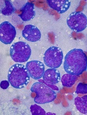
Researchers say they’ve discovered how the protein STAT3 interferes with an antitumor mechanism in cells, thereby promoting the growth of lymphoma and other cancers.
The group made their discovery using the Epstein-Barr virus (EBV) as a tool to investigate cancer development.
“Our findings add to the short list of known mechanisms by which a key cellular antitumor barrier is breached by STAT3 prior to cancer development,” said Sumita Bhaduri-McIntosh, MD, PhD, of Stony Brook University School of Medicine in New York.
“Because STAT3 interferes with this innate antitumor mechanism in cells, the opposite occurs when blood cells are infected in the lab with the cancer-causing virus EBV, and the cells continue to divide—a necessary step in cancer development.”
Dr Bhaduri-McIntosh and her colleagues described their research in PNAS.
The team explained that STAT3 inhibits a cancer-suppressing cellular activity called the DNA damage response (DDR). Normally, this response pauses cell division, allowing for the repair of damaged DNA.
But this study showed that EBV not only causes DNA damage when it infects and replicates in cells; it also activates and increases STAT3 expression. This starts a chain reaction that leads to an “un-pause” in cell division, thereby promoting cell proliferation. This, in combination with other pro-proliferative effects of the virus, can lead to cancers.
The researchers found that DDR does detect replication stress-associated DNA damage resulting from EBV infection. But signaling downstream of ATR proteins is impaired by STAT3. And this leads to relaxation of the intra-S phase checkpoint of the cell cycle.
STAT3 interrupts signaling from ATR to the protein Chk1 by promoting the loss of Claspin, a protein that assists ATR to phosphorylate Chk1. The loss of Claspin, which facilitates cell proliferation, is mediated by caspase 7.
Previous research suggested that STAT3 and Chk1 are potential targets for cancer therapies. Dr Bhaduri-McIntosh’s team said their results provide new insight into anticancer drug development because they reveal a mechanistic link between these 2 proteins.
Dr Bhaduri-McIntosh emphasized that, because STAT3 is involved in most cancers, her group’s findings could potentially impact the prevention or treatment of several types of cancer—something her lab is investigating.
Credit: Ed Uthman
Researchers say they’ve discovered how the protein STAT3 interferes with an antitumor mechanism in cells, thereby promoting the growth of lymphoma and other cancers.
The group made their discovery using the Epstein-Barr virus (EBV) as a tool to investigate cancer development.
“Our findings add to the short list of known mechanisms by which a key cellular antitumor barrier is breached by STAT3 prior to cancer development,” said Sumita Bhaduri-McIntosh, MD, PhD, of Stony Brook University School of Medicine in New York.
“Because STAT3 interferes with this innate antitumor mechanism in cells, the opposite occurs when blood cells are infected in the lab with the cancer-causing virus EBV, and the cells continue to divide—a necessary step in cancer development.”
Dr Bhaduri-McIntosh and her colleagues described their research in PNAS.
The team explained that STAT3 inhibits a cancer-suppressing cellular activity called the DNA damage response (DDR). Normally, this response pauses cell division, allowing for the repair of damaged DNA.
But this study showed that EBV not only causes DNA damage when it infects and replicates in cells; it also activates and increases STAT3 expression. This starts a chain reaction that leads to an “un-pause” in cell division, thereby promoting cell proliferation. This, in combination with other pro-proliferative effects of the virus, can lead to cancers.
The researchers found that DDR does detect replication stress-associated DNA damage resulting from EBV infection. But signaling downstream of ATR proteins is impaired by STAT3. And this leads to relaxation of the intra-S phase checkpoint of the cell cycle.
STAT3 interrupts signaling from ATR to the protein Chk1 by promoting the loss of Claspin, a protein that assists ATR to phosphorylate Chk1. The loss of Claspin, which facilitates cell proliferation, is mediated by caspase 7.
Previous research suggested that STAT3 and Chk1 are potential targets for cancer therapies. Dr Bhaduri-McIntosh’s team said their results provide new insight into anticancer drug development because they reveal a mechanistic link between these 2 proteins.
Dr Bhaduri-McIntosh emphasized that, because STAT3 is involved in most cancers, her group’s findings could potentially impact the prevention or treatment of several types of cancer—something her lab is investigating.
Credit: Ed Uthman
Researchers say they’ve discovered how the protein STAT3 interferes with an antitumor mechanism in cells, thereby promoting the growth of lymphoma and other cancers.
The group made their discovery using the Epstein-Barr virus (EBV) as a tool to investigate cancer development.
“Our findings add to the short list of known mechanisms by which a key cellular antitumor barrier is breached by STAT3 prior to cancer development,” said Sumita Bhaduri-McIntosh, MD, PhD, of Stony Brook University School of Medicine in New York.
“Because STAT3 interferes with this innate antitumor mechanism in cells, the opposite occurs when blood cells are infected in the lab with the cancer-causing virus EBV, and the cells continue to divide—a necessary step in cancer development.”
Dr Bhaduri-McIntosh and her colleagues described their research in PNAS.
The team explained that STAT3 inhibits a cancer-suppressing cellular activity called the DNA damage response (DDR). Normally, this response pauses cell division, allowing for the repair of damaged DNA.
But this study showed that EBV not only causes DNA damage when it infects and replicates in cells; it also activates and increases STAT3 expression. This starts a chain reaction that leads to an “un-pause” in cell division, thereby promoting cell proliferation. This, in combination with other pro-proliferative effects of the virus, can lead to cancers.
The researchers found that DDR does detect replication stress-associated DNA damage resulting from EBV infection. But signaling downstream of ATR proteins is impaired by STAT3. And this leads to relaxation of the intra-S phase checkpoint of the cell cycle.
STAT3 interrupts signaling from ATR to the protein Chk1 by promoting the loss of Claspin, a protein that assists ATR to phosphorylate Chk1. The loss of Claspin, which facilitates cell proliferation, is mediated by caspase 7.
Previous research suggested that STAT3 and Chk1 are potential targets for cancer therapies. Dr Bhaduri-McIntosh’s team said their results provide new insight into anticancer drug development because they reveal a mechanistic link between these 2 proteins.
Dr Bhaduri-McIntosh emphasized that, because STAT3 is involved in most cancers, her group’s findings could potentially impact the prevention or treatment of several types of cancer—something her lab is investigating.
Mediterranean diet tied to decreased platelets, WBCs

part of a Mediterranean diet
In a large study, individuals who strictly followed a Mediterranean diet had lower levels of platelets and white blood cells (WBCs) than those who deviated from the diet.
And the lower cell counts were associated with lower levels of inflammation.
The research also suggested the diet as a whole, and not just certain components, was responsible for these markers of improved health.
Marialaura Bonaccio, PhD, of the IRCCS Istituto Neurologico Mediterraneo NEUROMED in Italy, and her colleagues reported these findings in Blood.
The team noted that the Mediterranean diet—which is characterized by a wide consumption of plant foods, cereals, legumes, fish, and olive oil, as well as moderate wine consumption—has long been hailed as a heart-healthy eating plan. And previous research suggested the diet can reduce inflammation.
But the connection between the diet and levels of platelets and WBCs, 2 specific inflammatory markers in the body, has remained unclear.
“We undertook this study to understand the correlation between consuming a Mediterranean diet and specific health markers, including platelet levels and white blood cell counts, which can more specifically explain the diet’s benefits in reducing the long-term risk of cerebral and heart disease or other chronic conditions,” Dr Bonaccio said.
To do this, she and her colleagues analyzed the eating habits of 14,586 healthy Italian men and women aged 35 and older. At baseline, all subjects were healthy.
The researchers measured total platelet and WBC counts and grouped participants according to their levels (low, normal, or high), based on age- and gender-specific cut-offs.
Participants with high platelet levels were younger and had a greater incidence of high cholesterol and increased levels of common inflammation marker C-reactive protein when compared to subjects in the normal or low-platelet categories.
Individuals in the high-WBC category were mainly younger, male, and smokers. They had a higher body-mass index and higher levels of C-reactive protein and blood glucose than subjects in the other groups. They also showed higher prevalence of high blood pressure and high cholesterol.
The researchers determined participants’ adherence to a Mediterranean using 2 dietary scoring systems, the Mediterranean diet score or the Italian Mediterranean Index, which helped to accurately determine intake levels and portion sizes.
Results of these analyses revealed that adherence to the Mediterranean diet was directly related to lower levels of platelets and WBCs (P<0.0001 and P=0.008, respectively), which was correlated with lower levels of inflammation.
When compared with participants who did not follow the eating plan as closely, subjects who strictly followed the diet were less likely to belong to the group with the highest platelet counts (odds ratio=0.50) and more likely to belong to the group with the lowest WBC counts (odds ratio=1.41).
“Because the study included healthy participants, the lower levels of platelets and white blood cells in those who were more strictly consuming a Mediterranean diet indicate that this eating plan could account for substantial changes within normal ranges of variability,” Dr Bonaccio said.
“This is an important finding that has implications for how these anti-inflammatory markers are tracked among the general population.”
The researchers also evaluated the role of specific components of the diet to help clarify the observed correlation, including food antioxidant content and fiber intake, both of which have previously been connected to cardiovascular benefits.
These components only partially accounted for the link between the diet and WBC count. And they did not fully explain the correlation to platelet levels.
“An important finding of this study is that it indicates that the Mediterranean diet as a whole, and not just a few specific ingredients, is likely responsible for the beneficial health outcomes among the healthy population and should be encouraged as part of healthy eating habits,” Dr Bonaccio said.
“Building on these important findings, we continue to study this population to determine if the dietary habits may have an influence on cardiovascular disease-related mortality.”
part of a Mediterranean diet
In a large study, individuals who strictly followed a Mediterranean diet had lower levels of platelets and white blood cells (WBCs) than those who deviated from the diet.
And the lower cell counts were associated with lower levels of inflammation.
The research also suggested the diet as a whole, and not just certain components, was responsible for these markers of improved health.
Marialaura Bonaccio, PhD, of the IRCCS Istituto Neurologico Mediterraneo NEUROMED in Italy, and her colleagues reported these findings in Blood.
The team noted that the Mediterranean diet—which is characterized by a wide consumption of plant foods, cereals, legumes, fish, and olive oil, as well as moderate wine consumption—has long been hailed as a heart-healthy eating plan. And previous research suggested the diet can reduce inflammation.
But the connection between the diet and levels of platelets and WBCs, 2 specific inflammatory markers in the body, has remained unclear.
“We undertook this study to understand the correlation between consuming a Mediterranean diet and specific health markers, including platelet levels and white blood cell counts, which can more specifically explain the diet’s benefits in reducing the long-term risk of cerebral and heart disease or other chronic conditions,” Dr Bonaccio said.
To do this, she and her colleagues analyzed the eating habits of 14,586 healthy Italian men and women aged 35 and older. At baseline, all subjects were healthy.
The researchers measured total platelet and WBC counts and grouped participants according to their levels (low, normal, or high), based on age- and gender-specific cut-offs.
Participants with high platelet levels were younger and had a greater incidence of high cholesterol and increased levels of common inflammation marker C-reactive protein when compared to subjects in the normal or low-platelet categories.
Individuals in the high-WBC category were mainly younger, male, and smokers. They had a higher body-mass index and higher levels of C-reactive protein and blood glucose than subjects in the other groups. They also showed higher prevalence of high blood pressure and high cholesterol.
The researchers determined participants’ adherence to a Mediterranean using 2 dietary scoring systems, the Mediterranean diet score or the Italian Mediterranean Index, which helped to accurately determine intake levels and portion sizes.
Results of these analyses revealed that adherence to the Mediterranean diet was directly related to lower levels of platelets and WBCs (P<0.0001 and P=0.008, respectively), which was correlated with lower levels of inflammation.
When compared with participants who did not follow the eating plan as closely, subjects who strictly followed the diet were less likely to belong to the group with the highest platelet counts (odds ratio=0.50) and more likely to belong to the group with the lowest WBC counts (odds ratio=1.41).
“Because the study included healthy participants, the lower levels of platelets and white blood cells in those who were more strictly consuming a Mediterranean diet indicate that this eating plan could account for substantial changes within normal ranges of variability,” Dr Bonaccio said.
“This is an important finding that has implications for how these anti-inflammatory markers are tracked among the general population.”
The researchers also evaluated the role of specific components of the diet to help clarify the observed correlation, including food antioxidant content and fiber intake, both of which have previously been connected to cardiovascular benefits.
These components only partially accounted for the link between the diet and WBC count. And they did not fully explain the correlation to platelet levels.
“An important finding of this study is that it indicates that the Mediterranean diet as a whole, and not just a few specific ingredients, is likely responsible for the beneficial health outcomes among the healthy population and should be encouraged as part of healthy eating habits,” Dr Bonaccio said.
“Building on these important findings, we continue to study this population to determine if the dietary habits may have an influence on cardiovascular disease-related mortality.”
part of a Mediterranean diet
In a large study, individuals who strictly followed a Mediterranean diet had lower levels of platelets and white blood cells (WBCs) than those who deviated from the diet.
And the lower cell counts were associated with lower levels of inflammation.
The research also suggested the diet as a whole, and not just certain components, was responsible for these markers of improved health.
Marialaura Bonaccio, PhD, of the IRCCS Istituto Neurologico Mediterraneo NEUROMED in Italy, and her colleagues reported these findings in Blood.
The team noted that the Mediterranean diet—which is characterized by a wide consumption of plant foods, cereals, legumes, fish, and olive oil, as well as moderate wine consumption—has long been hailed as a heart-healthy eating plan. And previous research suggested the diet can reduce inflammation.
But the connection between the diet and levels of platelets and WBCs, 2 specific inflammatory markers in the body, has remained unclear.
“We undertook this study to understand the correlation between consuming a Mediterranean diet and specific health markers, including platelet levels and white blood cell counts, which can more specifically explain the diet’s benefits in reducing the long-term risk of cerebral and heart disease or other chronic conditions,” Dr Bonaccio said.
To do this, she and her colleagues analyzed the eating habits of 14,586 healthy Italian men and women aged 35 and older. At baseline, all subjects were healthy.
The researchers measured total platelet and WBC counts and grouped participants according to their levels (low, normal, or high), based on age- and gender-specific cut-offs.
Participants with high platelet levels were younger and had a greater incidence of high cholesterol and increased levels of common inflammation marker C-reactive protein when compared to subjects in the normal or low-platelet categories.
Individuals in the high-WBC category were mainly younger, male, and smokers. They had a higher body-mass index and higher levels of C-reactive protein and blood glucose than subjects in the other groups. They also showed higher prevalence of high blood pressure and high cholesterol.
The researchers determined participants’ adherence to a Mediterranean using 2 dietary scoring systems, the Mediterranean diet score or the Italian Mediterranean Index, which helped to accurately determine intake levels and portion sizes.
Results of these analyses revealed that adherence to the Mediterranean diet was directly related to lower levels of platelets and WBCs (P<0.0001 and P=0.008, respectively), which was correlated with lower levels of inflammation.
When compared with participants who did not follow the eating plan as closely, subjects who strictly followed the diet were less likely to belong to the group with the highest platelet counts (odds ratio=0.50) and more likely to belong to the group with the lowest WBC counts (odds ratio=1.41).
“Because the study included healthy participants, the lower levels of platelets and white blood cells in those who were more strictly consuming a Mediterranean diet indicate that this eating plan could account for substantial changes within normal ranges of variability,” Dr Bonaccio said.
“This is an important finding that has implications for how these anti-inflammatory markers are tracked among the general population.”
The researchers also evaluated the role of specific components of the diet to help clarify the observed correlation, including food antioxidant content and fiber intake, both of which have previously been connected to cardiovascular benefits.
These components only partially accounted for the link between the diet and WBC count. And they did not fully explain the correlation to platelet levels.
“An important finding of this study is that it indicates that the Mediterranean diet as a whole, and not just a few specific ingredients, is likely responsible for the beneficial health outcomes among the healthy population and should be encouraged as part of healthy eating habits,” Dr Bonaccio said.
“Building on these important findings, we continue to study this population to determine if the dietary habits may have an influence on cardiovascular disease-related mortality.”