User login
Hypercalcemia may be indicator for hematologic cancers
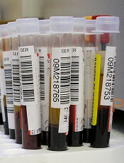
Credit: Graham Colm
Hypercalcemia could be an early indication of cancer, according to a study published in the British Journal of Cancer.
The connection between hypercalcemia and cancer is well known, but this study shows the condition can predate cancer diagnosis in primary care.
The association between hypercalcemia and cancers was particularly strong in men. And myeloma and other hematologic malignancies were among the most common cancers associated with hypercalcemia.
“All previous studies on hypercalcemia and cancer had been carried out with patients who had already been diagnosed with cancer; hypercalcemia was seen as a late effect of the cancer,” said study author Fergus Hamilton, of the University of Bristol in the UK.
“We wanted to look at the issue from a different perspective and find out if high calcium levels in blood could be used as an early indicator of cancer and, therefore, in the diagnosis of cancer.”
So the researchers analyzed the electronic records of 54,267 patients with elevated calcium levels and found that hypercalcemia was strongly associated with cancer, especially in males.
The positive predictive values for cancer in men were 11.5% for calcium levels between 2.60 and 2.79 mmol l-1, 27.9% for 2.8-2.99 mmol l-1, and 50% for >3.0 mmol l-1. In women, the corresponding values were 4.1%, 8.7%, and 16.7%, respectively.
In men, the most common cancers associated with hypercalcemia were lung (34%), prostate (21%), colorectal (8%), myeloma (8%), and other hematologic cancers (8%). There were 12 other cancer types recorded as well (19%).
In women, the most common cancers were myeloma (24%), breast (18%), other hematologic cancers (10%), lung (8%), and metastatic cancer with unknown primary (8%). There were 16 other cancers recorded among women (32%).
The researchers found no difference in calcium levels among the different cancers.
“We were surprised by the gender difference,” Dr Hamilton said. “There are a number of possible explanations for this, but we think it might be because women are much more likely to have hyperparathyroidism, another cause of hypercalcemia. Men rarely get this condition, so their hypercalcemia is more likely to be due to cancer.” 
Credit: Graham Colm
Hypercalcemia could be an early indication of cancer, according to a study published in the British Journal of Cancer.
The connection between hypercalcemia and cancer is well known, but this study shows the condition can predate cancer diagnosis in primary care.
The association between hypercalcemia and cancers was particularly strong in men. And myeloma and other hematologic malignancies were among the most common cancers associated with hypercalcemia.
“All previous studies on hypercalcemia and cancer had been carried out with patients who had already been diagnosed with cancer; hypercalcemia was seen as a late effect of the cancer,” said study author Fergus Hamilton, of the University of Bristol in the UK.
“We wanted to look at the issue from a different perspective and find out if high calcium levels in blood could be used as an early indicator of cancer and, therefore, in the diagnosis of cancer.”
So the researchers analyzed the electronic records of 54,267 patients with elevated calcium levels and found that hypercalcemia was strongly associated with cancer, especially in males.
The positive predictive values for cancer in men were 11.5% for calcium levels between 2.60 and 2.79 mmol l-1, 27.9% for 2.8-2.99 mmol l-1, and 50% for >3.0 mmol l-1. In women, the corresponding values were 4.1%, 8.7%, and 16.7%, respectively.
In men, the most common cancers associated with hypercalcemia were lung (34%), prostate (21%), colorectal (8%), myeloma (8%), and other hematologic cancers (8%). There were 12 other cancer types recorded as well (19%).
In women, the most common cancers were myeloma (24%), breast (18%), other hematologic cancers (10%), lung (8%), and metastatic cancer with unknown primary (8%). There were 16 other cancers recorded among women (32%).
The researchers found no difference in calcium levels among the different cancers.
“We were surprised by the gender difference,” Dr Hamilton said. “There are a number of possible explanations for this, but we think it might be because women are much more likely to have hyperparathyroidism, another cause of hypercalcemia. Men rarely get this condition, so their hypercalcemia is more likely to be due to cancer.”
Credit: Graham Colm
Hypercalcemia could be an early indication of cancer, according to a study published in the British Journal of Cancer.
The connection between hypercalcemia and cancer is well known, but this study shows the condition can predate cancer diagnosis in primary care.
The association between hypercalcemia and cancers was particularly strong in men. And myeloma and other hematologic malignancies were among the most common cancers associated with hypercalcemia.
“All previous studies on hypercalcemia and cancer had been carried out with patients who had already been diagnosed with cancer; hypercalcemia was seen as a late effect of the cancer,” said study author Fergus Hamilton, of the University of Bristol in the UK.
“We wanted to look at the issue from a different perspective and find out if high calcium levels in blood could be used as an early indicator of cancer and, therefore, in the diagnosis of cancer.”
So the researchers analyzed the electronic records of 54,267 patients with elevated calcium levels and found that hypercalcemia was strongly associated with cancer, especially in males.
The positive predictive values for cancer in men were 11.5% for calcium levels between 2.60 and 2.79 mmol l-1, 27.9% for 2.8-2.99 mmol l-1, and 50% for >3.0 mmol l-1. In women, the corresponding values were 4.1%, 8.7%, and 16.7%, respectively.
In men, the most common cancers associated with hypercalcemia were lung (34%), prostate (21%), colorectal (8%), myeloma (8%), and other hematologic cancers (8%). There were 12 other cancer types recorded as well (19%).
In women, the most common cancers were myeloma (24%), breast (18%), other hematologic cancers (10%), lung (8%), and metastatic cancer with unknown primary (8%). There were 16 other cancers recorded among women (32%).
The researchers found no difference in calcium levels among the different cancers.
“We were surprised by the gender difference,” Dr Hamilton said. “There are a number of possible explanations for this, but we think it might be because women are much more likely to have hyperparathyroidism, another cause of hypercalcemia. Men rarely get this condition, so their hypercalcemia is more likely to be due to cancer.”
Rivaroxaban rivals standard for VTE in cancer

Credit: CDC
MADRID—A pooled analysis of 2 studies suggests rivaroxaban prevents recurrent venous thromboembolism (VTE) in cancer patients as effectively as standard therapy, while conferring a lower risk of major bleeding.
The analysis included data from the EINSTEIN-DVT and EINSTEIN-PE trials, which were funded by the companies developing rivaroxaban (Xarelto), Bayer HealthCare Pharmaceuticals and Janssen Research & Development, LLC.
The results were published in Lancet Haematology and presented at the ESMO 2014 Congress (abstract LBA48).
The EINSTEIN-PE study included 3449 subjects with acute symptomatic pulmonary embolism (PE), with or without symptomatic deep vein thrombosis (DVT), who received anticoagulant therapy for 6 or 12 months.
The EINSTEIN-DVT study included 4833 patients who had acute symptomatic DVT, but no symptoms of PE, and received treatment for 3, 6, or 12 months.
In both studies, patients received either oral rivaroxaban (15 mg twice daily for 3 weeks, followed by 20 mg once daily) or standard therapy—enoxaparin (1.0 mg/kg twice daily) followed by a vitamin K antagonist (warfarin or acenocoumarol).
A total of 8281 patients were randomized in these studies—4150 to rivaroxaban and 4131 to standard therapy. Of the 655 patients (7.9%) with active cancer, 462 (5.6%) had cancer at baseline, and 193 (2.3%) were diagnosed during the study.
For this analysis, Martin H. Prins, MD, PhD, of Maastricht University Medical Center in The Netherlands, and his colleagues compared the 2 treatments in patients with active cancer.
VTE recurred in 4.5% (16/354) of the patients who were randomized to rivaroxaban and 6.6% (20/301) of patients randomized to standard therapy (hazard ratio [HR]=0.67).
The mortality rate was 16.4% (58/354) among patients randomized to rivaroxaban and 17.6% (53/301) among those randomized to standard therapy (HR=0.93).
Major bleeding occurred in 2.3% (8/353) of patients who received rivaroxaban and 5% (15/298) who received standard therapy (HR=0.42). And clinically relevant bleeding occurred in 13.6% (48/353) and 16.4% (49/298), respectively (HR=0.80).
Given these results, Dr Prins and his colleagues concluded that rivaroxaban can be considered an alternative to standard therapy in patients with cancer-associated VTE.
The team also said there is a need for a head-to-head comparison of rivaroxaban and long-term low-molecular-weight heparin in this patient population.
Credit: CDC
MADRID—A pooled analysis of 2 studies suggests rivaroxaban prevents recurrent venous thromboembolism (VTE) in cancer patients as effectively as standard therapy, while conferring a lower risk of major bleeding.
The analysis included data from the EINSTEIN-DVT and EINSTEIN-PE trials, which were funded by the companies developing rivaroxaban (Xarelto), Bayer HealthCare Pharmaceuticals and Janssen Research & Development, LLC.
The results were published in Lancet Haematology and presented at the ESMO 2014 Congress (abstract LBA48).
The EINSTEIN-PE study included 3449 subjects with acute symptomatic pulmonary embolism (PE), with or without symptomatic deep vein thrombosis (DVT), who received anticoagulant therapy for 6 or 12 months.
The EINSTEIN-DVT study included 4833 patients who had acute symptomatic DVT, but no symptoms of PE, and received treatment for 3, 6, or 12 months.
In both studies, patients received either oral rivaroxaban (15 mg twice daily for 3 weeks, followed by 20 mg once daily) or standard therapy—enoxaparin (1.0 mg/kg twice daily) followed by a vitamin K antagonist (warfarin or acenocoumarol).
A total of 8281 patients were randomized in these studies—4150 to rivaroxaban and 4131 to standard therapy. Of the 655 patients (7.9%) with active cancer, 462 (5.6%) had cancer at baseline, and 193 (2.3%) were diagnosed during the study.
For this analysis, Martin H. Prins, MD, PhD, of Maastricht University Medical Center in The Netherlands, and his colleagues compared the 2 treatments in patients with active cancer.
VTE recurred in 4.5% (16/354) of the patients who were randomized to rivaroxaban and 6.6% (20/301) of patients randomized to standard therapy (hazard ratio [HR]=0.67).
The mortality rate was 16.4% (58/354) among patients randomized to rivaroxaban and 17.6% (53/301) among those randomized to standard therapy (HR=0.93).
Major bleeding occurred in 2.3% (8/353) of patients who received rivaroxaban and 5% (15/298) who received standard therapy (HR=0.42). And clinically relevant bleeding occurred in 13.6% (48/353) and 16.4% (49/298), respectively (HR=0.80).
Given these results, Dr Prins and his colleagues concluded that rivaroxaban can be considered an alternative to standard therapy in patients with cancer-associated VTE.
The team also said there is a need for a head-to-head comparison of rivaroxaban and long-term low-molecular-weight heparin in this patient population.
Credit: CDC
MADRID—A pooled analysis of 2 studies suggests rivaroxaban prevents recurrent venous thromboembolism (VTE) in cancer patients as effectively as standard therapy, while conferring a lower risk of major bleeding.
The analysis included data from the EINSTEIN-DVT and EINSTEIN-PE trials, which were funded by the companies developing rivaroxaban (Xarelto), Bayer HealthCare Pharmaceuticals and Janssen Research & Development, LLC.
The results were published in Lancet Haematology and presented at the ESMO 2014 Congress (abstract LBA48).
The EINSTEIN-PE study included 3449 subjects with acute symptomatic pulmonary embolism (PE), with or without symptomatic deep vein thrombosis (DVT), who received anticoagulant therapy for 6 or 12 months.
The EINSTEIN-DVT study included 4833 patients who had acute symptomatic DVT, but no symptoms of PE, and received treatment for 3, 6, or 12 months.
In both studies, patients received either oral rivaroxaban (15 mg twice daily for 3 weeks, followed by 20 mg once daily) or standard therapy—enoxaparin (1.0 mg/kg twice daily) followed by a vitamin K antagonist (warfarin or acenocoumarol).
A total of 8281 patients were randomized in these studies—4150 to rivaroxaban and 4131 to standard therapy. Of the 655 patients (7.9%) with active cancer, 462 (5.6%) had cancer at baseline, and 193 (2.3%) were diagnosed during the study.
For this analysis, Martin H. Prins, MD, PhD, of Maastricht University Medical Center in The Netherlands, and his colleagues compared the 2 treatments in patients with active cancer.
VTE recurred in 4.5% (16/354) of the patients who were randomized to rivaroxaban and 6.6% (20/301) of patients randomized to standard therapy (hazard ratio [HR]=0.67).
The mortality rate was 16.4% (58/354) among patients randomized to rivaroxaban and 17.6% (53/301) among those randomized to standard therapy (HR=0.93).
Major bleeding occurred in 2.3% (8/353) of patients who received rivaroxaban and 5% (15/298) who received standard therapy (HR=0.42). And clinically relevant bleeding occurred in 13.6% (48/353) and 16.4% (49/298), respectively (HR=0.80).
Given these results, Dr Prins and his colleagues concluded that rivaroxaban can be considered an alternative to standard therapy in patients with cancer-associated VTE.
The team also said there is a need for a head-to-head comparison of rivaroxaban and long-term low-molecular-weight heparin in this patient population.
FDA approves cancer drugs faster, study shows

MADRID—The US Food and Drug Administration (FDA) tends to approve cancer drugs faster than Health Canada and the European Medicines Agency (EMA),
according to a study presented at the ESMO 2014 Congress.
On average, the FDA approved antineoplastic agents about 6 to 8 months faster than the EMA and Health Canada, researchers found.
One of the drugs studied had been FDA-approved for more than 4.5 years before the EMA and Health Canada authorized its use.
The researchers said these results suggest a need for a coordinated international approach to reduce the disparity in approval times.
“There needs to be a dialogue amongst industry, regulatory agencies, patient bodies, [the] research community, and oncology professionals on how best we can reduce the time to approval while ensuring safety for approved drugs,” said study investigator Sunil Verma, MD, of Sunnybrook Odette Cancer Center in Toronto, Canada.
A previous study, published in NEJM in 2012, showed that, between 2001 and 2010, the FDA tended to approve all types of drugs faster than the EMA and Health Canada.
Dr Verma and Nardin Samuel, an MD/PhD student at the University of Toronto, focused their study on cancer drugs and presented their findings at ESMO as abstract 1036O_PR.
The pair analyzed approval data for 41 antineoplastic agents and found the average time to FDA approval for these drugs was 6 months shorter than for the EMA and 7.6 months shorter than for Health Canada.
Azacitidine, which is approved to treat hematologic malignancies, had the greatest delay between FDA and Health Canada approval, at 66.1 months. The EMA approved azacitidine 10.3 months earlier than Health Canada but 55.8 months after the FDA.
The fastest approval time among the drugs studied was for cabazitaxel, which was approved for metastatic prostate cancer by the FDA just 17 days after the drug’s manufacturer filed for approval. In Canada and the European Union, the times to approval for cabazitaxel were 11.63 months and 11.03 months, respectively.
“It is not clear why there were these differences, but they are of some concern . . . ,” said David Cameron, MD, of the Edinburgh Cancer Research Centre in the UK, who was not involved in this research.
“[T]hey suggest that, in the absence of data to the contrary, there may be bureaucratic rather than medical/scientific reasons for differential geographical approval timelines, which, of course, will lead to differential geographical benefits from new agents.”
Dr Cameron added that more work is needed to understand the reasons for these differences, as well as assess any potential impact on patients.
MADRID—The US Food and Drug Administration (FDA) tends to approve cancer drugs faster than Health Canada and the European Medicines Agency (EMA),
according to a study presented at the ESMO 2014 Congress.
On average, the FDA approved antineoplastic agents about 6 to 8 months faster than the EMA and Health Canada, researchers found.
One of the drugs studied had been FDA-approved for more than 4.5 years before the EMA and Health Canada authorized its use.
The researchers said these results suggest a need for a coordinated international approach to reduce the disparity in approval times.
“There needs to be a dialogue amongst industry, regulatory agencies, patient bodies, [the] research community, and oncology professionals on how best we can reduce the time to approval while ensuring safety for approved drugs,” said study investigator Sunil Verma, MD, of Sunnybrook Odette Cancer Center in Toronto, Canada.
A previous study, published in NEJM in 2012, showed that, between 2001 and 2010, the FDA tended to approve all types of drugs faster than the EMA and Health Canada.
Dr Verma and Nardin Samuel, an MD/PhD student at the University of Toronto, focused their study on cancer drugs and presented their findings at ESMO as abstract 1036O_PR.
The pair analyzed approval data for 41 antineoplastic agents and found the average time to FDA approval for these drugs was 6 months shorter than for the EMA and 7.6 months shorter than for Health Canada.
Azacitidine, which is approved to treat hematologic malignancies, had the greatest delay between FDA and Health Canada approval, at 66.1 months. The EMA approved azacitidine 10.3 months earlier than Health Canada but 55.8 months after the FDA.
The fastest approval time among the drugs studied was for cabazitaxel, which was approved for metastatic prostate cancer by the FDA just 17 days after the drug’s manufacturer filed for approval. In Canada and the European Union, the times to approval for cabazitaxel were 11.63 months and 11.03 months, respectively.
“It is not clear why there were these differences, but they are of some concern . . . ,” said David Cameron, MD, of the Edinburgh Cancer Research Centre in the UK, who was not involved in this research.
“[T]hey suggest that, in the absence of data to the contrary, there may be bureaucratic rather than medical/scientific reasons for differential geographical approval timelines, which, of course, will lead to differential geographical benefits from new agents.”
Dr Cameron added that more work is needed to understand the reasons for these differences, as well as assess any potential impact on patients.
MADRID—The US Food and Drug Administration (FDA) tends to approve cancer drugs faster than Health Canada and the European Medicines Agency (EMA),
according to a study presented at the ESMO 2014 Congress.
On average, the FDA approved antineoplastic agents about 6 to 8 months faster than the EMA and Health Canada, researchers found.
One of the drugs studied had been FDA-approved for more than 4.5 years before the EMA and Health Canada authorized its use.
The researchers said these results suggest a need for a coordinated international approach to reduce the disparity in approval times.
“There needs to be a dialogue amongst industry, regulatory agencies, patient bodies, [the] research community, and oncology professionals on how best we can reduce the time to approval while ensuring safety for approved drugs,” said study investigator Sunil Verma, MD, of Sunnybrook Odette Cancer Center in Toronto, Canada.
A previous study, published in NEJM in 2012, showed that, between 2001 and 2010, the FDA tended to approve all types of drugs faster than the EMA and Health Canada.
Dr Verma and Nardin Samuel, an MD/PhD student at the University of Toronto, focused their study on cancer drugs and presented their findings at ESMO as abstract 1036O_PR.
The pair analyzed approval data for 41 antineoplastic agents and found the average time to FDA approval for these drugs was 6 months shorter than for the EMA and 7.6 months shorter than for Health Canada.
Azacitidine, which is approved to treat hematologic malignancies, had the greatest delay between FDA and Health Canada approval, at 66.1 months. The EMA approved azacitidine 10.3 months earlier than Health Canada but 55.8 months after the FDA.
The fastest approval time among the drugs studied was for cabazitaxel, which was approved for metastatic prostate cancer by the FDA just 17 days after the drug’s manufacturer filed for approval. In Canada and the European Union, the times to approval for cabazitaxel were 11.63 months and 11.03 months, respectively.
“It is not clear why there were these differences, but they are of some concern . . . ,” said David Cameron, MD, of the Edinburgh Cancer Research Centre in the UK, who was not involved in this research.
“[T]hey suggest that, in the absence of data to the contrary, there may be bureaucratic rather than medical/scientific reasons for differential geographical approval timelines, which, of course, will lead to differential geographical benefits from new agents.”
Dr Cameron added that more work is needed to understand the reasons for these differences, as well as assess any potential impact on patients.
Compound can inhibit metastasis in multiple myeloma

Credit: Daniel E. Sabath
A novel compound can prevent metastasis in mouse models of multiple myeloma (MM), according to research published in Cell Reports.
Investigators discovered that this compound, olaptesed pegol, can inhibit stromal cell-derived factor-1 (SDF-1), which attracts certain cells to new locations within the bone marrow.
By blocking the activity of SDF-1, olaptesed pegol renders the bone marrow uninviting to MM cells and prevents metastasis.
“Metastasis remains one of the most formidable complications we face as cancer researchers and physicians,” said study author Irene Ghobrial, MD, of the Dana-Farber Cancer Institute in Boston.
“Improvements in the treatment of metastatic cancers have, for the most part, not been nearly as dramatic as in primary disease.”
Dr Ghobrial and her colleagues studied MM because it is metastatic by nature. Myeloma cells originate in the bone marrow, depart for the bloodstream, and eventually return to the bones, where they form numerous colonies.
The team found that mice with advanced stages of MM had higher levels of SDF-1 at sites in the bones where metastasis had occurred.
“We reasoned that by neutralizing SDF-1, we could change the bone marrow environment to make it less receptive for multiple myeloma cells, reduce myeloma cells’ affinity for the marrow, and thereby inhibit the progression of the disease,” said Aldo Roccaro, MD, PhD, also of Dana-Farber.
Working with the German biotechnology company NOXXON Pharma, the investigators tested olaptesed pegol (a PEGylated mirror-image L-oligonucleotide), which binds to SDF-1.
The team found that olaptesed pegol modulates bone marrow niches and prevents MM cells from homing and engrafting to bone.
This slowed disease progression and prolonged survival in the animals, both compared to control mice and mice treated with AMD3100.
The investigators said it isn’t completely clear what becomes of the blood-borne MM cells that are prevented from metastasizing.
“We know that myeloma cells can’t survive for long if they’re circulating in the blood and can’t adhere to other tissue,” Dr Ghobrial said. “We saw no evidence that they had metastasized and begun to grow in other tissue either.”
“Our findings clearly document a therapeutic effect of olaptesed pegol in a mouse model of advanced myeloma. It is now being tested in a clinical trial of multiple myeloma patients, with more trials to come.”
Credit: Daniel E. Sabath
A novel compound can prevent metastasis in mouse models of multiple myeloma (MM), according to research published in Cell Reports.
Investigators discovered that this compound, olaptesed pegol, can inhibit stromal cell-derived factor-1 (SDF-1), which attracts certain cells to new locations within the bone marrow.
By blocking the activity of SDF-1, olaptesed pegol renders the bone marrow uninviting to MM cells and prevents metastasis.
“Metastasis remains one of the most formidable complications we face as cancer researchers and physicians,” said study author Irene Ghobrial, MD, of the Dana-Farber Cancer Institute in Boston.
“Improvements in the treatment of metastatic cancers have, for the most part, not been nearly as dramatic as in primary disease.”
Dr Ghobrial and her colleagues studied MM because it is metastatic by nature. Myeloma cells originate in the bone marrow, depart for the bloodstream, and eventually return to the bones, where they form numerous colonies.
The team found that mice with advanced stages of MM had higher levels of SDF-1 at sites in the bones where metastasis had occurred.
“We reasoned that by neutralizing SDF-1, we could change the bone marrow environment to make it less receptive for multiple myeloma cells, reduce myeloma cells’ affinity for the marrow, and thereby inhibit the progression of the disease,” said Aldo Roccaro, MD, PhD, also of Dana-Farber.
Working with the German biotechnology company NOXXON Pharma, the investigators tested olaptesed pegol (a PEGylated mirror-image L-oligonucleotide), which binds to SDF-1.
The team found that olaptesed pegol modulates bone marrow niches and prevents MM cells from homing and engrafting to bone.
This slowed disease progression and prolonged survival in the animals, both compared to control mice and mice treated with AMD3100.
The investigators said it isn’t completely clear what becomes of the blood-borne MM cells that are prevented from metastasizing.
“We know that myeloma cells can’t survive for long if they’re circulating in the blood and can’t adhere to other tissue,” Dr Ghobrial said. “We saw no evidence that they had metastasized and begun to grow in other tissue either.”
“Our findings clearly document a therapeutic effect of olaptesed pegol in a mouse model of advanced myeloma. It is now being tested in a clinical trial of multiple myeloma patients, with more trials to come.”
Credit: Daniel E. Sabath
A novel compound can prevent metastasis in mouse models of multiple myeloma (MM), according to research published in Cell Reports.
Investigators discovered that this compound, olaptesed pegol, can inhibit stromal cell-derived factor-1 (SDF-1), which attracts certain cells to new locations within the bone marrow.
By blocking the activity of SDF-1, olaptesed pegol renders the bone marrow uninviting to MM cells and prevents metastasis.
“Metastasis remains one of the most formidable complications we face as cancer researchers and physicians,” said study author Irene Ghobrial, MD, of the Dana-Farber Cancer Institute in Boston.
“Improvements in the treatment of metastatic cancers have, for the most part, not been nearly as dramatic as in primary disease.”
Dr Ghobrial and her colleagues studied MM because it is metastatic by nature. Myeloma cells originate in the bone marrow, depart for the bloodstream, and eventually return to the bones, where they form numerous colonies.
The team found that mice with advanced stages of MM had higher levels of SDF-1 at sites in the bones where metastasis had occurred.
“We reasoned that by neutralizing SDF-1, we could change the bone marrow environment to make it less receptive for multiple myeloma cells, reduce myeloma cells’ affinity for the marrow, and thereby inhibit the progression of the disease,” said Aldo Roccaro, MD, PhD, also of Dana-Farber.
Working with the German biotechnology company NOXXON Pharma, the investigators tested olaptesed pegol (a PEGylated mirror-image L-oligonucleotide), which binds to SDF-1.
The team found that olaptesed pegol modulates bone marrow niches and prevents MM cells from homing and engrafting to bone.
This slowed disease progression and prolonged survival in the animals, both compared to control mice and mice treated with AMD3100.
The investigators said it isn’t completely clear what becomes of the blood-borne MM cells that are prevented from metastasizing.
“We know that myeloma cells can’t survive for long if they’re circulating in the blood and can’t adhere to other tissue,” Dr Ghobrial said. “We saw no evidence that they had metastasized and begun to grow in other tissue either.”
“Our findings clearly document a therapeutic effect of olaptesed pegol in a mouse model of advanced myeloma. It is now being tested in a clinical trial of multiple myeloma patients, with more trials to come.”
Drug could treat a range of blood cancers
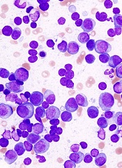
PHILADELPHIA—A drug that targets the ribosome may be active in a broad range of hematologic malignancies, researchers say.
The drug, CX-5461, inhibits the protein RNA polymerase I (Pol I), which is consistently upregulated in hematologic and other cancers.
CX-5461 significantly prolonged survival in mouse models of refractory acute myeloid leukemia (AML) and multiple myeloma (MM). It also synergized with everolimus to extend survival in mice with B-cell lymphoma.
Furthermore, the drug did not elicit severe adverse effects.
“We were excited to find that therapeutic doses of CX-5461 had little effect on normal cells in our experiments,” said Ross D. Hannan, PhD, of the Peter MacCallum Cancer Centre in Melbourne, Australia,
“Prior to these studies, few people would have guessed that such a therapeutic window could be obtained by targeting a so-called house-keeping protein that is essential to all cells for survival.”
Dr Hannan and his colleagues presented these findings in a poster at the AACR conference Hematologic Malignancies: Translating Discoveries to Novel Therapies.
The researchers previously showed that cancer cells are much more dependent on ribosome biogenesis than normal cells. And blocking the accelerated reading of ribosomal genes in mice—using CX5461—can cause lymphoma and leukemia cells to die, while sparing normal cells.
With their latest research, the group expanded upon these findings by testing CX-5461 in MLL-driven AML, V*κ-Myc-driven MM, and Eμ-Myc lymphoma.
They found that CX-5461 improved overall survival in MLL/ENL Nras leukemic mice, compared to placebo and standard therapy. The median survival was 17 days for vehicle-treated mice, 21 days for mice treated with cytarabine and doxorubicin, and 36 days for mice that received CX-5461 (P<0.0001 for vehicle vs CX-5461).
CX-5461 treated MLL-driven AML by inducing apoptosis, delaying cell-cycle progression, and promoting differentiation.
The researchers also found the therapeutic benefit of CX-5461 is not p53-dependent, which contradicts their previous findings. Human AML cell lines and primary patient samples were sensitive to CX-5461 independent of p53 status.
And MLL/ENL Nras p53-/- leukemic mice had significantly prolonged survival when treated with CX-5461, compared to vehicle-treated controls. The median survival was 11 days and 24 days, respectively (P<0.0001).
Likewise, CX-5461 significantly prolonged survival in mice bearing V*κ-Myc MM. The median survival was 103.5 days for controls and 175 days for mice that received CX-5461 (P<0.0001).
Finally, the researchers showed that CX-5461 synergizes with everolimus to treat Eμ-Myc lymphoma. The median survival was 15 days in control mice, 18 days in mice that received everolimus, 32 days in mice treated with CX-5461, and 54 days in mice that received both drugs (P<0.0001 for CX-5461 vs the combination).
“These results provide further rationale for the first-in-human phase 1 clinical trial that we initiated in July 2013 testing CX-5461 for patients with advanced hematological malignancies, including AML and multiple myeloma,” Dr Hannan said.
His group’s preclinical research was funded by the National Health and Medical Research Council, Australia; the Leukaemia Foundation of Australia; and Cancer Council Victoria, Melbourne, Australia. Senhwa Biosciences, the makers of CX-5461, provided the drug.
PHILADELPHIA—A drug that targets the ribosome may be active in a broad range of hematologic malignancies, researchers say.
The drug, CX-5461, inhibits the protein RNA polymerase I (Pol I), which is consistently upregulated in hematologic and other cancers.
CX-5461 significantly prolonged survival in mouse models of refractory acute myeloid leukemia (AML) and multiple myeloma (MM). It also synergized with everolimus to extend survival in mice with B-cell lymphoma.
Furthermore, the drug did not elicit severe adverse effects.
“We were excited to find that therapeutic doses of CX-5461 had little effect on normal cells in our experiments,” said Ross D. Hannan, PhD, of the Peter MacCallum Cancer Centre in Melbourne, Australia,
“Prior to these studies, few people would have guessed that such a therapeutic window could be obtained by targeting a so-called house-keeping protein that is essential to all cells for survival.”
Dr Hannan and his colleagues presented these findings in a poster at the AACR conference Hematologic Malignancies: Translating Discoveries to Novel Therapies.
The researchers previously showed that cancer cells are much more dependent on ribosome biogenesis than normal cells. And blocking the accelerated reading of ribosomal genes in mice—using CX5461—can cause lymphoma and leukemia cells to die, while sparing normal cells.
With their latest research, the group expanded upon these findings by testing CX-5461 in MLL-driven AML, V*κ-Myc-driven MM, and Eμ-Myc lymphoma.
They found that CX-5461 improved overall survival in MLL/ENL Nras leukemic mice, compared to placebo and standard therapy. The median survival was 17 days for vehicle-treated mice, 21 days for mice treated with cytarabine and doxorubicin, and 36 days for mice that received CX-5461 (P<0.0001 for vehicle vs CX-5461).
CX-5461 treated MLL-driven AML by inducing apoptosis, delaying cell-cycle progression, and promoting differentiation.
The researchers also found the therapeutic benefit of CX-5461 is not p53-dependent, which contradicts their previous findings. Human AML cell lines and primary patient samples were sensitive to CX-5461 independent of p53 status.
And MLL/ENL Nras p53-/- leukemic mice had significantly prolonged survival when treated with CX-5461, compared to vehicle-treated controls. The median survival was 11 days and 24 days, respectively (P<0.0001).
Likewise, CX-5461 significantly prolonged survival in mice bearing V*κ-Myc MM. The median survival was 103.5 days for controls and 175 days for mice that received CX-5461 (P<0.0001).
Finally, the researchers showed that CX-5461 synergizes with everolimus to treat Eμ-Myc lymphoma. The median survival was 15 days in control mice, 18 days in mice that received everolimus, 32 days in mice treated with CX-5461, and 54 days in mice that received both drugs (P<0.0001 for CX-5461 vs the combination).
“These results provide further rationale for the first-in-human phase 1 clinical trial that we initiated in July 2013 testing CX-5461 for patients with advanced hematological malignancies, including AML and multiple myeloma,” Dr Hannan said.
His group’s preclinical research was funded by the National Health and Medical Research Council, Australia; the Leukaemia Foundation of Australia; and Cancer Council Victoria, Melbourne, Australia. Senhwa Biosciences, the makers of CX-5461, provided the drug.
PHILADELPHIA—A drug that targets the ribosome may be active in a broad range of hematologic malignancies, researchers say.
The drug, CX-5461, inhibits the protein RNA polymerase I (Pol I), which is consistently upregulated in hematologic and other cancers.
CX-5461 significantly prolonged survival in mouse models of refractory acute myeloid leukemia (AML) and multiple myeloma (MM). It also synergized with everolimus to extend survival in mice with B-cell lymphoma.
Furthermore, the drug did not elicit severe adverse effects.
“We were excited to find that therapeutic doses of CX-5461 had little effect on normal cells in our experiments,” said Ross D. Hannan, PhD, of the Peter MacCallum Cancer Centre in Melbourne, Australia,
“Prior to these studies, few people would have guessed that such a therapeutic window could be obtained by targeting a so-called house-keeping protein that is essential to all cells for survival.”
Dr Hannan and his colleagues presented these findings in a poster at the AACR conference Hematologic Malignancies: Translating Discoveries to Novel Therapies.
The researchers previously showed that cancer cells are much more dependent on ribosome biogenesis than normal cells. And blocking the accelerated reading of ribosomal genes in mice—using CX5461—can cause lymphoma and leukemia cells to die, while sparing normal cells.
With their latest research, the group expanded upon these findings by testing CX-5461 in MLL-driven AML, V*κ-Myc-driven MM, and Eμ-Myc lymphoma.
They found that CX-5461 improved overall survival in MLL/ENL Nras leukemic mice, compared to placebo and standard therapy. The median survival was 17 days for vehicle-treated mice, 21 days for mice treated with cytarabine and doxorubicin, and 36 days for mice that received CX-5461 (P<0.0001 for vehicle vs CX-5461).
CX-5461 treated MLL-driven AML by inducing apoptosis, delaying cell-cycle progression, and promoting differentiation.
The researchers also found the therapeutic benefit of CX-5461 is not p53-dependent, which contradicts their previous findings. Human AML cell lines and primary patient samples were sensitive to CX-5461 independent of p53 status.
And MLL/ENL Nras p53-/- leukemic mice had significantly prolonged survival when treated with CX-5461, compared to vehicle-treated controls. The median survival was 11 days and 24 days, respectively (P<0.0001).
Likewise, CX-5461 significantly prolonged survival in mice bearing V*κ-Myc MM. The median survival was 103.5 days for controls and 175 days for mice that received CX-5461 (P<0.0001).
Finally, the researchers showed that CX-5461 synergizes with everolimus to treat Eμ-Myc lymphoma. The median survival was 15 days in control mice, 18 days in mice that received everolimus, 32 days in mice treated with CX-5461, and 54 days in mice that received both drugs (P<0.0001 for CX-5461 vs the combination).
“These results provide further rationale for the first-in-human phase 1 clinical trial that we initiated in July 2013 testing CX-5461 for patients with advanced hematological malignancies, including AML and multiple myeloma,” Dr Hannan said.
His group’s preclinical research was funded by the National Health and Medical Research Council, Australia; the Leukaemia Foundation of Australia; and Cancer Council Victoria, Melbourne, Australia. Senhwa Biosciences, the makers of CX-5461, provided the drug.
Study reveals mutation that causes aplastic anemia

of women in a family
By studying 3 generations of a family plagued by blood disorders, researchers discovered a genetic mutation that causes aplastic anemia.
The team performed whole-exome sequencing on DNA from the families and identified an inherited mutation on the ACD gene, which codes for the telomere-binding protein TPP1.
The mutation disrupts the interactions between telomeres and telomerase, which causes blood cells to die and results in aplastic anemia.
“Identifying this causal defect may help suggest future molecular-based treatments that bypass the gene defect and restore blood cell production,” said Hakon Hakonarson, MD, PhD, of The Children’s Hospital of Philadelphia in Pennsylvania.
Dr Hakonarson and his colleagues described this research in Blood.
The team studied an Australian family with aplastic anemia and other hematopoietic disorders, including leukemia. Whole-exome sequencing of the family’s DNA revealed an inherited mutation on the ACD gene.
The mutation is an amino acid deletion in the TEL patch of TPP1 (ΔK170). All of the family members with this mutation had short telomeres, and those with wild-type TPP1 did not.
The researchers introduced TPP1 with the ΔK170 mutation into 293T cells and found the protein could localize to telomeres but failed to recruit telomerase. The team said this indicates a causal relationship between the mutation and bone marrow disorders.
Without access to telomerase to help maintain telomeres, blood cells lose their structural integrity and die, resulting in bone marrow failure and aplastic anemia.
Nine other genes were previously found to play a role in bone marrow failure disorders. The current study adds ACD to the list and is the first time the gene has been shown to have a disease-causing role.
“This improved understanding of the underlying molecular mechanisms may suggest new approaches to treating disorders such as aplastic anemia,” Dr Hakonarson said. “For instance, investigators may identify other avenues for recruiting telomerase to telomeres to restore its protective function.”
of women in a family
By studying 3 generations of a family plagued by blood disorders, researchers discovered a genetic mutation that causes aplastic anemia.
The team performed whole-exome sequencing on DNA from the families and identified an inherited mutation on the ACD gene, which codes for the telomere-binding protein TPP1.
The mutation disrupts the interactions between telomeres and telomerase, which causes blood cells to die and results in aplastic anemia.
“Identifying this causal defect may help suggest future molecular-based treatments that bypass the gene defect and restore blood cell production,” said Hakon Hakonarson, MD, PhD, of The Children’s Hospital of Philadelphia in Pennsylvania.
Dr Hakonarson and his colleagues described this research in Blood.
The team studied an Australian family with aplastic anemia and other hematopoietic disorders, including leukemia. Whole-exome sequencing of the family’s DNA revealed an inherited mutation on the ACD gene.
The mutation is an amino acid deletion in the TEL patch of TPP1 (ΔK170). All of the family members with this mutation had short telomeres, and those with wild-type TPP1 did not.
The researchers introduced TPP1 with the ΔK170 mutation into 293T cells and found the protein could localize to telomeres but failed to recruit telomerase. The team said this indicates a causal relationship between the mutation and bone marrow disorders.
Without access to telomerase to help maintain telomeres, blood cells lose their structural integrity and die, resulting in bone marrow failure and aplastic anemia.
Nine other genes were previously found to play a role in bone marrow failure disorders. The current study adds ACD to the list and is the first time the gene has been shown to have a disease-causing role.
“This improved understanding of the underlying molecular mechanisms may suggest new approaches to treating disorders such as aplastic anemia,” Dr Hakonarson said. “For instance, investigators may identify other avenues for recruiting telomerase to telomeres to restore its protective function.”
of women in a family
By studying 3 generations of a family plagued by blood disorders, researchers discovered a genetic mutation that causes aplastic anemia.
The team performed whole-exome sequencing on DNA from the families and identified an inherited mutation on the ACD gene, which codes for the telomere-binding protein TPP1.
The mutation disrupts the interactions between telomeres and telomerase, which causes blood cells to die and results in aplastic anemia.
“Identifying this causal defect may help suggest future molecular-based treatments that bypass the gene defect and restore blood cell production,” said Hakon Hakonarson, MD, PhD, of The Children’s Hospital of Philadelphia in Pennsylvania.
Dr Hakonarson and his colleagues described this research in Blood.
The team studied an Australian family with aplastic anemia and other hematopoietic disorders, including leukemia. Whole-exome sequencing of the family’s DNA revealed an inherited mutation on the ACD gene.
The mutation is an amino acid deletion in the TEL patch of TPP1 (ΔK170). All of the family members with this mutation had short telomeres, and those with wild-type TPP1 did not.
The researchers introduced TPP1 with the ΔK170 mutation into 293T cells and found the protein could localize to telomeres but failed to recruit telomerase. The team said this indicates a causal relationship between the mutation and bone marrow disorders.
Without access to telomerase to help maintain telomeres, blood cells lose their structural integrity and die, resulting in bone marrow failure and aplastic anemia.
Nine other genes were previously found to play a role in bone marrow failure disorders. The current study adds ACD to the list and is the first time the gene has been shown to have a disease-causing role.
“This improved understanding of the underlying molecular mechanisms may suggest new approaches to treating disorders such as aplastic anemia,” Dr Hakonarson said. “For instance, investigators may identify other avenues for recruiting telomerase to telomeres to restore its protective function.”
Self-monitoring coagulometers get thumbs-up from NICE

The UK’s National Institute for Health and Care Excellence (NICE) has published a guidance recommending 2 technologies that enable patients on long-term anticoagulant therapy to self-monitor their clotting time.
The guidance supports use of the Coaguchek XS system (Roche Diagnostics) and the InRatio2 PT/INR Monitor (Alere) as options for some adults with atrial
fibrillation or heart valve disease who are on long-term anticoagulant therapy.
“The evidence shows that greater use of self-monitoring offers clinical and patient benefit and, over time, is likely to result in reductions in heart attacks and strokes caused by blood clots,” said Carole Longson, NICE Health Technology Evaluation Centre Director.
“Because self-monitoring provides almost instant results, self-monitoring can reduce anxiety, provide a sense of control for the patient, and remove the need to frequently attend clinics or hospitals.”
About the Coaguchek XS system
The Coaguchek XS system (Roche Diagnostics) consists of a meter and specifically designed test strips that can analyze a blood sample (fresh capillary blood or fresh untreated whole venous blood) and calculate the prothrombin time (PT) and the international normalized ratio (INR).
A code chip, which contains calibration data and the expiration date of the test strips, is inserted into the meter before it is switched on. Once the device is switched on, a test strip is inserted, and the blood sample is applied.
The test result is displayed approximately 1 minute after application of the sample, and the device automatically stores the result in its memory. The user is guided through the process by on-screen graphical instructions.
About the InRatio2 PT/INR Monitor
The INRatio2 PT/INR monitor (Alere) does a modified version of the 1-stage PT test using a recombinant human thromboplastin reagent. The clot formed in the reaction is detected by the change in the electrical impedance of the sample during the coagulation process.
The system consists of a monitor and disposable test strips. The monitor provides a user interface, heats the test strip to the appropriate reaction temperature, measures the impedance of blood samples, and calculates and reports PT and INR results.
Instructions and test results are displayed on an LCD. The monitor can store test results so that past results can be reviewed.
The UK’s National Institute for Health and Care Excellence (NICE) has published a guidance recommending 2 technologies that enable patients on long-term anticoagulant therapy to self-monitor their clotting time.
The guidance supports use of the Coaguchek XS system (Roche Diagnostics) and the InRatio2 PT/INR Monitor (Alere) as options for some adults with atrial
fibrillation or heart valve disease who are on long-term anticoagulant therapy.
“The evidence shows that greater use of self-monitoring offers clinical and patient benefit and, over time, is likely to result in reductions in heart attacks and strokes caused by blood clots,” said Carole Longson, NICE Health Technology Evaluation Centre Director.
“Because self-monitoring provides almost instant results, self-monitoring can reduce anxiety, provide a sense of control for the patient, and remove the need to frequently attend clinics or hospitals.”
About the Coaguchek XS system
The Coaguchek XS system (Roche Diagnostics) consists of a meter and specifically designed test strips that can analyze a blood sample (fresh capillary blood or fresh untreated whole venous blood) and calculate the prothrombin time (PT) and the international normalized ratio (INR).
A code chip, which contains calibration data and the expiration date of the test strips, is inserted into the meter before it is switched on. Once the device is switched on, a test strip is inserted, and the blood sample is applied.
The test result is displayed approximately 1 minute after application of the sample, and the device automatically stores the result in its memory. The user is guided through the process by on-screen graphical instructions.
About the InRatio2 PT/INR Monitor
The INRatio2 PT/INR monitor (Alere) does a modified version of the 1-stage PT test using a recombinant human thromboplastin reagent. The clot formed in the reaction is detected by the change in the electrical impedance of the sample during the coagulation process.
The system consists of a monitor and disposable test strips. The monitor provides a user interface, heats the test strip to the appropriate reaction temperature, measures the impedance of blood samples, and calculates and reports PT and INR results.
Instructions and test results are displayed on an LCD. The monitor can store test results so that past results can be reviewed.
The UK’s National Institute for Health and Care Excellence (NICE) has published a guidance recommending 2 technologies that enable patients on long-term anticoagulant therapy to self-monitor their clotting time.
The guidance supports use of the Coaguchek XS system (Roche Diagnostics) and the InRatio2 PT/INR Monitor (Alere) as options for some adults with atrial
fibrillation or heart valve disease who are on long-term anticoagulant therapy.
“The evidence shows that greater use of self-monitoring offers clinical and patient benefit and, over time, is likely to result in reductions in heart attacks and strokes caused by blood clots,” said Carole Longson, NICE Health Technology Evaluation Centre Director.
“Because self-monitoring provides almost instant results, self-monitoring can reduce anxiety, provide a sense of control for the patient, and remove the need to frequently attend clinics or hospitals.”
About the Coaguchek XS system
The Coaguchek XS system (Roche Diagnostics) consists of a meter and specifically designed test strips that can analyze a blood sample (fresh capillary blood or fresh untreated whole venous blood) and calculate the prothrombin time (PT) and the international normalized ratio (INR).
A code chip, which contains calibration data and the expiration date of the test strips, is inserted into the meter before it is switched on. Once the device is switched on, a test strip is inserted, and the blood sample is applied.
The test result is displayed approximately 1 minute after application of the sample, and the device automatically stores the result in its memory. The user is guided through the process by on-screen graphical instructions.
About the InRatio2 PT/INR Monitor
The INRatio2 PT/INR monitor (Alere) does a modified version of the 1-stage PT test using a recombinant human thromboplastin reagent. The clot formed in the reaction is detected by the change in the electrical impedance of the sample during the coagulation process.
The system consists of a monitor and disposable test strips. The monitor provides a user interface, heats the test strip to the appropriate reaction temperature, measures the impedance of blood samples, and calculates and reports PT and INR results.
Instructions and test results are displayed on an LCD. The monitor can store test results so that past results can be reviewed.
Murine studies support use of TKIs in ALL subtype

PHILADELPHIA—Experiments in mice reinforce the idea that tyrosine kinase inhibitors (TKIs) can treat patients with Ph-like acute lymphoblastic leukemia (ALL).
Investigators recently identified genomic alterations in Ph-like ALL that suggest these patients might respond to TKIs, and tests in a small number of patients supported this theory.
Now, preclinical results show that kinase fusions in Ph-like ALL activate signaling pathways differently, and this affects sensitivity to TKIs.
Kathryn Roberts, PhD, of St Jude Children’s Research Hospital in Memphis, Tennessee, and her colleagues presented these results at the AACR conference Hematologic Malignancies: Translating Discoveries to Novel Therapies.
“We recently described a subtype of B-cell acute lymphoblastic leukemia with very poor outcome that is characterized by genetic alterations involving tyrosine kinases, termed Ph-like ALL,” Dr Roberts said. “We wanted to examine whether these alterations contribute to the development of Ph-like ALL and determine if they could be targeted with tyrosine kinase inhibitors.”
“We showed, for the first time, that the kinase alterations we tested contribute to the development of Ph-like ALL, and that Ph-like ALL can be treated effectively with tyrosine kinase inhibitors in animal models. These findings provide a strong rationale for treating Ph-like ALL patients with targeted therapies to improve their survival.”
Dr Roberts and her colleagues first introduced kinase alterations—RCSD1-ABL2, SSBP2-CSF1R, or PAX5-JAK2—in IL-7-dependent, Arf-/- mouse pre-B cells expressing IK6.
They found that each fusion conferred cytokine-independent growth in vitro. And mice that received transplants of pre-B cells expressing RCSD1-ABL2 or SSBP2-CSF1R developed ALL with a pre-B immunophenotype.
The investigators then assessed the activation of kinase signaling pathways and TKI sensitivity in Arf-/- pre-B cells and human leukemic cells harvested from xenografted mice expressing ETV6-ABL1, RANBP2-ABL1, PAG1-ABL2, RCSD1-ABL2, SSBP2-CSF1R, IGH-EPOR, ATF7IP-JAK2, and PAX5-JAK2.
In both cell types, signaling pathway activation and TKI sensitivity differed according to the kinase fusion.
Cells expressing ABL1-class kinase fusions (ABL1, ABL2, CSF1R, and PDGFRB) exhibited pSTAT5 activation that was inhibited by imatinib or dasatinib. But in cells expressing ATF7IP-JAK2, PAX5-JAK2, or IGH-EPOR, pSTAT5 activation was only inhibited by ruxolitinib.
Finally, the investigators tested dasatinib in xenograft models of ETV6-ABL1, RCSD1-ABL2, PAG1-ABL2, or SSBP2-CSF1R ALL.
They found that treated mice had significantly lower leukemic burdens and splenic weights than control mice. And STAT5 phosphorylation was attenuated in cells from treated mice.
“Our studies show that different FDA-approved TKIs such as imatinib, dasatinib, ruxolitinib, or crizotinib could potentially be used to treat Ph-like ALL patients, depending on the type of kinase alterations their tumors bear,” Dr Roberts said.
“We were able to gain a better understanding of the genetics underlying Ph-like ALL, and our studies could help identify patients who will not respond optimally to current therapy. By knowing the exact genetic alteration upfront, we may be able to implement different therapeutic strategies to improve the survival rate of future patients with ALL.”
PHILADELPHIA—Experiments in mice reinforce the idea that tyrosine kinase inhibitors (TKIs) can treat patients with Ph-like acute lymphoblastic leukemia (ALL).
Investigators recently identified genomic alterations in Ph-like ALL that suggest these patients might respond to TKIs, and tests in a small number of patients supported this theory.
Now, preclinical results show that kinase fusions in Ph-like ALL activate signaling pathways differently, and this affects sensitivity to TKIs.
Kathryn Roberts, PhD, of St Jude Children’s Research Hospital in Memphis, Tennessee, and her colleagues presented these results at the AACR conference Hematologic Malignancies: Translating Discoveries to Novel Therapies.
“We recently described a subtype of B-cell acute lymphoblastic leukemia with very poor outcome that is characterized by genetic alterations involving tyrosine kinases, termed Ph-like ALL,” Dr Roberts said. “We wanted to examine whether these alterations contribute to the development of Ph-like ALL and determine if they could be targeted with tyrosine kinase inhibitors.”
“We showed, for the first time, that the kinase alterations we tested contribute to the development of Ph-like ALL, and that Ph-like ALL can be treated effectively with tyrosine kinase inhibitors in animal models. These findings provide a strong rationale for treating Ph-like ALL patients with targeted therapies to improve their survival.”
Dr Roberts and her colleagues first introduced kinase alterations—RCSD1-ABL2, SSBP2-CSF1R, or PAX5-JAK2—in IL-7-dependent, Arf-/- mouse pre-B cells expressing IK6.
They found that each fusion conferred cytokine-independent growth in vitro. And mice that received transplants of pre-B cells expressing RCSD1-ABL2 or SSBP2-CSF1R developed ALL with a pre-B immunophenotype.
The investigators then assessed the activation of kinase signaling pathways and TKI sensitivity in Arf-/- pre-B cells and human leukemic cells harvested from xenografted mice expressing ETV6-ABL1, RANBP2-ABL1, PAG1-ABL2, RCSD1-ABL2, SSBP2-CSF1R, IGH-EPOR, ATF7IP-JAK2, and PAX5-JAK2.
In both cell types, signaling pathway activation and TKI sensitivity differed according to the kinase fusion.
Cells expressing ABL1-class kinase fusions (ABL1, ABL2, CSF1R, and PDGFRB) exhibited pSTAT5 activation that was inhibited by imatinib or dasatinib. But in cells expressing ATF7IP-JAK2, PAX5-JAK2, or IGH-EPOR, pSTAT5 activation was only inhibited by ruxolitinib.
Finally, the investigators tested dasatinib in xenograft models of ETV6-ABL1, RCSD1-ABL2, PAG1-ABL2, or SSBP2-CSF1R ALL.
They found that treated mice had significantly lower leukemic burdens and splenic weights than control mice. And STAT5 phosphorylation was attenuated in cells from treated mice.
“Our studies show that different FDA-approved TKIs such as imatinib, dasatinib, ruxolitinib, or crizotinib could potentially be used to treat Ph-like ALL patients, depending on the type of kinase alterations their tumors bear,” Dr Roberts said.
“We were able to gain a better understanding of the genetics underlying Ph-like ALL, and our studies could help identify patients who will not respond optimally to current therapy. By knowing the exact genetic alteration upfront, we may be able to implement different therapeutic strategies to improve the survival rate of future patients with ALL.”
PHILADELPHIA—Experiments in mice reinforce the idea that tyrosine kinase inhibitors (TKIs) can treat patients with Ph-like acute lymphoblastic leukemia (ALL).
Investigators recently identified genomic alterations in Ph-like ALL that suggest these patients might respond to TKIs, and tests in a small number of patients supported this theory.
Now, preclinical results show that kinase fusions in Ph-like ALL activate signaling pathways differently, and this affects sensitivity to TKIs.
Kathryn Roberts, PhD, of St Jude Children’s Research Hospital in Memphis, Tennessee, and her colleagues presented these results at the AACR conference Hematologic Malignancies: Translating Discoveries to Novel Therapies.
“We recently described a subtype of B-cell acute lymphoblastic leukemia with very poor outcome that is characterized by genetic alterations involving tyrosine kinases, termed Ph-like ALL,” Dr Roberts said. “We wanted to examine whether these alterations contribute to the development of Ph-like ALL and determine if they could be targeted with tyrosine kinase inhibitors.”
“We showed, for the first time, that the kinase alterations we tested contribute to the development of Ph-like ALL, and that Ph-like ALL can be treated effectively with tyrosine kinase inhibitors in animal models. These findings provide a strong rationale for treating Ph-like ALL patients with targeted therapies to improve their survival.”
Dr Roberts and her colleagues first introduced kinase alterations—RCSD1-ABL2, SSBP2-CSF1R, or PAX5-JAK2—in IL-7-dependent, Arf-/- mouse pre-B cells expressing IK6.
They found that each fusion conferred cytokine-independent growth in vitro. And mice that received transplants of pre-B cells expressing RCSD1-ABL2 or SSBP2-CSF1R developed ALL with a pre-B immunophenotype.
The investigators then assessed the activation of kinase signaling pathways and TKI sensitivity in Arf-/- pre-B cells and human leukemic cells harvested from xenografted mice expressing ETV6-ABL1, RANBP2-ABL1, PAG1-ABL2, RCSD1-ABL2, SSBP2-CSF1R, IGH-EPOR, ATF7IP-JAK2, and PAX5-JAK2.
In both cell types, signaling pathway activation and TKI sensitivity differed according to the kinase fusion.
Cells expressing ABL1-class kinase fusions (ABL1, ABL2, CSF1R, and PDGFRB) exhibited pSTAT5 activation that was inhibited by imatinib or dasatinib. But in cells expressing ATF7IP-JAK2, PAX5-JAK2, or IGH-EPOR, pSTAT5 activation was only inhibited by ruxolitinib.
Finally, the investigators tested dasatinib in xenograft models of ETV6-ABL1, RCSD1-ABL2, PAG1-ABL2, or SSBP2-CSF1R ALL.
They found that treated mice had significantly lower leukemic burdens and splenic weights than control mice. And STAT5 phosphorylation was attenuated in cells from treated mice.
“Our studies show that different FDA-approved TKIs such as imatinib, dasatinib, ruxolitinib, or crizotinib could potentially be used to treat Ph-like ALL patients, depending on the type of kinase alterations their tumors bear,” Dr Roberts said.
“We were able to gain a better understanding of the genetics underlying Ph-like ALL, and our studies could help identify patients who will not respond optimally to current therapy. By knowing the exact genetic alteration upfront, we may be able to implement different therapeutic strategies to improve the survival rate of future patients with ALL.”
ETBs prove effective against lymphoma and myeloma

Credit: Rhoda Baer
PHILADELPHIA—A pair of engineered toxin bodies (ETBs) can successfully treat Burkitt lymphoma and multiple myeloma, according to preclinical research presented at the AACR conference Hematologic Malignancies: Translating Discoveries to Novel Therapies.
The ETBs, known as MT-4007 and MT-4007-D, work by targeting CD38.
They greatly reduced tumor burden and improved survival in mouse models. And they were well-tolerated, even at the highest doses administered.
“In this study, we found that the growth of human cancer cells in mice was substantially decreased, or the cells were even eliminated, following treatment with our investigational CD38-targeted therapy,” said Erin K. Willert, PhD, of Molecular Templates Inc., in Georgetown, Texas.
Dr Willert and her colleagues explained that ETBs are derived from the ribosome-inactivating alpha subunit of Shiga-like toxin 1 (SLT-1A). They have been engineered to contain a target binding domain fused to a modified SLT-1A protein, which allows for delivery to a cell surface target—in this case, CD38.
Upon binding to a CD38-expressing cell, the ETB enters the cell, routes to the cytosol, halts protein synthesis, and kills the cell.
The researchers first tested MT-4007 and MT-4007-D in a range of human cell lines. The agents exhibited cytotoxicity in CD38+ Burkitt lymphoma and multiple myeloma cell lines (H929, Daudi, ST486, and Raji). But neither agent proved cytotoxic in CD38- cell lines (U266, SKBR3, and HCC1954).
The team then moved on to test MT-4007 in a mouse model of Burkitt lymphoma. Following injection with Daudi-Luc cells, mice received no treatment or MT-4007 at 0.05 mg/kg, 0.5 mg/kg, or 2 mg/kg on days 3, 5, 8, 10, and 12.
Treated mice exhibited significantly reduced tumor burden compared to controls. The mean tumor burden for mice that received MT-4007 at 0.05 mg/kg was 29% of the control tumor burden (P<0.0001). It was 0.4% for mice that received 0.50 mg/kg (P<0.0001) and 0.02% for mice that received 2 mg/kg (P<0.0001).
In a model of multiple myeloma, MT-4007-D provided a dose-dependent delay in tumor growth. After receiving injections of H929 cells, mice received no treatment or MT-4007-D at 0.5 mg/kg, 2 mg/kg, or 3 mg/kg on days 1, 3, 5, 8, 10, and 12.
The researchers assessed efficacy by measuring the time to endpoint, which was a tumor volume of 2000 mm3. The median time to endpoint was 22.3 days in controls, 21.2 days in the 0.5 mg/kg arm (not significant), 24.5 days in the 2 mg/kg arm (P=0.004), and 26.2 days in the 3 mg/kg arm (P=0.04).
The team assessed safety using body weight. They found that all treated groups of mice maintained a stable weight, suggesting MT-4007-D is well-tolerated.
The researchers also noted that, in a previous dose-finding study, the maximum-tolerated dose of MT-4007 was not reached at the highest dose administered to mice (2 mg/kg), which suggests MT-4007 is well tolerated as well.
Finally, Dr Willert and her colleagues found that MT-4007 extends survival in models of Burkitt lymphoma. The team euthanized mice if they had a greater than 20% loss in body weight or symptoms such as hind limb paralysis.
In the control group, all 10 mice died, and the median survival was 34 days. In the 0.5 mg/kg treatment group, 5 mice died, and the median survival was 59.5 days (P=0.0002).
One mouse died in the 0.5 mg/kg group (P<0.0001), and none of the mice died in the 2 mg/kg group (P<0.0001). The median survival was undefined for both groups.
Dr Willert said these results suggest the ETBs should be moved forward to clinical trials in CD38+ B-cell malignancies such as multiple myeloma. And because the ETBs work differently from other treatments, they might prove effective in relapsed or refractory patients.
However, more preclinical research is needed before the ETBs can be tested in patients. MT-4007-D is under investigation in preclinical studies now.
This research was funded by Molecular Templates Inc., makers of MT-4007 and MT-4007-D.
Credit: Rhoda Baer
PHILADELPHIA—A pair of engineered toxin bodies (ETBs) can successfully treat Burkitt lymphoma and multiple myeloma, according to preclinical research presented at the AACR conference Hematologic Malignancies: Translating Discoveries to Novel Therapies.
The ETBs, known as MT-4007 and MT-4007-D, work by targeting CD38.
They greatly reduced tumor burden and improved survival in mouse models. And they were well-tolerated, even at the highest doses administered.
“In this study, we found that the growth of human cancer cells in mice was substantially decreased, or the cells were even eliminated, following treatment with our investigational CD38-targeted therapy,” said Erin K. Willert, PhD, of Molecular Templates Inc., in Georgetown, Texas.
Dr Willert and her colleagues explained that ETBs are derived from the ribosome-inactivating alpha subunit of Shiga-like toxin 1 (SLT-1A). They have been engineered to contain a target binding domain fused to a modified SLT-1A protein, which allows for delivery to a cell surface target—in this case, CD38.
Upon binding to a CD38-expressing cell, the ETB enters the cell, routes to the cytosol, halts protein synthesis, and kills the cell.
The researchers first tested MT-4007 and MT-4007-D in a range of human cell lines. The agents exhibited cytotoxicity in CD38+ Burkitt lymphoma and multiple myeloma cell lines (H929, Daudi, ST486, and Raji). But neither agent proved cytotoxic in CD38- cell lines (U266, SKBR3, and HCC1954).
The team then moved on to test MT-4007 in a mouse model of Burkitt lymphoma. Following injection with Daudi-Luc cells, mice received no treatment or MT-4007 at 0.05 mg/kg, 0.5 mg/kg, or 2 mg/kg on days 3, 5, 8, 10, and 12.
Treated mice exhibited significantly reduced tumor burden compared to controls. The mean tumor burden for mice that received MT-4007 at 0.05 mg/kg was 29% of the control tumor burden (P<0.0001). It was 0.4% for mice that received 0.50 mg/kg (P<0.0001) and 0.02% for mice that received 2 mg/kg (P<0.0001).
In a model of multiple myeloma, MT-4007-D provided a dose-dependent delay in tumor growth. After receiving injections of H929 cells, mice received no treatment or MT-4007-D at 0.5 mg/kg, 2 mg/kg, or 3 mg/kg on days 1, 3, 5, 8, 10, and 12.
The researchers assessed efficacy by measuring the time to endpoint, which was a tumor volume of 2000 mm3. The median time to endpoint was 22.3 days in controls, 21.2 days in the 0.5 mg/kg arm (not significant), 24.5 days in the 2 mg/kg arm (P=0.004), and 26.2 days in the 3 mg/kg arm (P=0.04).
The team assessed safety using body weight. They found that all treated groups of mice maintained a stable weight, suggesting MT-4007-D is well-tolerated.
The researchers also noted that, in a previous dose-finding study, the maximum-tolerated dose of MT-4007 was not reached at the highest dose administered to mice (2 mg/kg), which suggests MT-4007 is well tolerated as well.
Finally, Dr Willert and her colleagues found that MT-4007 extends survival in models of Burkitt lymphoma. The team euthanized mice if they had a greater than 20% loss in body weight or symptoms such as hind limb paralysis.
In the control group, all 10 mice died, and the median survival was 34 days. In the 0.5 mg/kg treatment group, 5 mice died, and the median survival was 59.5 days (P=0.0002).
One mouse died in the 0.5 mg/kg group (P<0.0001), and none of the mice died in the 2 mg/kg group (P<0.0001). The median survival was undefined for both groups.
Dr Willert said these results suggest the ETBs should be moved forward to clinical trials in CD38+ B-cell malignancies such as multiple myeloma. And because the ETBs work differently from other treatments, they might prove effective in relapsed or refractory patients.
However, more preclinical research is needed before the ETBs can be tested in patients. MT-4007-D is under investigation in preclinical studies now.
This research was funded by Molecular Templates Inc., makers of MT-4007 and MT-4007-D.
Credit: Rhoda Baer
PHILADELPHIA—A pair of engineered toxin bodies (ETBs) can successfully treat Burkitt lymphoma and multiple myeloma, according to preclinical research presented at the AACR conference Hematologic Malignancies: Translating Discoveries to Novel Therapies.
The ETBs, known as MT-4007 and MT-4007-D, work by targeting CD38.
They greatly reduced tumor burden and improved survival in mouse models. And they were well-tolerated, even at the highest doses administered.
“In this study, we found that the growth of human cancer cells in mice was substantially decreased, or the cells were even eliminated, following treatment with our investigational CD38-targeted therapy,” said Erin K. Willert, PhD, of Molecular Templates Inc., in Georgetown, Texas.
Dr Willert and her colleagues explained that ETBs are derived from the ribosome-inactivating alpha subunit of Shiga-like toxin 1 (SLT-1A). They have been engineered to contain a target binding domain fused to a modified SLT-1A protein, which allows for delivery to a cell surface target—in this case, CD38.
Upon binding to a CD38-expressing cell, the ETB enters the cell, routes to the cytosol, halts protein synthesis, and kills the cell.
The researchers first tested MT-4007 and MT-4007-D in a range of human cell lines. The agents exhibited cytotoxicity in CD38+ Burkitt lymphoma and multiple myeloma cell lines (H929, Daudi, ST486, and Raji). But neither agent proved cytotoxic in CD38- cell lines (U266, SKBR3, and HCC1954).
The team then moved on to test MT-4007 in a mouse model of Burkitt lymphoma. Following injection with Daudi-Luc cells, mice received no treatment or MT-4007 at 0.05 mg/kg, 0.5 mg/kg, or 2 mg/kg on days 3, 5, 8, 10, and 12.
Treated mice exhibited significantly reduced tumor burden compared to controls. The mean tumor burden for mice that received MT-4007 at 0.05 mg/kg was 29% of the control tumor burden (P<0.0001). It was 0.4% for mice that received 0.50 mg/kg (P<0.0001) and 0.02% for mice that received 2 mg/kg (P<0.0001).
In a model of multiple myeloma, MT-4007-D provided a dose-dependent delay in tumor growth. After receiving injections of H929 cells, mice received no treatment or MT-4007-D at 0.5 mg/kg, 2 mg/kg, or 3 mg/kg on days 1, 3, 5, 8, 10, and 12.
The researchers assessed efficacy by measuring the time to endpoint, which was a tumor volume of 2000 mm3. The median time to endpoint was 22.3 days in controls, 21.2 days in the 0.5 mg/kg arm (not significant), 24.5 days in the 2 mg/kg arm (P=0.004), and 26.2 days in the 3 mg/kg arm (P=0.04).
The team assessed safety using body weight. They found that all treated groups of mice maintained a stable weight, suggesting MT-4007-D is well-tolerated.
The researchers also noted that, in a previous dose-finding study, the maximum-tolerated dose of MT-4007 was not reached at the highest dose administered to mice (2 mg/kg), which suggests MT-4007 is well tolerated as well.
Finally, Dr Willert and her colleagues found that MT-4007 extends survival in models of Burkitt lymphoma. The team euthanized mice if they had a greater than 20% loss in body weight or symptoms such as hind limb paralysis.
In the control group, all 10 mice died, and the median survival was 34 days. In the 0.5 mg/kg treatment group, 5 mice died, and the median survival was 59.5 days (P=0.0002).
One mouse died in the 0.5 mg/kg group (P<0.0001), and none of the mice died in the 2 mg/kg group (P<0.0001). The median survival was undefined for both groups.
Dr Willert said these results suggest the ETBs should be moved forward to clinical trials in CD38+ B-cell malignancies such as multiple myeloma. And because the ETBs work differently from other treatments, they might prove effective in relapsed or refractory patients.
However, more preclinical research is needed before the ETBs can be tested in patients. MT-4007-D is under investigation in preclinical studies now.
This research was funded by Molecular Templates Inc., makers of MT-4007 and MT-4007-D.
Platelets respond to their surroundings, study shows
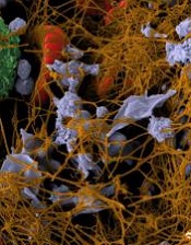
Credit: Andre E.X. Brown
Platelets can sense and respond to their surroundings, according to research published in PNAS.
Researchers reported that platelets can detect mechanical aspects of their environment and transduce those cues into biological signals.
Experiments showed that platelets could sense the stiffness of a fibrin/fibrinogen substrate, and increasing stiffness was associated with increased platelet adhesion, spreading, and activation.
“Platelets are smarter than we give them credit for, in that they are able to sense the physical characteristics of their environment and respond in a graduated way,” said study author Wilbur Lam, MD, PhD, of the Emory University School of Medicine in Atlanta, Georgia.
He and his colleagues were able to separate physical and biochemical effects on platelet behavior by forming polymer gels with different degrees of stiffness and then overlaying each with the same coating of fibrinogen.
With stiffer gels, the researchers observed an increase in platelet adhesion, spreading, and activation. This behavior was most pronounced when the concentration of fibrinogen was relatively low.
“This variability helps to explain platelet behavior in the 3D context of a clot in the body, which can be quite heterogenous in makeup,” Dr Lam said.
The researchers were also able to dissect platelet biochemistry by allowing the platelets to adhere and then spread on the various gels under the influence of drugs that interfere with different biochemical steps.
The team found that integrins, which engage the fibrinogen, and the protein Rac1 are involved in the initial mechanical sensing during adhesion. Myosin and actin, components of the cytoskeleton, are responsible for platelet spreading.
“We found that the initial adhesion and later spreading are separable, because different biochemical pathways are involved in each step,” Dr Lam said. “Our data show that mechanosensing can occur and plays important roles even when the cellular structural building blocks are fairly basic, even when the nucleus is absent.”
The researchers believe these findings could influence the design of medical devices, as modifying the stiffness of materials used in these devices might reduce the formation of blood clots. The results could also guide the refinement of anticoagulant therapy.
Credit: Andre E.X. Brown
Platelets can sense and respond to their surroundings, according to research published in PNAS.
Researchers reported that platelets can detect mechanical aspects of their environment and transduce those cues into biological signals.
Experiments showed that platelets could sense the stiffness of a fibrin/fibrinogen substrate, and increasing stiffness was associated with increased platelet adhesion, spreading, and activation.
“Platelets are smarter than we give them credit for, in that they are able to sense the physical characteristics of their environment and respond in a graduated way,” said study author Wilbur Lam, MD, PhD, of the Emory University School of Medicine in Atlanta, Georgia.
He and his colleagues were able to separate physical and biochemical effects on platelet behavior by forming polymer gels with different degrees of stiffness and then overlaying each with the same coating of fibrinogen.
With stiffer gels, the researchers observed an increase in platelet adhesion, spreading, and activation. This behavior was most pronounced when the concentration of fibrinogen was relatively low.
“This variability helps to explain platelet behavior in the 3D context of a clot in the body, which can be quite heterogenous in makeup,” Dr Lam said.
The researchers were also able to dissect platelet biochemistry by allowing the platelets to adhere and then spread on the various gels under the influence of drugs that interfere with different biochemical steps.
The team found that integrins, which engage the fibrinogen, and the protein Rac1 are involved in the initial mechanical sensing during adhesion. Myosin and actin, components of the cytoskeleton, are responsible for platelet spreading.
“We found that the initial adhesion and later spreading are separable, because different biochemical pathways are involved in each step,” Dr Lam said. “Our data show that mechanosensing can occur and plays important roles even when the cellular structural building blocks are fairly basic, even when the nucleus is absent.”
The researchers believe these findings could influence the design of medical devices, as modifying the stiffness of materials used in these devices might reduce the formation of blood clots. The results could also guide the refinement of anticoagulant therapy.
Credit: Andre E.X. Brown
Platelets can sense and respond to their surroundings, according to research published in PNAS.
Researchers reported that platelets can detect mechanical aspects of their environment and transduce those cues into biological signals.
Experiments showed that platelets could sense the stiffness of a fibrin/fibrinogen substrate, and increasing stiffness was associated with increased platelet adhesion, spreading, and activation.
“Platelets are smarter than we give them credit for, in that they are able to sense the physical characteristics of their environment and respond in a graduated way,” said study author Wilbur Lam, MD, PhD, of the Emory University School of Medicine in Atlanta, Georgia.
He and his colleagues were able to separate physical and biochemical effects on platelet behavior by forming polymer gels with different degrees of stiffness and then overlaying each with the same coating of fibrinogen.
With stiffer gels, the researchers observed an increase in platelet adhesion, spreading, and activation. This behavior was most pronounced when the concentration of fibrinogen was relatively low.
“This variability helps to explain platelet behavior in the 3D context of a clot in the body, which can be quite heterogenous in makeup,” Dr Lam said.
The researchers were also able to dissect platelet biochemistry by allowing the platelets to adhere and then spread on the various gels under the influence of drugs that interfere with different biochemical steps.
The team found that integrins, which engage the fibrinogen, and the protein Rac1 are involved in the initial mechanical sensing during adhesion. Myosin and actin, components of the cytoskeleton, are responsible for platelet spreading.
“We found that the initial adhesion and later spreading are separable, because different biochemical pathways are involved in each step,” Dr Lam said. “Our data show that mechanosensing can occur and plays important roles even when the cellular structural building blocks are fairly basic, even when the nucleus is absent.”
The researchers believe these findings could influence the design of medical devices, as modifying the stiffness of materials used in these devices might reduce the formation of blood clots. The results could also guide the refinement of anticoagulant therapy.