User login
Doug Brunk is a San Diego-based award-winning reporter who began covering health care in 1991. Before joining the company, he wrote for the health sciences division of Columbia University and was an associate editor at Contemporary Long Term Care magazine when it won a Jesse H. Neal Award. His work has been syndicated by the Los Angeles Times and he is the author of two books related to the University of Kentucky Wildcats men's basketball program. Doug has a master’s degree in magazine journalism from the S.I. Newhouse School of Public Communications at Syracuse University. Follow him on Twitter @dougbrunk.
Primary care physicians face vitamin D conundrum
“I don’t know.”
That’s what family physician Michael L. LeFevre tells patients who ask him if they should be screened for vitamin D levels or if they could benefit from vitamin D supplementation.

“We can say that vitamin D is related to health, but we’re really not sure what the optimal blood level is for good health, ” he said at a public conference on vitamin D sponsored by the National Institutes of Health.
“If I have a limited amount of time and a limited capacity to engage in shared decision making with my patients, and my patient is a good candidate for a statin, I’m going to talk about statins long before I’m going to talk about vitamin D. That’s a no-brainer,” explained Dr. LeFevre of the department of family and community medicine at the University of Missouri–Columbia.
He was one of many speakers over the course of the day-and-a-half-long meeting who acknowledged the lack of evidence for prescribing vitamin D for ailments ranging from depression to nonspecific pain.
At this point, there are three decisions about vitamin D that a primary care physician has to make, Dr. LeFevre said.
First, “do I choose to either bring up screening or bring up supplementation for the general population that I’m seeing one on one, in the relatively small window of time I have to address preventive services with my patients?” asked Dr. LeFevre, who also chairs the U.S. Preventive Services Task Force. “Does vitamin D rise to that level?”
Second, “if my patients ask me if they should have their vitamin D level checked or if they should be taking vitamin D supplements, what can I tell them at this point?”
Finally, “when my patients present with nonspecific symptoms such as fatigue, weakness, pain without specific pathology, or a clinical picture such as depression or fibromyalgia, is there a role for me to check their vitamin D level – and then, at a particular threshold, prescribe vitamin D to make them feel better?”
That’s the vitamin D conundrum facing today’s primary care physicians. “I facetiously teach my residents that if something good happens in medicine, take credit for it, even if you had absolutely no influence over the outcome,” Dr. LeFevre said. “If I give somebody vitamin D and they feel better, they think I’m a great doctor, even if that had nothing to do with whether their pain improved.”
For now, unknowns about vitamin D are plentiful, Dr. LeFevre cautioned.
“What’s different about people who have a vitamin D level of 40 ng/mL from those with a vitamin D level of 20 ng/mL?” he asked.
“Is it just what they’re eating? Is it how much time they’re spending in the sun? Are there other things? Can we really be sure that giving them a drug (vitamin D) and getting their level up, either with a fixed-dose approach or a treat-to-target approach [will help]? Do they get better? Do they feel better? Do they avoid feeling worse later on? Do they have fewer fractures? Do they die of fewer heart attacks?” he asked.
“We need those studies to be sure that what we’re doing is good,” Dr. LeFevre noted.
Another speaker, Dr. Bess Dawson-Hughes, called for research efforts to better understand the role of 25-hydroxyvitamin D, and she highlighted the need for standardized vitamin D assays.
“We don’t really know what we can promise or expect from supplements with a lot of the chronic diseases that are being ‘treated’ with vitamin D today,” added Dr. Dawson-Hughes, who directs the bone metabolism laboratory at the Tufts University U.S. Department of Agriculture Human Nutrition Research Center on Aging. “It’s created an expensive and excessive [practice of] measuring and treating.”
Dr. LeFevre and Dr. Dawson-Hughes reported having no relevant financial disclosures.
On Twitter @dougbrunk
“I don’t know.”
That’s what family physician Michael L. LeFevre tells patients who ask him if they should be screened for vitamin D levels or if they could benefit from vitamin D supplementation.
“We can say that vitamin D is related to health, but we’re really not sure what the optimal blood level is for good health, ” he said at a public conference on vitamin D sponsored by the National Institutes of Health.
“If I have a limited amount of time and a limited capacity to engage in shared decision making with my patients, and my patient is a good candidate for a statin, I’m going to talk about statins long before I’m going to talk about vitamin D. That’s a no-brainer,” explained Dr. LeFevre of the department of family and community medicine at the University of Missouri–Columbia.
He was one of many speakers over the course of the day-and-a-half-long meeting who acknowledged the lack of evidence for prescribing vitamin D for ailments ranging from depression to nonspecific pain.
At this point, there are three decisions about vitamin D that a primary care physician has to make, Dr. LeFevre said.
First, “do I choose to either bring up screening or bring up supplementation for the general population that I’m seeing one on one, in the relatively small window of time I have to address preventive services with my patients?” asked Dr. LeFevre, who also chairs the U.S. Preventive Services Task Force. “Does vitamin D rise to that level?”
Second, “if my patients ask me if they should have their vitamin D level checked or if they should be taking vitamin D supplements, what can I tell them at this point?”
Finally, “when my patients present with nonspecific symptoms such as fatigue, weakness, pain without specific pathology, or a clinical picture such as depression or fibromyalgia, is there a role for me to check their vitamin D level – and then, at a particular threshold, prescribe vitamin D to make them feel better?”
That’s the vitamin D conundrum facing today’s primary care physicians. “I facetiously teach my residents that if something good happens in medicine, take credit for it, even if you had absolutely no influence over the outcome,” Dr. LeFevre said. “If I give somebody vitamin D and they feel better, they think I’m a great doctor, even if that had nothing to do with whether their pain improved.”
For now, unknowns about vitamin D are plentiful, Dr. LeFevre cautioned.
“What’s different about people who have a vitamin D level of 40 ng/mL from those with a vitamin D level of 20 ng/mL?” he asked.
“Is it just what they’re eating? Is it how much time they’re spending in the sun? Are there other things? Can we really be sure that giving them a drug (vitamin D) and getting their level up, either with a fixed-dose approach or a treat-to-target approach [will help]? Do they get better? Do they feel better? Do they avoid feeling worse later on? Do they have fewer fractures? Do they die of fewer heart attacks?” he asked.
“We need those studies to be sure that what we’re doing is good,” Dr. LeFevre noted.
Another speaker, Dr. Bess Dawson-Hughes, called for research efforts to better understand the role of 25-hydroxyvitamin D, and she highlighted the need for standardized vitamin D assays.
“We don’t really know what we can promise or expect from supplements with a lot of the chronic diseases that are being ‘treated’ with vitamin D today,” added Dr. Dawson-Hughes, who directs the bone metabolism laboratory at the Tufts University U.S. Department of Agriculture Human Nutrition Research Center on Aging. “It’s created an expensive and excessive [practice of] measuring and treating.”
Dr. LeFevre and Dr. Dawson-Hughes reported having no relevant financial disclosures.
On Twitter @dougbrunk

Autoantibodies not predictive of surgery for Graves’ disease
CORONADO, CALIF.– Measurement of thyroid antibodies had no impact on the clinical features of Graves’ disease best treated with surgery, results from a single-center retrospective study showed.
The findings are part of a larger study meant to develop ways to help patients decide between surgery and radioactive iodine for their Graves’ disease. “We know that certain clinical features make patients lean toward surgery instead of radioactive iodine, including severe ophthalmopathy and young women who have concerns about family planning,” Dr. Dawn M. Elfenbein said in an interview during the annual meeting of the American Thyroid Association. “In the long-term picture, I think that a lot of provider preferences go into this decision, but we’d like to move this to being more of a patient-centered decision. So we’re trying to see if there are biochemical factors about patients that can help us recommend one treatment versus the other.”

Dr. Elfenbein of the section of endocrine surgery in the department of surgery at the University of Wisconsin, Madison, and her associates reviewed the records of 469 patients treated with radioactive iodine (RAI) or surgery for Graves’ disease at the institution during August 2008-September 2013. They excluded patients with contraindications to RAI or those with other indications for surgery or who previously failed either treatment.
Of the 469 patients, 78% were women, and their mean age was 40 years. Most (71%) underwent RAI while 29% underwent surgery. The percentage of patients who opted for surgery increased each year of the study, from 14% in the first year to 52% in the final year.
More than half of the patients (52%) had thyroid peroxidase (TPO) measured prior to treatment, while 44% had thyroglobulin antibodies (TgAb) and 45% had either thyroid simulating immunoglobulin (TSIg) or thyrotropin receptor antibody (TRAb) measured.
Dr. Elfenbein and her associates found that clinical features such as ophthalmopathy or a compressive goiter appeared to influence a patient’s decision to choose surgery over RAI. On bivariate analysis, patients with a positive TSIg or TRAb were significantly more likely to have ophthalmopathy (41% vs. 25%) while patients with positive TgAb were significantly more likely to have goiter (71% vs. 55%). On multivariate analysis, however, antibody positivity was not predictive of ophthalmopathy or goiter.
“Surgery became the primary definitive treatment of choice over RAI for Graves’ disease at our institution over the study period, particularly for younger women,” the researchers wrote in their abstract. The concluded that measurement of thyroid-specific antibodies “does not independently predict those clinical features best treated with surgery. Clinical features and patient preferences should be considered independently from autoantibody levels.”
Dr. Elfenbein acknowledged certain limitations of the study, including its retrospective design and that fact that autoantibody levels were not measured after treatment. “What would be more helpful is to measure antibodies in everyone before they undergo treatment and measure those same antibodies after treatment to see what happens; to see if there are any differences in recurrence rates with higher antibodies,” she said. “We can be more deliberate moving forward.” She reported having no financial disclosures.
On Twitter @dougbrunk
CORONADO, CALIF.– Measurement of thyroid antibodies had no impact on the clinical features of Graves’ disease best treated with surgery, results from a single-center retrospective study showed.
The findings are part of a larger study meant to develop ways to help patients decide between surgery and radioactive iodine for their Graves’ disease. “We know that certain clinical features make patients lean toward surgery instead of radioactive iodine, including severe ophthalmopathy and young women who have concerns about family planning,” Dr. Dawn M. Elfenbein said in an interview during the annual meeting of the American Thyroid Association. “In the long-term picture, I think that a lot of provider preferences go into this decision, but we’d like to move this to being more of a patient-centered decision. So we’re trying to see if there are biochemical factors about patients that can help us recommend one treatment versus the other.”
Dr. Elfenbein of the section of endocrine surgery in the department of surgery at the University of Wisconsin, Madison, and her associates reviewed the records of 469 patients treated with radioactive iodine (RAI) or surgery for Graves’ disease at the institution during August 2008-September 2013. They excluded patients with contraindications to RAI or those with other indications for surgery or who previously failed either treatment.
Of the 469 patients, 78% were women, and their mean age was 40 years. Most (71%) underwent RAI while 29% underwent surgery. The percentage of patients who opted for surgery increased each year of the study, from 14% in the first year to 52% in the final year.
More than half of the patients (52%) had thyroid peroxidase (TPO) measured prior to treatment, while 44% had thyroglobulin antibodies (TgAb) and 45% had either thyroid simulating immunoglobulin (TSIg) or thyrotropin receptor antibody (TRAb) measured.
Dr. Elfenbein and her associates found that clinical features such as ophthalmopathy or a compressive goiter appeared to influence a patient’s decision to choose surgery over RAI. On bivariate analysis, patients with a positive TSIg or TRAb were significantly more likely to have ophthalmopathy (41% vs. 25%) while patients with positive TgAb were significantly more likely to have goiter (71% vs. 55%). On multivariate analysis, however, antibody positivity was not predictive of ophthalmopathy or goiter.
“Surgery became the primary definitive treatment of choice over RAI for Graves’ disease at our institution over the study period, particularly for younger women,” the researchers wrote in their abstract. The concluded that measurement of thyroid-specific antibodies “does not independently predict those clinical features best treated with surgery. Clinical features and patient preferences should be considered independently from autoantibody levels.”
Dr. Elfenbein acknowledged certain limitations of the study, including its retrospective design and that fact that autoantibody levels were not measured after treatment. “What would be more helpful is to measure antibodies in everyone before they undergo treatment and measure those same antibodies after treatment to see what happens; to see if there are any differences in recurrence rates with higher antibodies,” she said. “We can be more deliberate moving forward.” She reported having no financial disclosures.
On Twitter @dougbrunk
AT THE ATA ANNUAL MEETING
Key clinical point: Measuring thyroid antibodies adds no value in predicting clinical features of Graves’ disease best treated with surgery.
Major finding: On bivariate analysis, patients with a positive TSIg or TRAb were more likely to have ophthalmopathy (41% vs. 25%; P = .04) while patients with positive TgAb were more likely to have goiter (71% vs. 55%; P = .03). On multivariate analysis, however, antibody positivity was not predictive of ophthalmopathy or goiter.
Data source: A retrospective analysis of 469 patients treated with radioactive iodine (RAI) or surgery for Graves’ disease at the University of Wisconsin, Madison, during August 2008-September 2013.
Disclosures:Dr. Elfenbein reported having no financial disclosures.
Vitamin D landscape marked by lack of consensus
If you’re stumped about what to tell patients who ask you if they should be adding supplemental vitamin D to their diet, you’re not alone.
Speaker after speaker at a public conference on vitamin D sponsored by the National Institutes of Health acknowledged that there is general disagreement among well-respected scientists and medical organizations not only about recommended intakes, but about whether supplementation of vitamin D (25-hydroxyvitamin D) has any impact on ailments ranging from depression and nonspecific pain to hypertension and fall prevention.
“Most people agree that at least in high-risk individuals with osteoporosis, vitamin D has an impact on bone and skeletal health, but maybe not in those who are asymptomatic and in healthy individuals as a preventive tool,” said Dr. Clifford J. Rosen, director of clinical and translational research and a senior scientist at Maine Medical Center Research Institute, Scarborough, Me. “There seems to be growing evidence that in high-risk individuals, or in those who repeatedly fall, vitamin D may have an impact, particularly in those with very-low levels of 25-D.”
Other relationships lack conclusive randomized control data, although there are strong observational data for vitamin D’s role in preventing type 2 diabetes. Dr. Rosen is one of the investigators in a National Institute of Diabetes and Digestive and Kidney Diseases–funded clinical trial known as D2D: a study of 4,000 IU of vitamin D vs. placebo in high-risk individuals with obesity and prediabetes. The primary outcome is time to onset of type 2 diabetes. “Currently, that [trial is] in its second year and is about 30% recruited,” said Dr. Rosen, who is also a member of the FDA Advisory Panel on Endocrinologic and Metabolic Drugs. “One of the biggest obstacles to recruitment has been the constant use of vitamin D by people being screened. [They say] ‘Why should I go into a clinical trial when I’m taking vitamin D, and my doctor tells me that it will prevent diabetes?’”
The potential benefit of vitamin D intake on reducing the risk for developing cardiovascular disease, cancer, and stroke is being investigated in the NIH-funded VITAL trial. Clinicians involved in this project have enrolled more than 28,000 men and women with no prior history of these illnesses, investigating the impact of taking vitamin D3 supplements (2,000 IU/day) or omega 3 fatty acids (1 G/day).
In the meantime, current vitamin D guidance and conclusions differ among leading medical organizations. For example, the American Geriatrics Society (AGS) recommends a daily dose of 4,000 IU for fall prevention in elderly individuals. This differs from the daily dose for adults recommended by the Endocrine Society (1,500-2,000 IU), Institute of Medicine (an average requirement of 400 IU and 600-800 IU meeting the greatest need), the United States Preventive Services Task Force (600-800 IU as a fall-prevention strategy), the Standing Committee of European Doctors (600-800 IU), and the National Osteoporosis Foundation (400-1,000 IU). “How do we reconcile vitamin D intake with vitamin D levels?” asked Dr. Rosen, who is also a professor of medicine at Tufts University. “This is one of the hallmarks of the questions or problems we have, or the lack of consistency of data. We know that intakes do not reflect serum 25-D levels to a great extent.”
In addition, the terminology for serum 25-D is not clear. “Is it a deficiency? Is it a disease? What does that mean?” he asked. “More importantly, we don’t really understand what vitamin D insufficiency is. Is it a disease? Not a disease? Is it inadequate intake?”
The definition of optimal 25-D is also a matter of debate, he continued. “What’s the upper level? What does pharmacological treatment mean with respect to long-term outcomes. What is the tolerable upper limit? What is the potentially toxic limit?”
A lack of consensus also exists regarding one’s risk of vitamin D deficiency. For example, the AGS puts this risk at less than 30 ng/mL, the Endocrine Society at less than 20 ng/mL, and the Institute of Medicine at less than 12 ng/mL. “We have a lot of inconsistency in the data,” Dr. Rosen concluded. “There’s not unanimity in recommendations, even among so-called experts.”

During the same session, Dr. Peter Millard presented findings from a national analysis of vitamin D level testing in adult patients conducted from January 2013 to September 2014. The sample, drawn from Athenahealth integrated electronic health records (EHRs), included more than 6,000 internists and family physicians and 2,000 nonphysician clinicians, translating into an estimated 900,000 patient encounters per month. During that time period 4%-5% of all adult patient encounters were associated with a vitamin D test ordered.
“Curiously, the Sunbelt states of Arizona and Nevada have a high testing rate, in the 8.5%-10.7% range,” said Dr. Millard, a family physician who practices at Seaport Community Health Center in Belfast, Me. “Perhaps it’s because of snowbirds coming down from Canada to get tested. I don’t know. There are certainly lots of retirees in those two states. There are also high levels of testing in Illinois, Maryland, Rhode Island, and Delaware. I don’t have a hypothesis as to why there are variations, but that’s about 10% of all encounters associated with a vitamin D test in those states, which seems like quite a high number.”
The greatest proportion of tests occurred in patients over the age of 65 (39%) years, and about 70% of all vitamin D tests were conducted in females.
Fewer than 0.1% of tests were associated with a diagnosis of osteoporosis. The most common diagnoses associated with ordering of a vitamin D test were depression and falls. “This was particularly true in the elderly group, where falls became much more important, and depression slightly less,” Dr. Millard said. He noted that the EHR findings “don’t answer many questions but clearly the cost for [vitamin D] testing itself is significant. The amount of time my colleagues are spending doing tests and interpreting tests for patients [and] deciding what to do about those results is very considerable. In the long run, the research to resolve these issues may not be anywhere near as expensive as continuing to do what we’re doing now.”
Dr. Rosen and Dr. Millard reporting having no financial disclosures.
On Twitter @dougbrunk
If you’re stumped about what to tell patients who ask you if they should be adding supplemental vitamin D to their diet, you’re not alone.
Speaker after speaker at a public conference on vitamin D sponsored by the National Institutes of Health acknowledged that there is general disagreement among well-respected scientists and medical organizations not only about recommended intakes, but about whether supplementation of vitamin D (25-hydroxyvitamin D) has any impact on ailments ranging from depression and nonspecific pain to hypertension and fall prevention.
“Most people agree that at least in high-risk individuals with osteoporosis, vitamin D has an impact on bone and skeletal health, but maybe not in those who are asymptomatic and in healthy individuals as a preventive tool,” said Dr. Clifford J. Rosen, director of clinical and translational research and a senior scientist at Maine Medical Center Research Institute, Scarborough, Me. “There seems to be growing evidence that in high-risk individuals, or in those who repeatedly fall, vitamin D may have an impact, particularly in those with very-low levels of 25-D.”
Other relationships lack conclusive randomized control data, although there are strong observational data for vitamin D’s role in preventing type 2 diabetes. Dr. Rosen is one of the investigators in a National Institute of Diabetes and Digestive and Kidney Diseases–funded clinical trial known as D2D: a study of 4,000 IU of vitamin D vs. placebo in high-risk individuals with obesity and prediabetes. The primary outcome is time to onset of type 2 diabetes. “Currently, that [trial is] in its second year and is about 30% recruited,” said Dr. Rosen, who is also a member of the FDA Advisory Panel on Endocrinologic and Metabolic Drugs. “One of the biggest obstacles to recruitment has been the constant use of vitamin D by people being screened. [They say] ‘Why should I go into a clinical trial when I’m taking vitamin D, and my doctor tells me that it will prevent diabetes?’”
The potential benefit of vitamin D intake on reducing the risk for developing cardiovascular disease, cancer, and stroke is being investigated in the NIH-funded VITAL trial. Clinicians involved in this project have enrolled more than 28,000 men and women with no prior history of these illnesses, investigating the impact of taking vitamin D3 supplements (2,000 IU/day) or omega 3 fatty acids (1 G/day).
In the meantime, current vitamin D guidance and conclusions differ among leading medical organizations. For example, the American Geriatrics Society (AGS) recommends a daily dose of 4,000 IU for fall prevention in elderly individuals. This differs from the daily dose for adults recommended by the Endocrine Society (1,500-2,000 IU), Institute of Medicine (an average requirement of 400 IU and 600-800 IU meeting the greatest need), the United States Preventive Services Task Force (600-800 IU as a fall-prevention strategy), the Standing Committee of European Doctors (600-800 IU), and the National Osteoporosis Foundation (400-1,000 IU). “How do we reconcile vitamin D intake with vitamin D levels?” asked Dr. Rosen, who is also a professor of medicine at Tufts University. “This is one of the hallmarks of the questions or problems we have, or the lack of consistency of data. We know that intakes do not reflect serum 25-D levels to a great extent.”
In addition, the terminology for serum 25-D is not clear. “Is it a deficiency? Is it a disease? What does that mean?” he asked. “More importantly, we don’t really understand what vitamin D insufficiency is. Is it a disease? Not a disease? Is it inadequate intake?”
The definition of optimal 25-D is also a matter of debate, he continued. “What’s the upper level? What does pharmacological treatment mean with respect to long-term outcomes. What is the tolerable upper limit? What is the potentially toxic limit?”
A lack of consensus also exists regarding one’s risk of vitamin D deficiency. For example, the AGS puts this risk at less than 30 ng/mL, the Endocrine Society at less than 20 ng/mL, and the Institute of Medicine at less than 12 ng/mL. “We have a lot of inconsistency in the data,” Dr. Rosen concluded. “There’s not unanimity in recommendations, even among so-called experts.”
During the same session, Dr. Peter Millard presented findings from a national analysis of vitamin D level testing in adult patients conducted from January 2013 to September 2014. The sample, drawn from Athenahealth integrated electronic health records (EHRs), included more than 6,000 internists and family physicians and 2,000 nonphysician clinicians, translating into an estimated 900,000 patient encounters per month. During that time period 4%-5% of all adult patient encounters were associated with a vitamin D test ordered.
“Curiously, the Sunbelt states of Arizona and Nevada have a high testing rate, in the 8.5%-10.7% range,” said Dr. Millard, a family physician who practices at Seaport Community Health Center in Belfast, Me. “Perhaps it’s because of snowbirds coming down from Canada to get tested. I don’t know. There are certainly lots of retirees in those two states. There are also high levels of testing in Illinois, Maryland, Rhode Island, and Delaware. I don’t have a hypothesis as to why there are variations, but that’s about 10% of all encounters associated with a vitamin D test in those states, which seems like quite a high number.”
The greatest proportion of tests occurred in patients over the age of 65 (39%) years, and about 70% of all vitamin D tests were conducted in females.
Fewer than 0.1% of tests were associated with a diagnosis of osteoporosis. The most common diagnoses associated with ordering of a vitamin D test were depression and falls. “This was particularly true in the elderly group, where falls became much more important, and depression slightly less,” Dr. Millard said. He noted that the EHR findings “don’t answer many questions but clearly the cost for [vitamin D] testing itself is significant. The amount of time my colleagues are spending doing tests and interpreting tests for patients [and] deciding what to do about those results is very considerable. In the long run, the research to resolve these issues may not be anywhere near as expensive as continuing to do what we’re doing now.”
Dr. Rosen and Dr. Millard reporting having no financial disclosures.
On Twitter @dougbrunk

Ultrasound bests elastography for specificity of thyroid cancer diagnosis
CORONADO, CALIF.– Compared with elastography, ultrasound predictors of malignancy were more specific for presurgical diagnosis and in differentiating between benign and malignant thyroid nodules, results from a pooled analysis showed.
“Elastography is controversial,” Dr. Parisha Bhatia said in an interview during the annual meeting of the American Thyroid Association. “Some studies have reported that it has better sensitivity and specificity, compared with conventional ultrasound, but others have found it not to be helpful.”

In an effort to compare the efficacy of elastography and ultrasound in determining benign and malignant thyroid nodules, and to determine if elastography has a complementary role to fine needle aspiration (FNA), Dr. Bhatia and her associates searched Embase and PubMed databases for articles involving more than 50 nodules using specimen histology as the reference standard. They discovered 14 prospective studies and organized them into one of two groups. Group 1 included nodules with FNA cytology–proven “benign/malignant” result. Group 2 included nodules with FNA cytology as “intermediate.” The elasticity score was compared with ultrasound features such as taller than wide, irregular margins, internal vascularity, calcification, and absence of halo to determine validity measures and likelihood ratios.
Dr. Bhatia, an endocrine surgeon at Tulane University, New Orleans, reported on findings from 2,732 nodules in the pooled analysis. Of these, 64% were benign, 25% were malignant, and 11% were indeterminate.
In group 1, elastography showed a sensitivity of 75%, a specificity of 80%, a positive predictive value (PPV) of 61%, a negative predictive value (NPV) of 89%, and a likelihood ratio of 5.7, with a higher predictive value for nodules smaller than 1 cm in diameter (PPV of 79% and NPV of 83%). The strongest ultrasound-related predictor of malignancies was “taller than wide” (a specificity of 92%, a NPV of 51%, yet a sensitivity of only 24%), followed by irregular margins (a specificity of 91%, sensitivity of 48%, and a PPV of 64%). Combination of elastography and ultrasound had the highest sensitivity (96%) and NPV (96%), yet lower specificity (46%) and PPV (46%).
In group 2, elastography yielded a higher sensitivity (90%) and NPV (92%), while ultrasound features were highly specific, with the highest values for “taller than wide” shape (85%) in these thyroid nodules.
“Ultrasound predictors of malignancy prove to be more specific for presurgical diagnosis and differentiation of benign and malignant thyroid nodules,” the researchers wrote in their abstract. “When used as an adjunct, the higher sensitivity and NPV of combination of both the techniques can lead to better selection of candidates for FNA.”
Dr. Bhatia acknowledged certain limitations of the study, including the fact that the pooled data contained little information on indeterminate thyroid nodules. She reported having no financial disclosures.
On Twitter @dougbrunk
CORONADO, CALIF.– Compared with elastography, ultrasound predictors of malignancy were more specific for presurgical diagnosis and in differentiating between benign and malignant thyroid nodules, results from a pooled analysis showed.
“Elastography is controversial,” Dr. Parisha Bhatia said in an interview during the annual meeting of the American Thyroid Association. “Some studies have reported that it has better sensitivity and specificity, compared with conventional ultrasound, but others have found it not to be helpful.”
In an effort to compare the efficacy of elastography and ultrasound in determining benign and malignant thyroid nodules, and to determine if elastography has a complementary role to fine needle aspiration (FNA), Dr. Bhatia and her associates searched Embase and PubMed databases for articles involving more than 50 nodules using specimen histology as the reference standard. They discovered 14 prospective studies and organized them into one of two groups. Group 1 included nodules with FNA cytology–proven “benign/malignant” result. Group 2 included nodules with FNA cytology as “intermediate.” The elasticity score was compared with ultrasound features such as taller than wide, irregular margins, internal vascularity, calcification, and absence of halo to determine validity measures and likelihood ratios.
Dr. Bhatia, an endocrine surgeon at Tulane University, New Orleans, reported on findings from 2,732 nodules in the pooled analysis. Of these, 64% were benign, 25% were malignant, and 11% were indeterminate.
In group 1, elastography showed a sensitivity of 75%, a specificity of 80%, a positive predictive value (PPV) of 61%, a negative predictive value (NPV) of 89%, and a likelihood ratio of 5.7, with a higher predictive value for nodules smaller than 1 cm in diameter (PPV of 79% and NPV of 83%). The strongest ultrasound-related predictor of malignancies was “taller than wide” (a specificity of 92%, a NPV of 51%, yet a sensitivity of only 24%), followed by irregular margins (a specificity of 91%, sensitivity of 48%, and a PPV of 64%). Combination of elastography and ultrasound had the highest sensitivity (96%) and NPV (96%), yet lower specificity (46%) and PPV (46%).
In group 2, elastography yielded a higher sensitivity (90%) and NPV (92%), while ultrasound features were highly specific, with the highest values for “taller than wide” shape (85%) in these thyroid nodules.
“Ultrasound predictors of malignancy prove to be more specific for presurgical diagnosis and differentiation of benign and malignant thyroid nodules,” the researchers wrote in their abstract. “When used as an adjunct, the higher sensitivity and NPV of combination of both the techniques can lead to better selection of candidates for FNA.”
Dr. Bhatia acknowledged certain limitations of the study, including the fact that the pooled data contained little information on indeterminate thyroid nodules. She reported having no financial disclosures.
On Twitter @dougbrunk
AT THE ATA ANNUAL MEETING
Key clinical point: Ultrasound is more specific than elastography in helping clinicians make a presurgical diagnosis of thyroid cancer.
Major finding: Compared with elastography, ultrasound was more specific in presurgical diagnosis and in differentiating between benign and malignant thyroid nodules (specificity of 92% vs. 80%, respectively).
Data source: A pooled analysis of 14 prospective studies involving findings from 2,732 thyroid nodules.
Disclosures: Dr. Bhatia reported having no financial disclosures.

Expert dispels common strength-training myths
LAS VEGAS – In the opinion of Dr. Sally S. Harris, there are at least three myths associated with strength training by children and adolescents. One is that the practice is dangerous to the immature skeleton.
In fact, strength training is no riskier for injuries than are contact sports, such as soccer, football, and basketball, Dr. Harris said at a pediatric update sponsored by the American Academy of Pediatrics California District 9.
“There’s a theoretical concern than heavy weights are going to injure open growth plates,” she said. “That’s why one of the main restrictions is that kids do sets of multiple repetitions of weights 10-12 times as opposed to maximal lifts – something that you can lift only once. Proper equipment, supervision, and design are important to prevent injury.”

A second myth is that strength training “will somehow bulk you up and you’ll lose flexibility, which is primarily a concern of coaches,” said Dr. Harris, who practices in the departments of sports medicine and pediatrics at Palo Alto (Calif.) Medical Foundation. “This is not true; flexibility is actually improved if you incorporate stretching with a strength-training program.”
A third myth is that no improvement in strength can occur until adolescence. This is false, Dr. Harris said, referring to published studies demonstrating that strength gains of 30%-50% can occur in preadolescence. “Prepubescent children make the same relative strength gains as teenagers and adults do, it’s just that the absolute strength gains are less, because they’re starting with less muscle,” she explained.
General guidelines for weight training prior to skeletal maturity include no one-repetition maximal lifts because of the theoretical risk of overloading growth plates, and no ballistic maneuvers or Olympic-style lifts. “These are the jerky, bouncy things like dead lifts – competitive weight-training maneuvers,” she said. “It’s not recommended that children do competitive strength training although there are young body builders, and there don’t seem to be any medical issues with that.”
Dr. Harris, who founded the AAP section on sports medicine, listed several potential benefits of strength training, including improved self-esteem, cardiovascular fitness, increased bone density, improved lipid profiles, increased lean body mass, possible improved sports performance, and possible injury prevention. However, whether strength training directly translates to improved sports performance is a matter of debate.
“There’s no compelling evidence that’s shown that general sports performance is enhanced, but we do know that if you strengthen the quadriceps, for example, you’ll improve your vertical jump,” Dr. Harris said. “You would think that might correlate to sports that involve jumping, but it’s hard to demonstrate that it enhances sports performance itself.”
According the guidelines from the AAP (Pediatrics 2008;121:835-40), proper strength training involves six to eight exercises, including core musculature and all major muscle groups, done in sets of multiple repetitions, increasing resistance in increments of 5%-10%. The recommended frequency is two to three times per week for 20- to 30-minute sessions over a course of 8 weeks, with 10-minute warm-up and cool-down periods.
Common sense guidelines include the use of proper techniques such as a straight back and slightly flexed extremities, and a spotter for heavy lifts or free weights. “Most of the serious weight-training injuries have occurred in the home setting with kids dropping barbells on their chest using their parents’ equipment,” Dr. Harris. “That’s preventable.” She went on to note that strength-training exercises should be performed in slow, controlled motions in shoes with good traction. Participants should avoid hyperventilation, Valsalva maneuvers, and the use of anabolic steroids or hormone precursors.
Dr. Harris said that parents often ask her if it’s safe for their adolescent to use creatine, a source of protein energy containing glycine, arginine, and methionine. Theoretically, the more creatine available for immediate use in muscles, the more peak power produced, “so there’s potential benefit for short bursts of exercise lasting 30 seconds or less,” she said. “You spare yourself a lactate-generating mechanism, and you have an increase in lean tissue, which allows you to train longer and more intensely.”
Humans produce creatine naturally via the liver and kidneys, but the idea is that if you supply your body with extra creatine, “you’re going to reap benefits that creatine has on generating adenosine triphosphate (ATP),” she said. The creatine “donates its phosphors to adenosine diphosphate (ADP) to make ATP, which is what muscles use for energy. The ATP in a muscle is used for the first 30 seconds of energy, so it’s beneficial for things that last less than 30 seconds like a sprint, a power lift, or a tackle. It’s not helpful for endurance activities.”
Creatine is available in many forms, including as a pill, a chew, a powder, and as an ingredient in energy bars, sports drinks, and chewing gum. “The cellular uptake is enhanced if it’s in a liquid form with glucose in combination,” Dr. Harris said. “Uptake is decreased by caffeine.”
A loading dose of 5 g every 6 hours for 5 days is recommended. This increases creatine stores in muscles by 15%-30% and remains elevated for 2 weeks to 2 months. “That sounds impressive,” Dr. Harris said. “The problem is, most people don’t do a loading dose. There are a lot of GI side effects associated with that, so they just skip to the maintenance dose of 3-6 g per day. But slowly over time creatine decreases despite the supplementation, so you need to have times off, which is why you cycle. You go on for 5-8 weeks and off the 2-4 weeks.”
According to limited surveys of creatine use by high schoolers, 55% don’t know the dose they’re taking, and 23% report taking doses higher than recommended. Moreover, 13%-30% of people who take creatine are nonresponders, “so a lot of people are taking this and it’s no benefit to them,” Dr. Harris said.
Side effects include weight gain from water retention, anecdotal reports of muscle cramps, stiffness, muscle tension injury, dehydration, and heat illness. “The biggest concern is renal function, but we don’t know the long-term effects,” she said. “There are no effects on blood pressure, liver enzymes, electrolytes, uric acid, hematologic parameters, muscle enzymes, and lipid profiles. That’s reassuring.”
Even so, the use of creatine by adolescents hasn’t been formally studied, and its effects on the brain, cardiac muscle, testes, and other creatine-containing tissues is unknown, “so most medical organizations recommend against it, including the AAP and the American College of Sports Medicine,” Dr. Harris said.
Another concern about creatine is that adolescents “may assume that supplements can substitute for proper nutrition or good athletic training,” Dr. Harris said. “Some feel that it’s a slippery slope to the use of steroids and other harmful and banned substances. On the other hand, it’s not banned by any group and it’s not drug- tested because it’s a source of energy in the diet. It’s not an anabolic agent. Some liken it to caffeine; it’s part of the diet so only high levels should be banned.”
Dr. Harris reported having no financial disclosures.
On Twitter @dougbrunk
LAS VEGAS – In the opinion of Dr. Sally S. Harris, there are at least three myths associated with strength training by children and adolescents. One is that the practice is dangerous to the immature skeleton.
In fact, strength training is no riskier for injuries than are contact sports, such as soccer, football, and basketball, Dr. Harris said at a pediatric update sponsored by the American Academy of Pediatrics California District 9.
“There’s a theoretical concern than heavy weights are going to injure open growth plates,” she said. “That’s why one of the main restrictions is that kids do sets of multiple repetitions of weights 10-12 times as opposed to maximal lifts – something that you can lift only once. Proper equipment, supervision, and design are important to prevent injury.”
A second myth is that strength training “will somehow bulk you up and you’ll lose flexibility, which is primarily a concern of coaches,” said Dr. Harris, who practices in the departments of sports medicine and pediatrics at Palo Alto (Calif.) Medical Foundation. “This is not true; flexibility is actually improved if you incorporate stretching with a strength-training program.”
A third myth is that no improvement in strength can occur until adolescence. This is false, Dr. Harris said, referring to published studies demonstrating that strength gains of 30%-50% can occur in preadolescence. “Prepubescent children make the same relative strength gains as teenagers and adults do, it’s just that the absolute strength gains are less, because they’re starting with less muscle,” she explained.
General guidelines for weight training prior to skeletal maturity include no one-repetition maximal lifts because of the theoretical risk of overloading growth plates, and no ballistic maneuvers or Olympic-style lifts. “These are the jerky, bouncy things like dead lifts – competitive weight-training maneuvers,” she said. “It’s not recommended that children do competitive strength training although there are young body builders, and there don’t seem to be any medical issues with that.”
Dr. Harris, who founded the AAP section on sports medicine, listed several potential benefits of strength training, including improved self-esteem, cardiovascular fitness, increased bone density, improved lipid profiles, increased lean body mass, possible improved sports performance, and possible injury prevention. However, whether strength training directly translates to improved sports performance is a matter of debate.
“There’s no compelling evidence that’s shown that general sports performance is enhanced, but we do know that if you strengthen the quadriceps, for example, you’ll improve your vertical jump,” Dr. Harris said. “You would think that might correlate to sports that involve jumping, but it’s hard to demonstrate that it enhances sports performance itself.”
According the guidelines from the AAP (Pediatrics 2008;121:835-40), proper strength training involves six to eight exercises, including core musculature and all major muscle groups, done in sets of multiple repetitions, increasing resistance in increments of 5%-10%. The recommended frequency is two to three times per week for 20- to 30-minute sessions over a course of 8 weeks, with 10-minute warm-up and cool-down periods.
Common sense guidelines include the use of proper techniques such as a straight back and slightly flexed extremities, and a spotter for heavy lifts or free weights. “Most of the serious weight-training injuries have occurred in the home setting with kids dropping barbells on their chest using their parents’ equipment,” Dr. Harris. “That’s preventable.” She went on to note that strength-training exercises should be performed in slow, controlled motions in shoes with good traction. Participants should avoid hyperventilation, Valsalva maneuvers, and the use of anabolic steroids or hormone precursors.
Dr. Harris said that parents often ask her if it’s safe for their adolescent to use creatine, a source of protein energy containing glycine, arginine, and methionine. Theoretically, the more creatine available for immediate use in muscles, the more peak power produced, “so there’s potential benefit for short bursts of exercise lasting 30 seconds or less,” she said. “You spare yourself a lactate-generating mechanism, and you have an increase in lean tissue, which allows you to train longer and more intensely.”
Humans produce creatine naturally via the liver and kidneys, but the idea is that if you supply your body with extra creatine, “you’re going to reap benefits that creatine has on generating adenosine triphosphate (ATP),” she said. The creatine “donates its phosphors to adenosine diphosphate (ADP) to make ATP, which is what muscles use for energy. The ATP in a muscle is used for the first 30 seconds of energy, so it’s beneficial for things that last less than 30 seconds like a sprint, a power lift, or a tackle. It’s not helpful for endurance activities.”
Creatine is available in many forms, including as a pill, a chew, a powder, and as an ingredient in energy bars, sports drinks, and chewing gum. “The cellular uptake is enhanced if it’s in a liquid form with glucose in combination,” Dr. Harris said. “Uptake is decreased by caffeine.”
A loading dose of 5 g every 6 hours for 5 days is recommended. This increases creatine stores in muscles by 15%-30% and remains elevated for 2 weeks to 2 months. “That sounds impressive,” Dr. Harris said. “The problem is, most people don’t do a loading dose. There are a lot of GI side effects associated with that, so they just skip to the maintenance dose of 3-6 g per day. But slowly over time creatine decreases despite the supplementation, so you need to have times off, which is why you cycle. You go on for 5-8 weeks and off the 2-4 weeks.”
According to limited surveys of creatine use by high schoolers, 55% don’t know the dose they’re taking, and 23% report taking doses higher than recommended. Moreover, 13%-30% of people who take creatine are nonresponders, “so a lot of people are taking this and it’s no benefit to them,” Dr. Harris said.
Side effects include weight gain from water retention, anecdotal reports of muscle cramps, stiffness, muscle tension injury, dehydration, and heat illness. “The biggest concern is renal function, but we don’t know the long-term effects,” she said. “There are no effects on blood pressure, liver enzymes, electrolytes, uric acid, hematologic parameters, muscle enzymes, and lipid profiles. That’s reassuring.”
Even so, the use of creatine by adolescents hasn’t been formally studied, and its effects on the brain, cardiac muscle, testes, and other creatine-containing tissues is unknown, “so most medical organizations recommend against it, including the AAP and the American College of Sports Medicine,” Dr. Harris said.
Another concern about creatine is that adolescents “may assume that supplements can substitute for proper nutrition or good athletic training,” Dr. Harris said. “Some feel that it’s a slippery slope to the use of steroids and other harmful and banned substances. On the other hand, it’s not banned by any group and it’s not drug- tested because it’s a source of energy in the diet. It’s not an anabolic agent. Some liken it to caffeine; it’s part of the diet so only high levels should be banned.”
Dr. Harris reported having no financial disclosures.
On Twitter @dougbrunk
EXPERT ANALYSIS AT AAP PEDIATRIC UPDATE

Managing molluscum contagiosum: ‘The great imitator’
LAS VEGAS – Although molluscum contagiosum is harmless, it can be confused with warts or lesions commonly related to herpes and various types of acne.
“Molluscum contagiosum really is one of the great imitators,” Dr. James Treat said at a pediatric update sponsored by the American Academy of Pediatrics California District 9. “They can be great big cysts, and they can be tiny ditzels; you can barely tell they’re there. They also can be more classic, umbilicated papules.”

Clinicians can make a quick diagnosis by angling an otoscope to the side of the lesion, said Dr. Treat. Once illuminated, “You’ll see this tiny white spicule at the center of the lesion. That’s a great way of diagnosing it.”
Children and adolescents often present with inflamed, painful lesions, prompting concern from parents. In fact, Dr. Treat identified a recent article naming inflamed molluscum lesions as the BOTE sign: the beginning of the end (Pediatrics 2013; 131:e1650-3). “If your spots are getting red, your body knows the virus is there,” he said. “You don’t have to do anything else about it. They’re almost never infected. It’s just like an ingrown hair. It can look like a pus bump; you get inflammation; it’s a little bit painful. They’re usually just inflamed. If you think someone truly has cellulitis, of course, give them antibiotics. But the majority [of lesions] are not infected.”
When in doubt, culture the lesion. “If you grow streptococcus or staphylococcus, then you know you need to treat them,” he said. “If you grow normal skin flora, though, don’t be surprised. That’s what’s most commonly going to happen. It’s similar to acne or an ingrown hair.”
The time course for complete clearance of molluscum is usually 1-2 years. “It’s probably a bit shorter than that, but it’s better to underpromise,” said Dr. Treat, a pediatric dermatologist at Children’s Hospital of Philadelphia.
No Food and Drug Administration–approved treatment options for molluscum exist, Dr. Treat noted. For lesions under the armpit, for example, he often recommends treating associated dermatitis with hydrocortisone and applying moisturizer to the lesions until they resolve, he said. However, most parents want other treatment options, especially if their child competes in wrestling or other contact sports. His preferred treatment is cantharone, also known as “beetle juice.” Clinicians apply the liquid cantharone solution directly to the lesions in the office, and instruct the parents to help the child wash off the solution after 2 hours. The treatment causes blistering and ultimate breakdown of the molluscum.
“Some centers are not allowing clinicians to use cantharone, because it never went through FDA approval,” Dr. Treat said. “I never use it on the face, around the eye, or inside the diaper area. I put a tiny drop on, and instruct them to wash it off after 2 hours.”
Cryotherapy is another option, though Dr. Treat advised caution. “I think you can do it if you have a motivated teenager who wants their five spots gone very quickly, or if you have a young child that has a couple of spots on the cheek and you can hold them very still and freeze very lightly to get those spots to go away,” he said. “I would not perform cryotherapy around the eye. You have to be careful with cryotherapy because it hurts, and you can cause more inflammation than you really expected, or a blister, for something that’s benign and is going to go away on its own. But there’s definitely a role for cryotherapy for a kid who needs to wrestle very soon or get back to sports, or has lesions in areas where you can’t control the cantharone.”
Curettage also may be used, Dr. Treat said, although “it’s not something we commonly do unless you have a very motivated 10-, 12-, or 14-year-old who can stay still, or unless you have a child that has one or two spots located in a difficult area to treat otherwise,” he noted. Topical options include tretinoin. “I don’t think it works very well, but it’s something to do, and it’s been used on the faces of children,” said Dr. Treat. He recommended using a Q-tip swab to apply a pinhead-sized amount of tretinoin to individual lesions. “Try not to treat the skin around it,” he advised. “Your goal is irritation of the lesion; you are trying to irritate the skin to get the immune system to notice the molluscum.” He characterized sinecatechins as “expensive and often irritating” and considers oral cimetidine as a “last-ditch effort” for patients with molluscum lesions all over the body. “It works 20-30% of the time,” he said. “You have to take it two to three times a day, and you have to take it at the high end of the normal dosing range. But there are some data that show that in patients with bad atopic dermatitis who have terrible molluscum, it might work.”
Dr. Treat reported having no relevant financial disclosures.
On Twitter @dougbrunk
LAS VEGAS – Although molluscum contagiosum is harmless, it can be confused with warts or lesions commonly related to herpes and various types of acne.
“Molluscum contagiosum really is one of the great imitators,” Dr. James Treat said at a pediatric update sponsored by the American Academy of Pediatrics California District 9. “They can be great big cysts, and they can be tiny ditzels; you can barely tell they’re there. They also can be more classic, umbilicated papules.”
Clinicians can make a quick diagnosis by angling an otoscope to the side of the lesion, said Dr. Treat. Once illuminated, “You’ll see this tiny white spicule at the center of the lesion. That’s a great way of diagnosing it.”
Children and adolescents often present with inflamed, painful lesions, prompting concern from parents. In fact, Dr. Treat identified a recent article naming inflamed molluscum lesions as the BOTE sign: the beginning of the end (Pediatrics 2013; 131:e1650-3). “If your spots are getting red, your body knows the virus is there,” he said. “You don’t have to do anything else about it. They’re almost never infected. It’s just like an ingrown hair. It can look like a pus bump; you get inflammation; it’s a little bit painful. They’re usually just inflamed. If you think someone truly has cellulitis, of course, give them antibiotics. But the majority [of lesions] are not infected.”
When in doubt, culture the lesion. “If you grow streptococcus or staphylococcus, then you know you need to treat them,” he said. “If you grow normal skin flora, though, don’t be surprised. That’s what’s most commonly going to happen. It’s similar to acne or an ingrown hair.”
The time course for complete clearance of molluscum is usually 1-2 years. “It’s probably a bit shorter than that, but it’s better to underpromise,” said Dr. Treat, a pediatric dermatologist at Children’s Hospital of Philadelphia.
No Food and Drug Administration–approved treatment options for molluscum exist, Dr. Treat noted. For lesions under the armpit, for example, he often recommends treating associated dermatitis with hydrocortisone and applying moisturizer to the lesions until they resolve, he said. However, most parents want other treatment options, especially if their child competes in wrestling or other contact sports. His preferred treatment is cantharone, also known as “beetle juice.” Clinicians apply the liquid cantharone solution directly to the lesions in the office, and instruct the parents to help the child wash off the solution after 2 hours. The treatment causes blistering and ultimate breakdown of the molluscum.
“Some centers are not allowing clinicians to use cantharone, because it never went through FDA approval,” Dr. Treat said. “I never use it on the face, around the eye, or inside the diaper area. I put a tiny drop on, and instruct them to wash it off after 2 hours.”
Cryotherapy is another option, though Dr. Treat advised caution. “I think you can do it if you have a motivated teenager who wants their five spots gone very quickly, or if you have a young child that has a couple of spots on the cheek and you can hold them very still and freeze very lightly to get those spots to go away,” he said. “I would not perform cryotherapy around the eye. You have to be careful with cryotherapy because it hurts, and you can cause more inflammation than you really expected, or a blister, for something that’s benign and is going to go away on its own. But there’s definitely a role for cryotherapy for a kid who needs to wrestle very soon or get back to sports, or has lesions in areas where you can’t control the cantharone.”
Curettage also may be used, Dr. Treat said, although “it’s not something we commonly do unless you have a very motivated 10-, 12-, or 14-year-old who can stay still, or unless you have a child that has one or two spots located in a difficult area to treat otherwise,” he noted. Topical options include tretinoin. “I don’t think it works very well, but it’s something to do, and it’s been used on the faces of children,” said Dr. Treat. He recommended using a Q-tip swab to apply a pinhead-sized amount of tretinoin to individual lesions. “Try not to treat the skin around it,” he advised. “Your goal is irritation of the lesion; you are trying to irritate the skin to get the immune system to notice the molluscum.” He characterized sinecatechins as “expensive and often irritating” and considers oral cimetidine as a “last-ditch effort” for patients with molluscum lesions all over the body. “It works 20-30% of the time,” he said. “You have to take it two to three times a day, and you have to take it at the high end of the normal dosing range. But there are some data that show that in patients with bad atopic dermatitis who have terrible molluscum, it might work.”
Dr. Treat reported having no relevant financial disclosures.
On Twitter @dougbrunk
EXPERT ANALYSIS AT PEDIATRIC UPDATE
Shoulder morbidity common after thyroid cancer surgery
CORONADO, CALIF. – More than 50% of patients who underwent surgery for differentiated thyroid cancer experienced shoulder morbidity up to 10 years after the procedure, results from a Dutch study showed.
“What’s causing the pain?” Dr. Romana T. Netea-Maier asked in an interview at the annual meeting of the American Thyroid Association. “It may be that the spinal accessory nerve or other nerves have been injured during the surgery. We don’t know.”

In what she said is the first study of its kind, Dr. Netea-Maier and her associates compared the prevalence of shoulder morbidity and its relation to clinical characteristics and quality of life in 109 patients who underwent surgery for differentiated thyroid cancer at Radboud University Medical Center, Nijmegen, the Netherlands, with a group of 81 healthy controls and a group of 59 patients who underwent surgery for benign thyroid pathology. Main outcome measures were the prevalence of shoulder complaints based on results of the Disabilities of the Arm, Shoulder, and Hand (DASH) questionnaire and the European Organization for Research and Treatment of Cancer, Quality of Life Questionnaire-C-30 (EORTC QLQ-C30).
Dr. Netea-Maier, of the department of medicine at the university, reported that the mean age of patients in the two surgery groups was 46 years, and 73% were women. During an average of 10 years following surgery, 59% of patients in the thyroid cancer group and 49% of patients in the benign thyroid pathology group reported shoulder morbidity, compared with 14% of controls (P < .01). The chief complaints among patients in the thyroid cancer group were pain (25%), muscle weakness (8%), and tingling (8%), while the main complaints among those with benign thyroid pathology were pain (38%), and tingling (7%).
Compared with healthy controls, patients in the thyroid cancer group scored worse on all subscales of the DASH and the EORTC QLQ-C30. On bivariate analysis, level V neck dissection, spinal accessory nerve damage, and employment status were associated with the prevalence of shoulder complaints and DASH scores, while the prevalence of shoulder complaints and DASH scores correlated significantly with EORTC QLQ-C30 scores.
The researchers found that only 12% of patients in the thyroid cancer group received preoperative information on the potential for shoulder morbidity and 35% received additional care for postoperative shoulder complaints.
“The take-home message would be to inform your patients of the potential for shoulder comorbidity, because what we have shown here is that patients do not recall being informed about this possible complication before the surgery,” Dr. Netea-Maier said. “If they have complaints, start with physiotherapy early on.”
Dr. Netea-Maier reported having no financial disclosures.
On Twitter @dougbrunk
CORONADO, CALIF. – More than 50% of patients who underwent surgery for differentiated thyroid cancer experienced shoulder morbidity up to 10 years after the procedure, results from a Dutch study showed.
“What’s causing the pain?” Dr. Romana T. Netea-Maier asked in an interview at the annual meeting of the American Thyroid Association. “It may be that the spinal accessory nerve or other nerves have been injured during the surgery. We don’t know.”
In what she said is the first study of its kind, Dr. Netea-Maier and her associates compared the prevalence of shoulder morbidity and its relation to clinical characteristics and quality of life in 109 patients who underwent surgery for differentiated thyroid cancer at Radboud University Medical Center, Nijmegen, the Netherlands, with a group of 81 healthy controls and a group of 59 patients who underwent surgery for benign thyroid pathology. Main outcome measures were the prevalence of shoulder complaints based on results of the Disabilities of the Arm, Shoulder, and Hand (DASH) questionnaire and the European Organization for Research and Treatment of Cancer, Quality of Life Questionnaire-C-30 (EORTC QLQ-C30).
Dr. Netea-Maier, of the department of medicine at the university, reported that the mean age of patients in the two surgery groups was 46 years, and 73% were women. During an average of 10 years following surgery, 59% of patients in the thyroid cancer group and 49% of patients in the benign thyroid pathology group reported shoulder morbidity, compared with 14% of controls (P < .01). The chief complaints among patients in the thyroid cancer group were pain (25%), muscle weakness (8%), and tingling (8%), while the main complaints among those with benign thyroid pathology were pain (38%), and tingling (7%).
Compared with healthy controls, patients in the thyroid cancer group scored worse on all subscales of the DASH and the EORTC QLQ-C30. On bivariate analysis, level V neck dissection, spinal accessory nerve damage, and employment status were associated with the prevalence of shoulder complaints and DASH scores, while the prevalence of shoulder complaints and DASH scores correlated significantly with EORTC QLQ-C30 scores.
The researchers found that only 12% of patients in the thyroid cancer group received preoperative information on the potential for shoulder morbidity and 35% received additional care for postoperative shoulder complaints.
“The take-home message would be to inform your patients of the potential for shoulder comorbidity, because what we have shown here is that patients do not recall being informed about this possible complication before the surgery,” Dr. Netea-Maier said. “If they have complaints, start with physiotherapy early on.”
Dr. Netea-Maier reported having no financial disclosures.
On Twitter @dougbrunk
AT THE ATA ANNUAL MEETING
Key clinical point: Postoperative shoulder morbidity is highly prevalent in patients who undergo surgery for thyroid cancer.
Major finding: During an average of 10 years following surgery, 59% of patients in the thyroid cancer group and 49% of patients in the benign thyroid pathology group reported shoulder morbidity, compared with 14% of controls (P < .01).
Data source: A Dutch study of 109 patients who underwent surgery for differentiated thyroid cancer, compared with 81 healthy controls and 59 patients who underwent surgery for benign thyroid pathology.
Disclosures: Dr. Netea-Maier reported having no financial disclosures.

Can patients be desensitized to nickel?
VANCOUVER, B.C. – Nickel has long ranked at the top of common contact allergens. In fact, it earned the No. 1 spot in the North American Contact Dermatitis Group patch test results in 2009-2010.
A team of Canadian researchers began to wonder: Is it feasible to induce tolerance to nickel, or to desensitize patients to this substance found in everything from jewelry and orthodontic devices to vitamins and herbal remedies? At the annual meeting of the Pacific Dermatologic Association, Dr. Gillian C. de Gannes, a dermatologist who directs the University of British Columbia Contact Dermatitis Clinic in Vancouver, discussed preliminary findings from a proof-of-principle study designed to answer that question.
“We have a nickel detection kit for our patients – the dimethylglyoxime test – but at this point, allergen avoidance counseling is how we treat nickel allergy,” she said. “The regulatory CD4-positive CD25-positive T cells (Tregs) modulate nickel sensitivity in humans, and topical application of calcipotriol induces Treg cells to prevent both the induction and elicitation of contact hypersensitivity in mice. It’s [also] been shown that topical vitamin D analogues prevented topical sensitization to DNCB [dinitrochlorobenzene] in humans: a very potent sensitizer,” she noted (Arch. Dermatol. 2006;142:1332-4).
In an effort to investigate whether topical immune modulators can stop preestablished contact hypersensitivity to nickel, Dr. de Gannes and her associates recruited 24 volunteers to participate in a double-blind, controlled trial, and randomized them to one of four groups: petrolatum ointment, betamethasone dipropionate ointment, calcipotriol ointment, and the combination of betamethasone dipropionate and calcipotriol ointments. “We first do a nickel patch test on the distal forearm to confirm that this person is sensitized to nickel,” she explained. “That’s our confirmation stage. On the opposite arm, we randomize them to one of the four groups of ointment, and we instruct them to apply a measured amount of ointment in a defined area twice daily for a week. They come back to us at the end of that week, and we repeat the nickel patch test where they applied their ointment to see whether they react or not.”
So far, preliminary results from 13 patients showed that application of some of the topical products induced tolerance and decreased reactivity to nickel, “but we’ve not yet been able to desensitize patients,” Dr. de Gannes noted. “This is just an interim analysis, and we have not unblinded the study yet. Hopefully we’ll have more meaningful results within the next year.”
The strategy is clinically relevant, she continued, because “we have hairstylists, for example, who come to us, allergic to the chemicals that they’re using. They really want to get back to their job. If this is a hand dermatitis caused by nickel or other contact allergens, and I can possibly give that hairstylist an ointment to apply to her hands in the morning and night and get back to her job, that’s a happy worker. That scenario would be a successful clinical outcome of this research, but we have a lot more work to do.”
Dr. de Gannes said that she had no relevant financial conflicts of interest to disclose.
On Twitter @dougbrunk
EXPERT ANALYSIS AT PDA 2014

Radioactive iodine may boost survival in papillary thyroid cancer
CORONADO, CALIF. – The use of radioactive iodine in patients with papillary thyroid cancer showed a small but statistically significant survival benefit for all tumor size categories, a long-term analysis of national data suggested.
“The incidence of papillary thyroid carcinoma is rapidly rising, but the survival advantage of radioactive iodine ablation has not been confirmed,” Dr. Paritosh Suman said at the annual meeting of the American Thyroid Association.

To investigate the effect of radioactive iodine (RAI) on papillary thyroid cancer mortality, Dr. Suman and his associates identified 108,565 patients from the National Cancer Data Base who were diagnosed with papillary thyroid cancer and underwent total or near total thyroidectomy between 1998 and 2006.
The investigators classified tumors into one of four groups by diameter size: 10 mm or less, 11-20 mm, 21-40 mm, and greater than 40 mm. The study researchers used Cox regression analysis to quantify the effect of radioactive iodine, adjusting for clinicopathologic, demographic, and socioeconomic variables. A total of 52% of the patients were older than 45 years, 77% were female, and 54% received RAI.
Factors predicting the use of RAI were being male, having positive margins, having cervical lymph node involvement, and having a tumor size greater than 4 cm in diameter, said Dr. Suman, an endocrinology surgery fellow at North Shore University Health System in Evanston, Ill. The 10-year overall survival rate was 90% in those who received RAI, compared with 87.4% among those who did not, for a small but statistically significant survival advantage (P < .0001).
Among patients who received RAI, the 10-year survival advantage by diameter of tumor was 3.4% for those with tumors 10 mm or less; 2.8% with 11-20 mm; 3.3% with 21-40 mm, and 5.7% with greater than 40 mm.
Age also played a factor, with a 10-year survival advantage of 0.5% for those aged 18-35 years; 1.5% for those aged 36-45 years; 0.9% for those aged 46-55 years; 0.6% for those aged 56-65 years; and 2.1% for those older than 65 years. Both men and women who received RAI had a statistically significant survival advantage at 10 years (3.9% and 1.6%, respectively).
When the researchers assessed the 10-year survival advantage by lymph node category of patients who received RAI, the rates were 2.9% for N-0, 4.2% for N-1, 5.2% for N-1A, and 5.8% for N-1B. By margin status, the 10-year survival advantage was 3.1% for negative margins, 3.6% for positive margins, 3.5% for microscopic margins, and 8.8% for gross margins.
In an analysis of all patients with very low-risk, low-risk, and high-risk papillary thyroid carcinoma according to ATA guidelines for the use of RAI, the study authors found a 10-year survival advantage of RAI in each of the three categories.
Among patients with very low-risk papillary thyroid carcinoma, the rate of 10-year survival was 92.2% among those who received RAI, compared with 89% among those who did not (hazard ratio, 0.74). Similar associations were observed in those with low-risk carcinoma (91.8% vs. 89%; HR, 0.80) and in those with high-risk carcinoma (86.2% vs. 79.2%; HR, 0.71).
“RAI is associated with a statistically significant but small overall survival advantage for most patients,” Dr. Suman said. “High-risk patients, defined by large tumor size, lymph node involvement, and gross margins, achieve the greatest benefit with RAI ablation.”
Dr. Suman reported having no relevant financial disclosures.
On Twitter @dougbrunk
CORONADO, CALIF. – The use of radioactive iodine in patients with papillary thyroid cancer showed a small but statistically significant survival benefit for all tumor size categories, a long-term analysis of national data suggested.
“The incidence of papillary thyroid carcinoma is rapidly rising, but the survival advantage of radioactive iodine ablation has not been confirmed,” Dr. Paritosh Suman said at the annual meeting of the American Thyroid Association.
To investigate the effect of radioactive iodine (RAI) on papillary thyroid cancer mortality, Dr. Suman and his associates identified 108,565 patients from the National Cancer Data Base who were diagnosed with papillary thyroid cancer and underwent total or near total thyroidectomy between 1998 and 2006.
The investigators classified tumors into one of four groups by diameter size: 10 mm or less, 11-20 mm, 21-40 mm, and greater than 40 mm. The study researchers used Cox regression analysis to quantify the effect of radioactive iodine, adjusting for clinicopathologic, demographic, and socioeconomic variables. A total of 52% of the patients were older than 45 years, 77% were female, and 54% received RAI.
Factors predicting the use of RAI were being male, having positive margins, having cervical lymph node involvement, and having a tumor size greater than 4 cm in diameter, said Dr. Suman, an endocrinology surgery fellow at North Shore University Health System in Evanston, Ill. The 10-year overall survival rate was 90% in those who received RAI, compared with 87.4% among those who did not, for a small but statistically significant survival advantage (P < .0001).
Among patients who received RAI, the 10-year survival advantage by diameter of tumor was 3.4% for those with tumors 10 mm or less; 2.8% with 11-20 mm; 3.3% with 21-40 mm, and 5.7% with greater than 40 mm.
Age also played a factor, with a 10-year survival advantage of 0.5% for those aged 18-35 years; 1.5% for those aged 36-45 years; 0.9% for those aged 46-55 years; 0.6% for those aged 56-65 years; and 2.1% for those older than 65 years. Both men and women who received RAI had a statistically significant survival advantage at 10 years (3.9% and 1.6%, respectively).
When the researchers assessed the 10-year survival advantage by lymph node category of patients who received RAI, the rates were 2.9% for N-0, 4.2% for N-1, 5.2% for N-1A, and 5.8% for N-1B. By margin status, the 10-year survival advantage was 3.1% for negative margins, 3.6% for positive margins, 3.5% for microscopic margins, and 8.8% for gross margins.
In an analysis of all patients with very low-risk, low-risk, and high-risk papillary thyroid carcinoma according to ATA guidelines for the use of RAI, the study authors found a 10-year survival advantage of RAI in each of the three categories.
Among patients with very low-risk papillary thyroid carcinoma, the rate of 10-year survival was 92.2% among those who received RAI, compared with 89% among those who did not (hazard ratio, 0.74). Similar associations were observed in those with low-risk carcinoma (91.8% vs. 89%; HR, 0.80) and in those with high-risk carcinoma (86.2% vs. 79.2%; HR, 0.71).
“RAI is associated with a statistically significant but small overall survival advantage for most patients,” Dr. Suman said. “High-risk patients, defined by large tumor size, lymph node involvement, and gross margins, achieve the greatest benefit with RAI ablation.”
Dr. Suman reported having no relevant financial disclosures.
On Twitter @dougbrunk
AT THE ATA ANNUAL MEETING
Key clinical point: The use of radioactive iodine is associated with a small but statistically significant increase in overall survival in most patients with papillary thyroid carcinoma.
Major finding: The 10-year overall survival rate was 90% in patients who received RAI, compared with 87.4% among those who did not, a small but statistically significant difference (P < .0001).
Data source: An analysis of 108,565 patients from the National Cancer Data Base who were diagnosed with papillary thyroid cancer and underwent total or near total thyroidectomy between 1998 and 2006.
Disclosures: Dr. Suman reported having no relevant financial disclosures.
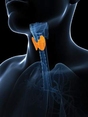
Beta-blockers expand treatment options for infantile hemangiomas
VANCOUVER, B.C. – Propranolol may have revolutionized the treatment of infantile hemangiomas over the past decade, but other beta-blockers are making inroads in the treatment of these and other pediatric dermatologic disorders.
One such beta-blocker is atenolol, a hydrophilic cardioselective agent that acts principally on beta1-adrenergic receptors, “and theoretically has less chance of hypoglycemia and bronchoconstriction,” Dr. Joseph M. Lam said at the annual meeting of the Pacific Dermatologic Association. Atenolol does not cross the blood-brain barrier and features once-daily dosing, he noted.

A recent noninferiority trial in 23 patients with infantile hemangiomas compared atenolol 1 mg/kg/day with propranolol 2 mg/kg/day, and followed the patients for 6 months (J. Am. Acad. Dermatol. 2014;70:1045-9). Patients in both groups achieved a similar rate of complete response (54% in the atenolol group, compared with 60% in the propranolol group) and there were no significant adverse effects in either group. “There was rebound growth in 26% of patients once the medication was withdrawn,” said Dr. Lam of the department of pediatrics at British Columbia Children’s Hospital, Vancouver, who was not affiliated with the study. “Initially there was more rebound growth in the propranolol group versus the atenolol group (four vs. two cases, respectively), but that was not significant.”
Another study compared oral nadolol with propranolol in patients aged 1-12 months: 10 on nadolol and 9 matched controls on propranolol (Br. J. Dermatol; 2013:168:222-4). After 24 weeks of treatment patients in the nadolol group had statistically superior lesion shrinkage, compared with those in the propranolol group (P less than .0001). Dr. Lam described nadolol as a nonselective beta-blocker that “has no intrinsic sympathomimetic activity, little myocardial depressant activity, and does not cross the blood-brain barrier. So theoretically, you have less sleep disturbance than with propranolol. Practically, it’s easier dosing than propranolol. It’s 2 mg/kg per day and you divide that b.i.d. The solution is 10 mg/mL.”
Researchers also are studying topical application of beta-blockers for hemangiomas and other skin conditions, Dr. Lam noted. A pilot study of six patients with infantile hemangiomas on the face and neck demonstrated that twice-daily topical administration of timolol maleate 0.5% was safe and effective (Arch. Dermatol. 2010;146:564-5). Dr. Lam characterized timolol as “a hydrophilic molecule that permeates poorly across intact skin, but doesn’t work great for deep hemangiomas. It’s a good choice for superficial hemangiomas, and anyone can use this.”
A larger, retrospective, multicenter cohort study of timolol maleate 0.5% or 0.1% gel-forming solution for infantile hemangiomas also demonstrated positive results (Pediatr. Dermatol. 2012; 29:28-31). In that study, 72 of 73 treated patients improved, with better response seen in those who received the 0.5% solution.
In another pilot study, Chinese researchers assessed the efficacy of fractional carbon dioxide laser–assisted drug delivery of topical timolol solution for the treatment of deep infantile hemangioma (Pediatr. Dermatol. 2014;31:286-91). The regimen consisted of one pass without overlap delivered from a 10,600-nm fractional CO2 laser to a .12-mm spot size with a single pulse of 25-30 mJ. The researchers rated the results as “excellent” in 44% of patients. “good” in 44%, and “moderate” in 11%. Plasma timolol levels were detectable in all patients.
Additional data from an unrelated case series found that use of topical timolol successfully treated pediatric pyogenic granulomas (Pediatric Dermatol. 2014; 31:203-7). The agent “doesn’t work as fast as with hemangiomas, but it works as well, which is great because most pyogenic granulomas in young children are on the face or neck area,” Dr. Lam noted. “Some took up to 6 months to respond, but all patients in this series had no further bleeding. That’s usually the main concern for families.”
Current evidence suggests that timolol does not work well for port wine stains and other vascular malformations. To date, it has shown mixed results for other vascular tumors such as kaposiform hemangioendothelioma, tufted angioma, and Kasabach-Merritt phenomenon.
Dr. Lam disclosed that he is a member of the scientific advisory board for Johnson & Johnson, and is a speaker for the company. He also is a member of the Eczema Society of Canada’s Board of Directors.
VANCOUVER, B.C. – Propranolol may have revolutionized the treatment of infantile hemangiomas over the past decade, but other beta-blockers are making inroads in the treatment of these and other pediatric dermatologic disorders.
One such beta-blocker is atenolol, a hydrophilic cardioselective agent that acts principally on beta1-adrenergic receptors, “and theoretically has less chance of hypoglycemia and bronchoconstriction,” Dr. Joseph M. Lam said at the annual meeting of the Pacific Dermatologic Association. Atenolol does not cross the blood-brain barrier and features once-daily dosing, he noted.
A recent noninferiority trial in 23 patients with infantile hemangiomas compared atenolol 1 mg/kg/day with propranolol 2 mg/kg/day, and followed the patients for 6 months (J. Am. Acad. Dermatol. 2014;70:1045-9). Patients in both groups achieved a similar rate of complete response (54% in the atenolol group, compared with 60% in the propranolol group) and there were no significant adverse effects in either group. “There was rebound growth in 26% of patients once the medication was withdrawn,” said Dr. Lam of the department of pediatrics at British Columbia Children’s Hospital, Vancouver, who was not affiliated with the study. “Initially there was more rebound growth in the propranolol group versus the atenolol group (four vs. two cases, respectively), but that was not significant.”
Another study compared oral nadolol with propranolol in patients aged 1-12 months: 10 on nadolol and 9 matched controls on propranolol (Br. J. Dermatol; 2013:168:222-4). After 24 weeks of treatment patients in the nadolol group had statistically superior lesion shrinkage, compared with those in the propranolol group (P less than .0001). Dr. Lam described nadolol as a nonselective beta-blocker that “has no intrinsic sympathomimetic activity, little myocardial depressant activity, and does not cross the blood-brain barrier. So theoretically, you have less sleep disturbance than with propranolol. Practically, it’s easier dosing than propranolol. It’s 2 mg/kg per day and you divide that b.i.d. The solution is 10 mg/mL.”
Researchers also are studying topical application of beta-blockers for hemangiomas and other skin conditions, Dr. Lam noted. A pilot study of six patients with infantile hemangiomas on the face and neck demonstrated that twice-daily topical administration of timolol maleate 0.5% was safe and effective (Arch. Dermatol. 2010;146:564-5). Dr. Lam characterized timolol as “a hydrophilic molecule that permeates poorly across intact skin, but doesn’t work great for deep hemangiomas. It’s a good choice for superficial hemangiomas, and anyone can use this.”
A larger, retrospective, multicenter cohort study of timolol maleate 0.5% or 0.1% gel-forming solution for infantile hemangiomas also demonstrated positive results (Pediatr. Dermatol. 2012; 29:28-31). In that study, 72 of 73 treated patients improved, with better response seen in those who received the 0.5% solution.
In another pilot study, Chinese researchers assessed the efficacy of fractional carbon dioxide laser–assisted drug delivery of topical timolol solution for the treatment of deep infantile hemangioma (Pediatr. Dermatol. 2014;31:286-91). The regimen consisted of one pass without overlap delivered from a 10,600-nm fractional CO2 laser to a .12-mm spot size with a single pulse of 25-30 mJ. The researchers rated the results as “excellent” in 44% of patients. “good” in 44%, and “moderate” in 11%. Plasma timolol levels were detectable in all patients.
Additional data from an unrelated case series found that use of topical timolol successfully treated pediatric pyogenic granulomas (Pediatric Dermatol. 2014; 31:203-7). The agent “doesn’t work as fast as with hemangiomas, but it works as well, which is great because most pyogenic granulomas in young children are on the face or neck area,” Dr. Lam noted. “Some took up to 6 months to respond, but all patients in this series had no further bleeding. That’s usually the main concern for families.”
Current evidence suggests that timolol does not work well for port wine stains and other vascular malformations. To date, it has shown mixed results for other vascular tumors such as kaposiform hemangioendothelioma, tufted angioma, and Kasabach-Merritt phenomenon.
Dr. Lam disclosed that he is a member of the scientific advisory board for Johnson & Johnson, and is a speaker for the company. He also is a member of the Eczema Society of Canada’s Board of Directors.