User login
‘Father of hematopoietic cytokines’ dies
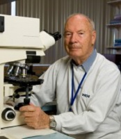
Photo courtesy of The Walter
and Eliza Hall Institute
of Medical Research
Donald Metcalf, MD, an Australian researcher who has been called “the father of hematopoietic cytokines,” has died at the age of 85.
Dr Metcalf’s studies of blood production led to his speculation that there must be a biological mechanism—one or more hormones—that control white blood cell production.
These substances, which he termed colony-stimulating factors (CSFs), were the focus of more than 50 years of research.
Over this time, Dr Metcalf led researchers to characterize and purify 4 separate CSFs—granulocyte CSF (G-CSF), granulocyte-macrophage CSF (GM-CSF), macrophage CSF (M-CSF), and multi-CSF (now called interleukin-3).
Dr Metcalf recognized that CSFs had a potential role in clinical medicine, and his team was among the first in the world to discover the genes for CSFs.
Dr Metcalf was a central figure in the international clinical trials of CSFs in the 1980s, assessing whether CSFs could boost immune cell numbers in cancer patients whose immune system was weakened as a side effect of chemotherapy. On the basis of these studies, G-CSF (Neupogen) was approved for clinical use in 1991.
Now, an estimated 20 million people have been treated with CSFs. As well as boosting the immune system in patients who receive chemotherapy or have other immune deficiencies, CSFs are thought to have revolutionized hematopoietic stem cell transplantation.
A man with many achievements
Dr Metcalf was born in 1929 and started school at the age of 3, by which time he was already reading. He entered university at the age of 16, ultimately obtaining bachelor’s and medical degrees from the University of Sydney.
After an internship at the Royal Prince Alfred Hospital in Sydney, Dr Metcalf joined the staff of the Walter and Eliza Hall Institute of Medical Research (WEHI) in Melbourne in 1954. He was supported by Cancer Council Victoria’s Carden Fellowship, an award he held until his retirement in 2014. (Dr Metcalf officially retired in 1996 but continued to do research until 2014.)
Dr Metcalf spent his early years at WEHI studying vaccinia virus. In 1965, he began studying blood cell formation and, by association, leukemia. In 1966, he became deputy director of WEHI and the head of its Cancer Research Unit.
Dr Metcalf took several sabbaticals from WEHI, serving as a visiting scientist at Harvard Medical School in Boston, Massachusetts; the Roswell Park Memorial Institute in Buffalo, New York; the Swiss Institute for Experimental Cancer Research in Lausanne, Switzerland; the Radiobiological Institute in Rijswijk, The Netherlands; and the University of Cambridge in the UK.
Among Dr Metcalf’s many honors and awards are the Companion of the Order of Australia (1993), the Albert Lasker Award for Clinical Medical Research (1993), the Gairdner Foundation International Award (1994), the Royal Medal of the Royal Society (1995), the Victoria Prize (2000), and the Prime Minister’s Prize for Science (2001).
Dr Metcalf is survived by his wife Jo; daughters Kate, Johanna, Penelope, and Mary-Ann; grandchildren James, Martin, Patrick, Elizabeth, Rose, and Robert; and their extended families. ![]()
Magnesium disappoints in sickle cell disease

sickle cell disease
Credit: St. Jude Hospital
SAN FRANCISCO—Magnesium does not improve outcomes in children hospitalized for sickle cell pain crises, results of the MAGiC study suggest.
Researchers hypothesized that magnesium—a known vasodilator, anti-inflammatory, and pain reliever—could alter the pathophysiology of pain crises.
However, when compared to normal saline, intravenous (IV) magnesium did not shorten hospital stays, lessen opioid use, or improve patients’ quality of life.
David C. Brousseau, MD, of the Medical College of Wisconsin and the Children’s Hospital of Wisconsin in Milwaukee, presented the results of this study at the 2014 ASH Annual Meeting (abstract 88).
Dr Brousseau noted that vasoocclusive crises are the most common acute complication of sickle cell disease and the most frequent cause of acute care or emergency department visits and hospitalizations. But recent changes in treatment have been minimal, with the judicious use of IV fluid and IV opioids being the mainstays of therapy.
“There have been few multicenter clinical trials evaluating new treatments, in part, due to a long history of difficulty with enrollment in interventional trials for sickle cell crises,” he continued. “These enrollment difficulties have been due to an inability to consent or to consent in a timely manner, leading to delayed initiation of study drug.”
With the MAGiC trial, Dr Brousseau and his colleagues sought to overcome this problem through a collaboration between pediatric emergency medicine physicians and pediatric hematologists.
In this randomized, double-blind trial, the researchers compared IV magnesium to normal saline. They enrolled children ages 4 to 21, with hemoglobin SS or hemoglobin SB° thalassemia, who were hospitalized after failing emergency department management for pain.
A total of 208 children were enrolled at 8 study sites over 3 years. Four children were excluded before receiving treatment, so 101 were randomized to receive magnesium and 103 to saline.
The children received 40 mg/kg of IV magnesium every 8 hours for a total of 6 doses or normal saline of an equivalent volume (1 mL/kg).
The treatment groups were well-balanced, with similar baseline age, sex, genotype, weight, history of acute chest syndrome or asthma, previous hospitalizations within the past 3 years, use of hydroxyurea, and days of pain prior to arrival.
The median time from the first emergency department opioid to the first study drug infusion was 7.3 hours in the magnesium group and 7.5 hours in the saline group.
For the study’s primary outcome, the researchers assessed patients’ length of stay from the first study drug infusion until 12 hours after the last IV opioid dose or the time of discharge, whichever came first.
“Approximately 50% of children [overall] met the study endpoint within 52 hours, and 25% met the study endpoint within 24 hours of the first drug infusion,” Dr Brousseau noted.
And there was no significant difference in the median length of stay between the treatment arms—56 hours in the magnesium arm and 47 hours in the placebo arm (P=0.264).
A secondary outcome was opioid use, recorded as morphine equivalents. There was no significant difference with this outcome, either. Patients in the magnesium arm received 1.46 mg/kg of morphine equivalents, compared to 1.28 mg/kg in the saline arm (P=0.12).
The researchers also assessed quality of life using the PedsQL sickle cell disease-specific module, fatigue module, and generic module. At 48 hours after the first infusion, there was no significant difference in quality of life scores between the treatment groups for any of the modules (P=0.17, 0.26, and 0.94, respectively). The same was true 1 week after discharge (P=0.55, 0.82, and 0.36, respectively).
As for safety, there was no significant difference between the treatment arms for most measures. However, patients in the magnesium arm were more likely to experience warmth upon infusion, at 26%, compared to 2% in the saline arm (P<0.01).
Acute chest syndrome occurred in 16% of patients in the magnesium arm and 14% in the saline arm (P=0.78). Hypotension occurred in 4% and 1%, respectively (P=0.39). And rehospitalization within 7 days occurred in 12% and 7%, respectively (P=0.11).
In closing, Dr Brousseau noted that, although the researchers did not prove their hypothesis correct, the MAGiC study was a success in one respect.
“Intravenous magnesium does not shorten length of stay, lessen opioid use, or improve quality of life in children hospitalized for sickle cell pain crises,” he said. “[However,] a collaboration between pediatric emergency department medicine physicians and pediatric hematologists allowed for successful enrollment in an acute intervention trial with a median time to first study drug of 7.5 hours.” ![]()
Reducing chemo drug’s cardiac side effects

Investigators have identified compounds that appear to prevent the cardiac damage caused by the chemotherapy drug doxorubicin.
The compounds target MDH2, an enzyme key to the generation of cellular energy in mitochondria.
And preclinical experiments showed that inhibiting MDH2 could prevent doxorubicin-induced damage to cardiac cells without reducing the drug’s antitumor effects.
The investigators detailed these experiments in Science Translational Medicine.
“Doxorubicin-induced cardiomyopathy limits the amount of the drug a patient can receive—which limits the ability to treat cancer—and even low, safer doses can lead to heart failure in up to 8% of patients,” explained study author Randall Peterson, PhD, of Massachusetts General Hospital in Charlestown.
“Finding an effective cardioprotective drug—essentially separating the good and bad effects of this form of chemotherapy—could increase the beneficial effects of doxorubicin against cancer while reducing the rate of heart failure in treated patients.”
To conduct a broad search for potential protective compounds, Dr Peterson and his colleagues developed a zebrafish model of doxorubicin-induced heart failure. They used this model to screen 3000 molecules from 2 chemical libraries for the ability to prevent the kind of cardiac damage caused by the drug.
Eight of the tested chemicals reduced damage to the hearts of zebrafish embryos, and two compounds—visnagin and diphenylurea—were the most potent in preventing both structural and functional damage.
Further in vitro and in vivo experiments revealed that either compound almost completely prevented the death of cardiac cells caused by doxorubicin. In mouse models of both high- and low-dose doxorubicin treatment, visnagin—a natural compound synthesized by the toothpick weed—was able to maintain cardiac function.
Investigation into the possible mechanism behind visnagin’s protective ability showed that the compound binds to and inhibits the action of MDH2, an enzyme essential to the generation of cellular energy by mitochondria.
Other agents that block MDH2 activity also protected zebrafish against doxorubicin-induced cardiac damage. And tests in both cellular and animal models of several types of cancer showed that neither visnagin nor diphenylurea reduced the antitumor action of doxorubicin.
“We are still trying to determine exactly how inhibition of MDH2 protects the heart, but one intriguing idea is that doxorubicin may kill cardiac and tumor cells in different ways,” Dr Peterson said. “Given the intense energy requirements of the beating heart, we speculate that cardiac cells may be especially susceptible to metabolic disturbance caused by doxorubicin and that inhibiting MDH2 may correct the metabolic imbalance and prevent the cells from dying.”
“It remains to be seen if visnagin’s protective effects are restricted to doxorubicin or if it can protect the heart from other kinds of damage. We are pursuing this question by testing its ability to protect heart muscle from oxygen deprivation during heart attacks and from the effects of other heart-damaging chemotherapy drugs.” ![]()
Weak magnetic fields not responsible for leukemia, study suggests

Research first carried out in the 1970s revealed an association between living near overhead power lines and an increased risk of childhood leukemia.
Although some later studies failed to find such a link, the International Agency for Research on Cancer has categorized low-frequency magnetic fields as “possibly carcinogenic.”
However, a mechanism for this association has never been found, and, now, researchers have ruled out one of the prime candidates.
The team studied the effects of weak magnetic fields (WMFs) on key human proteins, including those
crucial for health, and found they have no detectable impact.
The researchers detailed this discovery in the Journal of the Royal Society Interface.
Alex Jones, PhD, of The University of Manchester in the UK, and his colleagues looked at how WMFs affect flavoproteins, which are key to processes vital for healthy human function, such as the nervous system, DNA repair, and the biological clock.
If these proteins malfunction, there are serious knock-on effects for human health. But after subjecting flavoproteins to WMFs in the lab, the researchers found that WMFs have no detectable impact on these proteins.
“There is still some concern among the public about this potential link, which has been found in some studies into cases of childhood leukemia, but without any clear mechanism for why,” Dr Jones said.
“Flavoproteins transfer electrons from one place to another. Along the path the electrons take, very short-lived chemical species known as radical pairs are often created. Biochemical reactions involving radical pairs are considered the most plausible candidates for sensitivity to WMFs, but for them to be so, the reaction conditions have to be right. This research suggests that the correct conditions for biochemical effects of WMFs are likely to be rare in human biology.”
“More work on other possible links will need to be done,” noted study author Nigel Scrutton, PhD, also of the University of Manchester.
“But this study definitely takes us nearer to the point where we can say that power lines, mobile phones, and other similar devices are likely to be safe for humans.” ![]()
Product controls bleeding in kids with hemophilia A
the 2014 ASH Annual Meeting
SAN FRANCISCO—A recombinant factor VIII (FVIII) Fc fusion protein is effective for routine prophylaxis and control of bleeding in previously treated children with severe hemophilia A, according to the first phase 3 study of a long-acting FVIII in very young patients.
Prophylactic treatment of hemophilia A with recombinant FVIII requires frequent infusions, up to 3 to 4 per week.
Conventional FVIII replacement therapies have circulating half-lives of 8 to 12 hours.
And children exhibit faster clearance than adults, which may necessitate even more frequent infusions.
Recombinant FVIII Fc fusion protein (Eloctate) has been shown to have a 1.5-fold longer half-life when compared with recombinant FVIII (Advate) in a phase 3 study of adults and adolescents.
“In a pediatric population, we demonstrated similar pharmacokinetic safety and efficacy in terms of annualized bleeding rate, with no inhibitors and no adverse events related to the drug,” said Guy Young, MD, of the University of Southern California in Los Angeles.
At the 2014 ASH Annual Meeting, Dr Young and his colleagues reported results observed with recombinant FVIII Fc fusion protein in the KIDS A-LONG study (abstract 1494). This phase 3 trial was sponsored by Biogen Idec and Sobi, the companies developing recombinant FVIII Fc fusion protein.
The study enrolled 71 males under the age of 12 with severe hemophilia A (< 1 IU/dL endogenous FVIII activity), who had at least 50 prior exposure days to FVIII and no history of FVIII inhibitors.
The patients received twice-weekly prophylactic infusions of the drug, 25 IU/kg on day 1 and 50 IU/kg on day 4. Adjustments to dosing frequency up to once every 2 days and dose to ≤ 80 IU/kg were made as needed.
A subset of 25 patients under age 6 and 35 patients ages 6 to 11 underwent sequential pharmacokinetic evaluations with their prior FVIII therapy (50 IU/kg), followed by the recombinant FVIII Fc fusion protein (50 IU/kg).
“The recombinant factor VIII Fc fusion protein was effective for routine prophylaxis and for control of bleeding,” Dr Young said. “A low annualized bleeding rate was observed in both age cohorts.”
About three-quarters of the patients had a longer dosing interval with recombinant FVIII Fc fusion protein compared with their prior FVIII prophylactic dosing interval.
About 90% of the patients were on twice-weekly dosing at the end of the study compared with about 75% infusing at least 3 times a week pre-study, Dr Young said. Some 93% of bleeding episodes were controlled with 1 or 2 injections.
“The recombinant FVIII Fc fusion protein had a prolonged half-life and reduced clearance compared with conventional FVIII,” Dr Young noted.
Half-life extension was comparable to that observed in adults and adolescents.
Adverse events were generally similar to those expected for the pediatric hemophilia population.
Some 85.5% of subjects reported at least one adverse event, but no patient discontinued treatment due to an adverse event. Two non-serious events (myalgia and erythematous rash) were related to recombinant FVIII Fc fusion protein.
Five patients (7.2%) experienced a total of 7 serious adverse events, which were not related to treatment. There were no reports of anaphylaxis, vascular thrombotic events, or death.
Dr Young said the next step is to test the recombinant FVIII Fc fusion protein in previously untreated young hemophilia A patients to determine the rate of immunogenicity.
“We don’t expect to see antibodies in these patients,” he said. ![]()
Two activin receptor fusion proteins show promise in anemia
site of the ASH Annual Meeting
Photo courtesy of ASH
SAN FRANCISCO—Two activin receptor fusion proteins, luspatercept and sotatercept, increased hemoglobin levels and transfusion independence in patients with β-thalassemia and myelodysplastic syndromes (MDS)/chronic myelomonocytic leukemia (CMML), respectively, in phase 2 trials.
Luspatercept is a type IIB activin receptor, while sotatercept is type IIA. Both impact late-stage erythropoiesis and improve anemia.
Investigators reported the trial results at the 2014 ASH Annual Meeting.
Luspatercept in β-thalassemia
Antonio G. Piga, MD, of Turin University in Italy, explained that luspatercept binds to GDF11 and other ligands in the TGF-β superfamily and promotes late-stage erythroid maturation.
The study was designed in the US and conducted abroad, he said, because while β-thalassemia is rare in the US, it is not so in Europe.
Investigators evaluated whether luspatercept could increase hemoglobin levels 1.5 g/dL or more for at least 2 weeks in non-transfusion-dependent (NTD) patients.
And in transfusion-dependent (TD) patients, luspatercept was expected to decrease the transfusion burden by 20% or more over 12 weeks.
Thirty patients, 7 TD and 23 NTD, received an injection of luspatercept every 3 weeks for 3 months at doses ranging from 0.2 to 1.0 mg/kg.
The median age was 35, and 53% of patients were male. Eighty-three percent had had a splenectomy.
Luspatercept efficacy
Three-quarters of patients treated with 0.8 to 1.0 mg/kg increased their hemoglobin levels or reduced their transfusion burden.
Of the NTD patients, 8 of 12 with iron overload at baseline experienced a reduction in liver iron concentration of 1 mg or more at 16 weeks.
And in the TD group, “All patients had clinically improved reduction of transfusion dependence,” Dr Piga said.
They had a more than 60% reduction in transfusion burden over 12 weeks. This included 2 patients with β0 β0 genotype, who experienced a 79% and 75% reduction.
“There was a trend to lower liver iron concentration in TD patients,” Dr Piga noted, “except in 1 patient.”
And 5 of 5 TD patients experienced decreases in serum ferritin ranging from 12% to 60%.
Luspatercept safety
Luspatercept did not cause any treatment-related serious or severe adverse events. The most common adverse events were bone pain (20%), headache (17%), myalgia (13%), and asthenia (10%).
There was 1 grade 3 dose-limiting toxicity of worsening lumbar spine bone pain, and 3 patients discontinued early, 1 each with occipital headache, ankle pain, and back pain.
Luspatercept had beneficial effects on other complications of the disease, Dr Piga noted, such as the healing of leg ulcers in the 3 patients with this complication, 1 who is just ending the trial.
With these promising results, Dr Piga said the investigators are “anxious to start phase 3.”
Dr Piga reported the data as abstract 53. The study was supported by Acceleron Pharma and Celgene Corporation.
Sotatercept in MDS and CMML with anemia
Rami Komrokji, MD, of the Moffit Cancer Center in Tampa, Florida, explained that sotaterept increases the release of mature erythrocytes into circulation by a mechanism distinct from erythropoietin.
Sotatercept was shown to stimulate erythropoiesis and increase hemoglobin levels in healthy volunteers, so investigators undertook to study its potential to treat anemia.
They conducted a phase 2 dose-finding study to determine the best effective dose in patients with anemia and lower-risk MDS or nonproliferative CMML who were refractory to erythropoiesis-stimulating agents (ESAs).
Investigators evaluated 53 patients who had anemia of 9 g/dL or less requiring 2 or more units of red blood cells (RBCs) in the 12 weeks prior to enrollment.
Their white blood cell counts had to be under 13,000/μL, and they had to have no response, loss of response, or low chance of response to ESAs, reflected by serum erythropoietin of more than 500 mIU/mL.
Patients were a median age of 71, and 70% were male.
They received subcutaneous sotatercept at dose levels of 0.1, 0.3, 0.5, or 1.0 mg/kg once every 3 weeks for up to 24 months following the first treatment.
Sotatercept efficacy
The investigators evaluated efficacy for the entire cohort as well as in subgroups of patients with high transfusion burden (HTB) and low transfusion burden (LTB). Patients were defined as HTB if they required RBC transfusions of 4 or more units every 8 weeks and LTB as less than 4 units per 8 weeks.
Overall, 45% (24/53) of the evaluable patients achieved hematologic improvement as defined by IWG 2006 criteria.
Forty-two percent of HTB patients had a reduction in their transfusion burden of 4 or more RBC units per 8 weeks, with a median duration of longest response of 106 days (range, 62 to 345+). Eleven percent (5/44) achieved RBC transfusion independence of 56 days or more.
Sixty-three percent (5/8) of LTB patients achieved both RBC transfusion independence of 56 days or more and a mean hemoglobin increase of 1.5 mg/dL or more for at least 8 weeks.
Their maximum mean hemoglobin increase ranged from 1.9 to 4.4 g/dL, and the mean duration of RBC transfusion independence ranged from 76 to 233+ days. Of these 8 patients, 67% were in the 1.0 mg/kg cohort.
Sotatercept safety
“Most of the adverse events were not necessarily related to the treatment,” Dr Komrokji said, “and they were grade 1 or grade 2 toxicity.”
Twenty of 54* patients (37%) experienced 1 or more treatment-related adverse events, the most common of which were fatigue/asthenia (13%), headache (9%), decreased appetite (7%), nausea (7%), and dyspnea (6%).
Three patients discontinued the study due to treatment-emergent adverse events that were possibly related to sotatercept. One was for grade 2 hemolytic anemia, 1 for grade 3 hypertension, and 1 for grade 2 muscle weakness.
Dr Komrokji concluded saying the results showed “promising evidence of clinical activity” in these ESA-refractory, anemic, lower-risk MDS and CMML patients who have a “challenging and unmet need for treatment.”
He indicated that further exploration of sotatercept at higher dose levels and for longer treatment periods is planned and ongoing.
He presented the data as abstract 3251. The study was supported by Celgene Corporation. ![]()
*One patient was excluded from the efficacy analysis due to a protocol violation.
Discovery could help predict, prevent therapy-related AML

Credit: Rhoda Baer
A new study challenges the view that cancer treatment is a direct cause of therapy-related acute myeloid leukemia (AML).
The research suggests that mutations in p53 can accumulate in hematopoietic stem cells (HSCs) as a person ages, years before a cancer diagnosis.
If and when cancer develops, these mutated cells are more resistant to treatment and multiply at an accelerated pace after exposure to chemotherapy or radiation therapy, which can then lead to AML.
The findings, reported in Nature, open up new avenues for research to predict which patients are at risk of developing therapy-related AML and to find ways to prevent it.
“Until now, we’ve really understood very little about therapy-related AML and why it is so difficult to treat,” said study author Daniel Link, MD, of Washington University in St Louis, Missouri.
“This gives us some important clues for further studies aimed at treatment and prevention.”
Dr Link and his colleagues began this research by sequencing the genomes of 22 patients with therapy-related AML. The patients had similar numbers and types of mutations in their leukemia cells as other patients who developed AML without prior exposure to chemotherapy or radiation, an indication that cancer treatment does not cause widespread DNA damage.
“This is contrary to what physicians and scientists have long accepted as fact,” said study author Richard K. Wilson, PhD, of The Genome Institute at Washington University.
“It led us to consider a novel hypothesis: p53 mutations accumulate randomly as part of the aging process and are present in blood stem cells long before a patient is diagnosed with therapy-related AML.”
When therapy-related AML occurs, it typically develops 1 to 5 years after treatment with chemotherapy or radiation. Its incidence varies by cancer type. For example, 10% of lymphoma patients who relapse after chemotherapy go on to develop therapy-related AML, compared to 0.1% of breast cancer patients.
The researchers knew that patients with therapy-related AML are more likely than other AML patients to have a high rate of p53 mutations in their blood cells.
But the team was surprised to find that nearly 50% of 19 healthy subjects (aged 68 to 89 with no history of cancer or chemotherapy) had mutations in one copy of p53, an indicator that many people acquire mutations in this gene as they age.
The finding encouraged the researchers to dig further. They scoured the US to find bone marrow samples from patients with therapy-related AML that had been stored before the patients developed leukemia.
“We wanted to know whether we could go back in time—before a patient is diagnosed with therapy-related AML—to find the exact p53 mutation that caused them to develop leukemia years later,” Dr Link said.
The researchers found 7 bone marrow samples that fit the criteria. In 4 samples, they detected specific mutations in p53 that were present at very low rates in blood cells or bone marrow 3 to 6 years before the patients developed AML.
In the 3 cases in which p53 mutations could not be found, the researchers said it’s possible the mutations were present but at rates too low to be detected, or it may be that other age-related mutations contributed to the onset of therapy-related AML.
In related work in mice, the team showed that chemotherapy causes HSCs with mutations in p53 to divide rapidly, which gives them a competitive advantage. But that was not the case in HSCs with both copies of the gene intact.
The researchers suspect the early accumulation of p53 mutations in HSCs likely contributes to the frequent chromosomal and genetic abnormalities seen in patients with therapy-related AML and their poor responses to chemotherapy. The team believes other age-related mutations may be involved in the disease as well.
“We’re already conducting follow-up studies to look for other age-related mutations that may be at play in therapy-related AML,” Dr Link said. “As individuals, we’re not genetically homogeneous throughout our lives. Our DNA is constantly changing as we age, and we know this plays an important role in the development of cancer.”
“With advanced genomics, we can investigate the interplay between aging and the random accumulation of mutations, as a means to improve the diagnosis, treatment, and prevention of cancer.” ![]()
Ixazomib induces durable responses in hard-to-treat amyloidosis

Photo courtesy of ASH
SAN FRANCISCO—The oral proteasome inhibitor ixazomib induces high-quality hematologic responses in patients with relapsed/refractory systemic light-chain (AL) amyloidosis, with generally manageable side effects, a phase 1 study suggests.
Preliminary results from this study indicated that ixazomib had promise for treating AL amyloidosis.
Now, researchers have reported updated safety data and figures for hematologic and organ responses, progression-free survival, and overall survival.
The study was presented at the 2014 ASH Annual Meeting (abstract 3450) 5 days after the US Food and Drug Administration granted ixazomib breakthrough status for the treatment of relapsed/refractory AL amyloidosis.
“To have an oral drug that is well-tolerated and extremely effective in patients exposed to all lines of other therapy is remarkable in this disease,” said study investigator Giampaolo Merlini, MD, of the Amyloidosis Research and Treatment Center, Fondazione IRCCS Policlinico San Matteo, at the University of Pavia in Italy.
He and his colleagues evaluated ixazomib in 22 patients with a median age of 65 years. They were heavily pretreated, with 95% exposed to melphalan and 73% to bortezomib.
The patients received 4 mg of ixazomib on days 1, 8, and 15 of 28-day cycles for up to 12 cycles. Those who did not achieve a hematologic partial response after 3 cycles received added dexamethasone.
In 21 evaluable patients, the overall response rate was 52%, including a complete response and very good partial response in 43% of patients.
Dr Merlini noted that responses were deep and durable. And the high response rates translated into high organ response rates.
“End-organ damage is what kills patients,” he said. “In 18 evaluable patients, both the heart and kidney response rate was 45%.”
At 1 year, progression-free survival was 60%. At 2 years, overall survival was 63%.
The drug was well-tolerated as well. The 3 most common side effects—nausea, diarrhea, and fatigue—were mild and seen in about 30% of patients.
Severe grade 3 or higher side effects included thrombocytopenia, diarrhea, and rash, occurring in about 10% of patients.
The development program for ixazomib in AL amyloidosis progressed directly from this phase 1 trial to a phase 3 study, TOURMALINE-AL1. The trial is already underway, with 80 patients enrolled thus far.
TOURMALINE-AL1 investigators are comparing ixazomib plus dexamethasone to physician’s choice of treatment in patients with relapsed/refractory AL amyloidosis. Data from this trial are expected by the end of 2015.
“For the first time, we have evidence of an oral drug that is extremely effective,” Dr Merlini said. “We need to see the phase 3 data, but it could be a breakthrough.” ![]()
FDA approves first supplemental test for HTLV-I/II

Credit: Daniel Gay
The US Food and Drug Administration (FDA) has approved the first supplemental test for human T-cell lymphotropic virus-I/II (HTLV-I/II).
The test, MP Diagnostics HTLV Blot 2.4, is a qualitative enzyme immunoassay intended for use as an additional, more specific test for human serum or plasma specimens that have previously tested positive for HTLV-I/II.
MP Diagnostics HTLV Blot 2.4 can confirm infection with HTLV and differentiate between HTLV-I and HTLV-II.
The HTLVs are a group of human retroviruses known to cause diseases such as adult T-cell leukemia/lymphoma and myelopathy. HTLV can be transmitted from person to person through breastfeeding, unprotected sexual contact, or transfusion of blood from an infected donor.
Therefore, the FDA requires that donated blood be tested for HTLV-I/II antibodies. Currently, there are 2 FDA-licensed screening tests for HTLV-I/II. If a test is positive, the donation is discarded, and the donor is notified of his or her deferral.
The MP Diagnostics HTLV Blot 2.4 can provide blood establishments with additional information to convey to the donor. Specifically, the test can confirm HTLV infection and determine which virus type is causing the infection, HTLV-I or HTLV-II.
“The approval of MP Diagnostics HTLV Blot 2.4 will help blood establishments better counsel donors who have had positive results on an FDA-licensed HTLV-I/II screening test,” said Karen Midthun, MD, director of the FDA’s Center for Biologics Evaluation and Research.
MP Diagnostics HTLV Blot 2.4 is manufactured by MP Biomedicals Asia Pacific Pte. Ltd. in Singapore, a company of MP Biomedicals LLC in Santa Ana, California. For more information on the test, visit the MP Biomedicals website. ![]()
Credit: Daniel Gay
The US Food and Drug Administration (FDA) has approved the first supplemental test for human T-cell lymphotropic virus-I/II (HTLV-I/II).
The test, MP Diagnostics HTLV Blot 2.4, is a qualitative enzyme immunoassay intended for use as an additional, more specific test for human serum or plasma specimens that have previously tested positive for HTLV-I/II.
MP Diagnostics HTLV Blot 2.4 can confirm infection with HTLV and differentiate between HTLV-I and HTLV-II.
The HTLVs are a group of human retroviruses known to cause diseases such as adult T-cell leukemia/lymphoma and myelopathy. HTLV can be transmitted from person to person through breastfeeding, unprotected sexual contact, or transfusion of blood from an infected donor.
Therefore, the FDA requires that donated blood be tested for HTLV-I/II antibodies. Currently, there are 2 FDA-licensed screening tests for HTLV-I/II. If a test is positive, the donation is discarded, and the donor is notified of his or her deferral.
The MP Diagnostics HTLV Blot 2.4 can provide blood establishments with additional information to convey to the donor. Specifically, the test can confirm HTLV infection and determine which virus type is causing the infection, HTLV-I or HTLV-II.
“The approval of MP Diagnostics HTLV Blot 2.4 will help blood establishments better counsel donors who have had positive results on an FDA-licensed HTLV-I/II screening test,” said Karen Midthun, MD, director of the FDA’s Center for Biologics Evaluation and Research.
MP Diagnostics HTLV Blot 2.4 is manufactured by MP Biomedicals Asia Pacific Pte. Ltd. in Singapore, a company of MP Biomedicals LLC in Santa Ana, California. For more information on the test, visit the MP Biomedicals website.
First randomized evidence for kinase inhibitor use in AML
Photo courtesy of ASH
SAN FRANCISCO—Researchers have presented the first randomized evidence that kinase inhibitors are effective in the treatment of acute myeloid leukemia
(AML).
The multikinase inhibitor sorafenib improved event-free and relapse-free survival in younger patients.
“Interestingly, at this point in time, we can see no clear overall survival benefit for patients treated in the sorafenib arm,” said trial investigator Christoph Röllig, MD, of the Universitätsklinikum Dresden in Germany.
Dr Röllig, representing the Study Alliance Leukemia, presented data on sorafenib from the SORAML trial during the plenary session of the 2014 ASH Annual Meeting (abstract 6). Some funding for this trial was provided by Bayer Healthcare, the company developing sorafenib.
Dr Röllig explained that support for sorafenib’s clinical efficacy in AML was based primarily on case series and a few early phase clinical and nonrandomized
trials.
An earlier randomized study with sorafenib in older AML patients showed no beneficial antileukemic effect with the addition of the agent, and the treatment was associated with significant morbidity.
However, because the biology of AML and drug tolerance are different in younger people, the Study Alliance Leukemia decided to test the drug in a younger
patient population.
They randomized 276 newly diagnosed AML patients aged 60 years or younger to receive 2 cycles of induction chemotherapy with an anthracycline and cytarabine plus either sorafenib or placebo.
The sorafenib dose was 800 mg per day orally. All patients received at least one dose of study medication, forming the statistical analysis set.
Once in complete remission, intermediate-risk patients with a family donor and high-risk patients with a matched donor went on to stem cell transplant.
All other patients proceeded to high-dose cytarabine-based consolidation treatment plus sorafenib or placebo followed by 1 year of maintenance treatment with sorafenib or placebo.
The primary endpoint was event-free survival (EFS). An event was defined as primary treatment failure, relapse, or death.
Patients in each arm were a median age of 50 years, 17% were FLT3-ITD positive, and 33% were NPM1 mutated.
The complete response (CR) rate was 60% in the sorafenib arm and 59% in the placebo arm. Patients with FLT3-ITD mutation achieved a 57% CR rate with sorafenib and a 52% CR rate with placebo.
After a median follow-up of 3 years, investigators observed “significant prolongation of event-free survival in the sorafenib arm,” Dr Röllig said.
Patients on the sorafenib arm had a median EFS of 40% compared with 22% on the placebo arm, or 21 months compared with 9 months (P=0.013).
Patients were censored at the time of transplant. However, uncensored results were very similar, Dr Röllig noted, “with an even greater advantage for sorafenib.”
Fifty-six percent of sorafenib-treated patients were relapse-free and alive after 3 years, compared with 38% of patients on placebo (P=0.017).
The 3-year overall survival was 63% in the sorafenib arm and 56% in the placebo arm (P=0.382).
An exploratory analysis of FLT3-ITD patients revealed that 1-year EFS for sorafenib-treated patients was 54%, and, for placebo-treated patients, it was 50%.
“The reasons for potential efficacy of sorafenib in this mainly FLT3-ITD-negative population must remain speculative,” Dr Röllig said. “We can speculate that the inhibition of other kinases apart from FLT3 might be responsible for the efficacy of this drug in this patient population.”
Sorafenib significantly increased the risk of grade 3 or greater hand-foot syndrome (P<0.001), diarrhea (P=0.001), bleeding (P=0.016), rash (P=0.045), liver toxicity (P=0.048), and fever (P=0.035).
Dr Röllig indicated a confirmatory trial would be desirable in order to establish sorafenib in the AML treatment armamentarium.