User login
Study confirms IDH2 as therapeutic target in AML, MDS
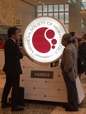
in the Moscone Center
SAN FRANCISCO—The first-in-human study of AG-221 has confirmed IDH2 as a therapeutic target in acute myeloid leukemia (AML) and myelodysplastic syndromes (MDS), according to investigators.
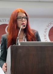
From the first dose of therapy, the high plasma level observed in patients “translates into a drastic decrease in 2-HG,” said Eytan M. Stein, MD, of Memorial Sloan Kettering Cancer Center in New York.
Mutations in IDH1 and IDH2 result in the accumulation of the oncometabolite 2-HG. And high levels of 2-HG prompt epigenetic changes to the cell, resulting in impaired cellular differentiation.
“By cycle 1, day 15, at all dose levels, there was fantastic inhibition of 2-HG,” Dr Stein said. “Indeed, this drastic inhibition of 2-HG led to profound clinical benefit.”
He presented interim results of the phase 1 study of AG-221 at the 2014 ASH Annual Meeting (abstract 115).*
The investigators have treated 73 patients with advanced IDH2-mutation-positive hematologic malignancies since the study began in September 2013.
Patients were a median age of 67 years (range, 33-90), and 74% had IDH2 R140 mutations. Most had relapsed/refractory AML (n=55), and the rest had MDS (n=6), untreated AML (n=5), chronic myelomonocytic leukemia (n=5), and myeloid sarcoma (n=1).
Patients received AG-221 as a single agent orally, once or twice a day, continuously, in 28-day cycles. Four dose-expansion cohorts at 100 mg each day were added last October.
To date, the maximum tolerated dose has not been reached, with the highest cumulative daily dose being 300 mg. Investigators observed a single dose-limiting toxicity in 1 patient: grade 5 hypoxia with fungal pneumonia and septic shock.
As of the data cutoff on October 1, 2014, 38 patients were still on therapy, and 35 had discontinued. Five patients withdrew after achieving complete remission (CR) in order to pursue allogeneic transplant.
Dr Stein pointed out that although the median number of prior chemotherapy regimens for the entire cohort was 2, “all of the patients who went on study were predicted to have dismal outcomes with conventional therapy.”
Safety
The therapy was well tolerated, with the most common adverse events (AEs) overall being typical for patients with advanced AML: nausea (23%), pyrexia (19%), and diarrhea (17%).
The majority of serious AEs were disease-related and unrelated to the study drug, Dr Stein said. Thirteen patients experienced 21 serious AEs that were possibly or probably related to treatment.
Investigators observed treatment-related leukocytosis in 3 patients, “but we think that is a differentiating effect of the study drug,” Dr Stein said.
He also pointed out that there was no increase in AEs with increased dose of the drug.
Of the 11 deaths reported, 9 were unrelated to the drug, and 2—sepsis/hypoxia and atrial flutter—were possibly related. The 30-day and 60-day all-cause mortality rates were 4.1% and 13.7%, respectively.
Efficacy
Of the 45 patients who were treated for at least 1 cycle and were evaluable for efficacy, 15 achieved a CR (n=6) or a CR with incomplete blood count recovery. The overall response rate is 56% (25/45).
“Responses appear durable,” Dr Stein commented, “and 90% of responders have had a response duration of at least 3 months, with the earliest patients on study having had durable responses for over 6 months.”
Many of the durable responses are partial remissions, he noted.
Seventeen patients have stable disease, and many of these patients remain on study. Two patients had progressive disease.
“Patients with stable disease have remained on study for a similar amount of time as the responders,” Dr Stein said, “suggesting that, despite the lack of a formal partial remission, many patients are deriving clinical benefit from study treatment.”
This study was sponsored by Agios Pharmaceuticals Inc., the company developing AG-221 in collaboration with Celgene.
Previous results from this study were presented at the 2014 EHA Annual Congress. Based on those results, AG-221 received fast track designation from the US Food and Drug Administration. ![]()
*Data in the presentation were updated from the abstract.
in the Moscone Center
SAN FRANCISCO—The first-in-human study of AG-221 has confirmed IDH2 as a therapeutic target in acute myeloid leukemia (AML) and myelodysplastic syndromes (MDS), according to investigators.
From the first dose of therapy, the high plasma level observed in patients “translates into a drastic decrease in 2-HG,” said Eytan M. Stein, MD, of Memorial Sloan Kettering Cancer Center in New York.
Mutations in IDH1 and IDH2 result in the accumulation of the oncometabolite 2-HG. And high levels of 2-HG prompt epigenetic changes to the cell, resulting in impaired cellular differentiation.
“By cycle 1, day 15, at all dose levels, there was fantastic inhibition of 2-HG,” Dr Stein said. “Indeed, this drastic inhibition of 2-HG led to profound clinical benefit.”
He presented interim results of the phase 1 study of AG-221 at the 2014 ASH Annual Meeting (abstract 115).*
The investigators have treated 73 patients with advanced IDH2-mutation-positive hematologic malignancies since the study began in September 2013.
Patients were a median age of 67 years (range, 33-90), and 74% had IDH2 R140 mutations. Most had relapsed/refractory AML (n=55), and the rest had MDS (n=6), untreated AML (n=5), chronic myelomonocytic leukemia (n=5), and myeloid sarcoma (n=1).
Patients received AG-221 as a single agent orally, once or twice a day, continuously, in 28-day cycles. Four dose-expansion cohorts at 100 mg each day were added last October.
To date, the maximum tolerated dose has not been reached, with the highest cumulative daily dose being 300 mg. Investigators observed a single dose-limiting toxicity in 1 patient: grade 5 hypoxia with fungal pneumonia and septic shock.
As of the data cutoff on October 1, 2014, 38 patients were still on therapy, and 35 had discontinued. Five patients withdrew after achieving complete remission (CR) in order to pursue allogeneic transplant.
Dr Stein pointed out that although the median number of prior chemotherapy regimens for the entire cohort was 2, “all of the patients who went on study were predicted to have dismal outcomes with conventional therapy.”
Safety
The therapy was well tolerated, with the most common adverse events (AEs) overall being typical for patients with advanced AML: nausea (23%), pyrexia (19%), and diarrhea (17%).
The majority of serious AEs were disease-related and unrelated to the study drug, Dr Stein said. Thirteen patients experienced 21 serious AEs that were possibly or probably related to treatment.
Investigators observed treatment-related leukocytosis in 3 patients, “but we think that is a differentiating effect of the study drug,” Dr Stein said.
He also pointed out that there was no increase in AEs with increased dose of the drug.
Of the 11 deaths reported, 9 were unrelated to the drug, and 2—sepsis/hypoxia and atrial flutter—were possibly related. The 30-day and 60-day all-cause mortality rates were 4.1% and 13.7%, respectively.
Efficacy
Of the 45 patients who were treated for at least 1 cycle and were evaluable for efficacy, 15 achieved a CR (n=6) or a CR with incomplete blood count recovery. The overall response rate is 56% (25/45).
“Responses appear durable,” Dr Stein commented, “and 90% of responders have had a response duration of at least 3 months, with the earliest patients on study having had durable responses for over 6 months.”
Many of the durable responses are partial remissions, he noted.
Seventeen patients have stable disease, and many of these patients remain on study. Two patients had progressive disease.
“Patients with stable disease have remained on study for a similar amount of time as the responders,” Dr Stein said, “suggesting that, despite the lack of a formal partial remission, many patients are deriving clinical benefit from study treatment.”
This study was sponsored by Agios Pharmaceuticals Inc., the company developing AG-221 in collaboration with Celgene.
Previous results from this study were presented at the 2014 EHA Annual Congress. Based on those results, AG-221 received fast track designation from the US Food and Drug Administration. ![]()
*Data in the presentation were updated from the abstract.
Amount of blood transfused doesn’t affect long-term mortality
![]()
Credit: UAB Hospital
Long-term mortality is not affected by the amount of blood a patient receives following surgery, according to research published in The Lancet.
Investigators compared a liberal transfusion strategy, in which patients received blood to maintain a hemoglobin level at 100 g/L or higher, and a restrictive strategy, in which patients received blood when hemoglobin levels were lower than 80 g/L or if they had symptoms of anemia.
And, at about 3 years of follow-up, there was no difference in mortality between the two groups.
Jeffrey L. Carson, MD, of Robert Wood Johnson Medical School in New Jersey, and his colleagues conducted this research, analyzing data from the FOCUS trial, which included patients from 47 hospitals in the US and Canada.
The trial enrolled 2016 adults age 50 and older, with a history of or risk factors for cardiovascular disease, who had postoperative hemoglobin concentrations lower than 100 g/L within 3 days of surgery to repair a hip fracture.
Patients were randomized by a central telephone system to the liberal (n=1007) or restrictive (n=1009) transfusion groups. The investigators analyzed the long-term mortality of these patients, which was established by linking participants to national death registries in the US and Canada.
The results revealed no difference in mortality from cardiovascular disease, cancer, or severe infection due to the amount of the blood given after surgery.
This supports the initial results of the FOCUS trial, which Dr Carson and his colleagues published in NEJM in 2011 and which demonstrated the safety of fewer transfusions in the short-term.
For the current analysis, the median duration of follow-up was 3.1 years. Eight hundred and forty-one patients (42%) died during that time—432 in the liberal transfusion group and 409 in the restrictive transfusion group. This difference was not statistically significant, with a hazard ratio of 1.09 and a P value of 0.21.
“There has been a steady decline in the amount of blood in transfusions given to patients in the past 3 to 5 years,” Dr Carson noted. “I think it is very reassuring that we have found that using less blood is okay, not just from a short-term perspective, but also a long-term perspective.”
Medical experts had worried that larger amounts of transfused blood might suppress immune function—which could lead to death from infection or cancer—or that smaller transfusions might worsen a patient’s chronic heart disease by depriving the heart of oxygen and other nutrients that it might have absorbed by pumping more blood.
But in both instances, Dr Carson and his colleagues found no difference in long-term death rates, regardless of the number of transfusions.
Dr Carson noted that there are health conditions, such as heart attacks, where the effects of the two transfusion strategies are less certain. Preliminary evidence suggests heart attack patients need more blood, not less. But additional studies are needed to confirm that. ![]()
Team identifies prognostic markers for CN-AML

Researchers say they’ve identified novel prognostic markers for older patients with cytogenetically normal acute myeloid leukemia (CN-AML).
The team examined the expression of long noncoding RNAs (lncRNAs) in patients with CN-AML who were at least 60 years of age.
This revealed a pattern of 48 lncRNAs that predicted both response to standard chemotherapy and overall survival.
The researchers described this discovery in PNAS.
“[Our findings] strongly suggest that lncRNA expression profiles can predict which patients will respond to standard therapy,” said principal investigator Clara D. Bloomfield, MD, of The Ohio State University Comprehensive Cancer Center in Columbus.
“That’s important because it would spare these patients from the toxic side effects of experimental therapies. Patients who are classified in the unfavorable group would receive different therapy: stem cell transplant or a clinical trial using new therapeutic approaches. Thus, this research will help to tailor leukemia therapy to each individual.”
In addition, she said, the study revealed novel targets for the development of new therapies.
Dr Bloomfield and her colleagues began this research by analyzing bone marrow samples from 148 older patients with CN-AML who were treated on Cancer and Leukemia Group B clinical trials. All had received similar chemotherapy regimens.
The researchers first identified the 48 lncRNAs that were most associated with survival. Using these 48 lncRNAs, the team divided patients into two groups: those with a favorable prognostic score and those with an unfavorable score.
The researchers then validated the scores in an independent matched set of 71 similarly treated CN-AML patients and compared patients with an unfavorable score to those with a favorable score.
Results showed that patients with an unfavorable score had a lower complete response (CR) rate—54%, compared to 89% for patients with a favorable score.
Three years after CR, only 7% of patients with an unfavorable score were disease-free, compared with 39% of patients with a favorable score.
Overall survival at 3 years for those with an unfavorable score was 10%, compared to 43% for patients with a favorable score.
Distinct lncRNA profiles were associated with mutations in 6 genes—FLT3, NPM1, CEBPA, IDH2, ASXL1, and RUNX1.
The researchers concluded that lncRNA expression profiles are associated with recurrent mutations, clinical features, and outcome in AML. A fraction of these lncRNAs may have a functional role in leukemogenesis. Furthermore, lncRNAs could be used as biomarkers for outcome in AML. ![]()
Blinatumomab confirmed as treatment option in MRD+ ALL
SAN FRANCISCO—The first international, multicenter trial in acute lymphoblastic leukemia (ALL) using minimal residual disease (MRD) as a criterion for inclusion has confirmed clinical benefit for patients using a non-chemotherapeutic approach.
In the BLAST trial, blinatumomab, a bispecific T-cell engager antibody that directs cytotoxic T cells to CD19-positive cells, produced a complete MRD response in 80% of patients who were in complete hematologic remission but had quantifiable MRD at the time of treatment.
This echoes results of an earlier phase 2 study, in which blinatumomab produced an 80% MRD response rate in 20 patients with B-precursor ALL and persistent or relapsed MRD.
Nearly all patients with persistent or recurrent MRD relapse despite continued chemotherapy, according to Nicola Gökbuget, MD, PhD, of Goethe University Hospital in Frankfurt, Germany.
“The question,” she said, “is how to treat these patients.”
She presented one effective method, as shown by the BLAST trial, at the 2014 ASH Annual Meeting (abstract 379).
The trial enrolled 116 patients aged 18 or older with B-precursor ALL in complete hematologic remission but with MRD ≥ 10-3.
Patients could be in second or later remission, but they were excluded if they had a prior allogeneic stem cell transplant, circulating blasts or extra medullary ALL involvement, CNS pathology, prior chemotherapy within 2 weeks, or radiotherapy within 4 weeks.
Enrolled patients were a median age of 45 years (range, 18-76), and 34% were 65 or older. Most patients had high baseline MRD levels: 39% were ≥ 10-2 to < 10-1, and 45% were ≥ 10-3 to < 10-2.
Investigators evaluated MRD levels after each cycle. Analysis was performed in a central lab in Kiel, Germany, and was based on amplification of immunoglobulin and/or T-cell receptor gene rearrangements by PCR.
The primary endpoint was the proportion of patients achieving a complete MRD response after 1 cycle of blinatumomab.
Patients received 15 μg/m2 of blinatumomab per day by continuous intravenous infusion for 4 weeks, followed by a treatment-free period of 2 weeks. Responders could receive up to 4 cycles of therapy or receive a transplant any time after the first cycle.
Results
In the efficacy-evaluable population, 80% of 103 patients achieved a complete MRD response after 1 cycle, defined as MRD negative with no amplification in PCR with a minimum sensitivity of 10-4. And 85% achieved an incomplete MRD response of <10-4 with a minimum sensitivity of 10-4.
Results were similar in the full analysis set of 113 patients: 78% achieved complete MRD response after 1 cycle, and 85% achieved an incomplete MRD response.
Dr Gökbuget noted that 2 patients who initially achieved an incomplete response achieved a complete response during continued treatment in cycle 2.
The investigators analyzed the clinical characteristics of the patients and found that no factor—age, treatment interruptions, neurologic events, relapse history, nor gender—correlated with MRD response.
“I think that the good news is that it means that the compound was active in all of the patients,” Dr Gökbuget commented.
Adverse events
All patients experienced at least one adverse event (AE), and 2 were fatal—subdural hemorrhage and treatment-related pneumonitis. Serious treatment-related AEs that occurred in 3% or more of the
patients included tremor (7%), aphasia (5%), and encephalopathy (5%).
Thirty-one percent of patients interrupted their treatment because of AEs, which were primarily due to cytokine-related symptoms like pyrexia and neurologic events, including tremor, aphasia, and dizziness. Seventeen percent of patients permanently discontinued treatment due to an AE.
However, Dr Gökbuget pointed out that most AEs were grade 1 or 2, and the treatment interruption did not correlate with response.
She said the next step for this trial is to investigate whether high MRD response translates into long-term clinical benefit such as continued molecular remission and long-term survival.
“For me personally, this trial is very important because it is an up-to-date trial where we used PCR-based methods to identify patients with a high risk of relapse and treat them before the relapse occurs,” Dr Gökbuget said. “[W]e used a new endpoint, which is also MRD based, and . . . we used a new, non-chemotherapy treatment to eradicate this highly resistant, persistent ALL subclone.”
Blinatumomab received US Food and Drug Administration approval a few days before the start of the ASH Annual Meeting.
The BLAST trial was funded by Amgen Inc., the company developing blinatumomab. ![]()
FDA approves pathogen inactivation system for plasma
The US Food and Drug Administration (FDA) has approved the INTERCEPT Blood System for plasma, the first pathogen inactivation system for the preparation of plasma to reduce the risk of transfusion-transmitted infections.
The system inactivates pathogens through a photochemical process involving controlled exposure to ultraviolet light and the chemical amotosalen. The plasma is then purified to remove the chemical and its byproducts.
The INTERCEPT Blood System for plasma can be used to reduce pathogens in plasma derived from whole blood and plasma obtained by apheresis.
The system has proven effective in reducing a broad spectrum of viruses, bacteria, spirochetes, and parasites. However, there is no pathogen inactivation process that has been shown to eliminate all pathogens. Certain viruses (eg, human parvovirus B19) and spores formed by certain bacteria are known to be resistant to the INTERCEPT process.
“The approval of devices like the INTERCEPT Blood System allows blood establishments to prepare plasma that carries a lower risk of transmitting infectious pathogens through transfusion,” said Karen Midthun, MD, director of the FDA’s Center for Biologics Evaluation and Research.
The INTERCEPT Blood System for plasma, which is marketed by Cerus Corporation, has been approved in Europe since 2006 and was recently made available in the US under an Investigational Device Exemption study. The system is being used to prepare Ebola convalescent plasma for transfusion into acutely infected patients.
Clinical studies
Researchers investigated the safety and effectiveness of plasma prepared with the INTERCEPT Blood System in 6 clinical studies and 2 post-marketing studies.
The research showed no significant difference in coagulation factor activity between INTERCEPT-processed plasma and unprocessed fresh-frozen plasma (FFP) in healthy subjects.
There was no significant difference in prothrombin time or activated partial thromboplastin time between INTERCEPT-processed plasma and FFP in patients with coagulation factor deficiencies or in patients with liver disease.
In patients who underwent liver transplant, there was no significant difference in plasma use, hepatic artery thrombosis, or mortality whether patients received INTERCEPT-process plasma or FFP.
In a prospective, phase 3 trial and a retrospective study of patients with thrombotic thrombocytopenic purpura (TTP), there was no significant difference in the percentage of patients who achieved remission with INTERCEPT-treated plasma or FFP.
Adverse events
In a post-marketing study*, researchers analyzed acute transfusion reactions (ATRs) after 57,171 INTERCEPT-processed plasma components were transfused to 9669 patients. Thirty-two subjects (0.3%) experienced an ATR after 41 transfusion episodes (0.2%), including 5 subjects (0.05%) who experienced an ATR after more than 1 transfusion.
Six ATRs were considered serious and possibly or probably related to transfusion. There were 3 instances of allergic reaction or symptoms of allergic reaction (eg, rash, tachycardia, hypotension, respiratory symptoms, and chills), 2 cases of fluid overload, and 1 report of respiratory distress.
In another hemovigilance study, researchers compared the frequency of adverse events due to any of the 4 types of FFP prepared and delivered by the French Blood Establishment over a 10-year period—methylene blue, amotosalen, quarantine, and solvent-detergent.
The study showed that all types of FFP were associated with a low rate of adverse events. Per 10,000 deliveries, there were 7.14 events in the quarantine group, 4.86 in the solvent-detergent group, 4.16 in the amotosalen group, and 1.05 in the methylene blue group.
The package insert of the INTERCEPT Blood System for plasma warns that amotosalen-treated plasma has been associated with cardiac events.
In the aforementioned prospective trial of the system in patients with TTP, 5 patients treated with INTERCEPT-processed plasma and none with conventional plasma had adverse events in the cardiac system organ class.
This included angina pectoris (n=3), cardiac arrest (n=1), bradycardia (n=1), tachycardia (n=1), and sinus arrhythmia (n=1). None of the events resulted in documented myocardial infarction or death. ![]()
*Results have been updated since publication.
Newborn screening test for SCID gets FDA approval

Credit: Vera Kratochvil
The US Food and Drug Administration (FDA) has approved marketing of the EnLite Neonatal TREC Kit, the first screening test for severe combined immunodeficiency (SCID) in newborns.
Using a few drops of blood from the newborn’s heel, which is dried on filter paper, the EnLite Neonatal TREC Kit can determine whether T-cell receptor excision circles (TREC) DNA is low or missing from the newborn’s blood.
Newborns with SCID typically have no or low amounts of TREC DNA compared to healthy infants.
Additional testing is required to obtain a SCID diagnosis once the EnLite Neonatal TREC Kit has suggested a child may have SCID.
“SCID is a fatal disease that can be treated with early intervention, including screening,” said Alberto Gutierrez, PhD, director of the Office of In Vitro Diagnostics and Radiological Health in the FDA’s Center for Devices and Radiological Health.
“For the first time, the FDA is allowing the marketing of a newborn screening test that will enable states to incorporate an FDA-reviewed SCID test into their standard newborn screening panels and allow earlier identification for affected individuals.”
The FDA reviewed the EnLite Neonatal TREC Kit through its de novo classification process, a regulatory pathway for some novel, low- to moderate-risk medical devices that are not substantially equivalent to an already legally marketed device.
The FDA’s review included a clinical study of approximately 6400 blood spot specimens from routine screening of newborns, 17 of which had a confirmed SCID diagnosis. The EnLite Neonatal TREC Kit correctly identified all 17 SCID cases.
The agency also evaluated the test’s ability to accurately distinguish low TREC DNA numbers that would be observed in newborns with SCID from high TREC DNA numbers that would be present in healthy newborns. The FDA found the EnLite Neonatal TREC Kit could adequately detect very low TREC DNA values that are associated with SCID.
The EnLite Neonatal TREC Kit is not intended for use as a diagnostic test or to screen for SCID-like syndromes, such as DiGeorge syndrome or Omenn syndrome. Likewise, it is not intended to screen for less acute SCID syndromes, such as leaky SCID or variant SCID.
The EnLite Neonatal TREC Kit is manufactured by Wallac Oy, a subsidiary of PerkinElmer, at its facility in Turku, Finland. PerkinElmer is based in Waltham, Massachusetts. ![]()
Study reveals superior antiviral drug for lymphoma

A study of lymphoma patients receiving R-CHOP chemotherapy showed that use of the antiviral drug entecavir resulted in a lower incidence of hepatitis B virus (HBV)-related hepatitis and HBV reactivation, when compared with the drug lamivudine.
HBV reactivation is a well-documented complication of chemotherapy that can result in life-threatening liver failure, as well as delays in chemotherapy or
premature termination of treatment.
However, researchers have not identified an optimal approach to prevent HBV reactivation.
So He Huang, MD, of the Sun Yat-sen University Cancer Center in Guangzhou, China, and colleagues set out to do that. They reported their findings in JAMA alongside a related editorial.
The team enrolled 121 patients with untreated diffuse large B-cell lymphoma who were receiving chemotherapy with rituximab, cyclophosphamide, doxorubicin, vincristine, and prednisone (R-CHOP) and were seropositive for the hepatitis B surface antigen.
Patients were randomized to receive entecavir (n=61) or lamivudine (n=60). They began taking these drugs 1 week before the initiation of R-CHOP and continued until 6 months after they completed chemotherapy.
The study was conducted from February 2008 through December 2012 at 10 medical centers in China. This trial was a substudy of a parent study designed to compare a 3-week R-CHOP regimen with a 2-week R-CHOP regimen.
Results showed that, compared to the lamivudine group, patients in the entecavir group had significantly lower rates of hepatitis (8.2% vs 23.3%, P=0.02), HBV-related hepatitis (0% vs 13.3%, P=0.003), HBV reactivation (6.6% vs 30%, P=0.001), delayed hepatitis B (0% vs 8.3%, P=0.03), and chemotherapy disruption (1.6% vs 18.3%, P=0.002).
The rate of treatment-related adverse events was not significantly different between the groups. Such events occurred in 24.6% of patients in the entecavir group and 30.0% of patients in the lamivudine group (P=0.50).
The researchers noted that entecavir is more expensive than lamivudine, so additional studies are needed to determine whether all patients who are seropositive for the hepatitis B surface antigen and are receiving rituximab-based immunosuppressive therapy should be given entecavir.
Additional studies could also help pinpoint which patients will benefit most from entecavir prophylaxis. However, if these findings are replicated, they support the use of entecavir in these patients. ![]()
A study of lymphoma patients receiving R-CHOP chemotherapy showed that use of the antiviral drug entecavir resulted in a lower incidence of hepatitis B virus (HBV)-related hepatitis and HBV reactivation, when compared with the drug lamivudine.
HBV reactivation is a well-documented complication of chemotherapy that can result in life-threatening liver failure, as well as delays in chemotherapy or
premature termination of treatment.
However, researchers have not identified an optimal approach to prevent HBV reactivation.
So He Huang, MD, of the Sun Yat-sen University Cancer Center in Guangzhou, China, and colleagues set out to do that. They reported their findings in JAMA alongside a related editorial.
The team enrolled 121 patients with untreated diffuse large B-cell lymphoma who were receiving chemotherapy with rituximab, cyclophosphamide, doxorubicin, vincristine, and prednisone (R-CHOP) and were seropositive for the hepatitis B surface antigen.
Patients were randomized to receive entecavir (n=61) or lamivudine (n=60). They began taking these drugs 1 week before the initiation of R-CHOP and continued until 6 months after they completed chemotherapy.
The study was conducted from February 2008 through December 2012 at 10 medical centers in China. This trial was a substudy of a parent study designed to compare a 3-week R-CHOP regimen with a 2-week R-CHOP regimen.
Results showed that, compared to the lamivudine group, patients in the entecavir group had significantly lower rates of hepatitis (8.2% vs 23.3%, P=0.02), HBV-related hepatitis (0% vs 13.3%, P=0.003), HBV reactivation (6.6% vs 30%, P=0.001), delayed hepatitis B (0% vs 8.3%, P=0.03), and chemotherapy disruption (1.6% vs 18.3%, P=0.002).
The rate of treatment-related adverse events was not significantly different between the groups. Such events occurred in 24.6% of patients in the entecavir group and 30.0% of patients in the lamivudine group (P=0.50).
The researchers noted that entecavir is more expensive than lamivudine, so additional studies are needed to determine whether all patients who are seropositive for the hepatitis B surface antigen and are receiving rituximab-based immunosuppressive therapy should be given entecavir.
Additional studies could also help pinpoint which patients will benefit most from entecavir prophylaxis. However, if these findings are replicated, they support the use of entecavir in these patients.
Researchers show CTL019 cells proliferate and persist
SAN FRANCISCO—Two goals for cell therapy with chimeric antigen receptor (CAR) T cells are significant levels of in vivo proliferation and persistence after the cells are infused.
Researchers at the University of Pennsylvania, working with CTL019 cells, are beginning to see both of these phenomena in children with relapsed, refractory acute lymphoblastic leukemia (ALL).
Stephan Grupp, MD, PhD, described these results at the 2014 ASH Annual Meeting (abstract 380).*
CTL019 is a second-generation chimeric protein engineered using a single-chain variable fragment of an antibody that targets CD19 on B cells. It is combined with the intracellular signaling domains 4-1BB and CD3 zeta and expanded ex vivo with anti-CD3/anti-CD28.
“We take T cells from the patient—this is an individualized or personalized product,” Dr Grupp explained. “We transfect the T cells with a virus, and, in our case, we are using a lentiviral vector. This permanently modifies the T cells.”
“And this allows the expression of the CAR protein in the T cells, which then drives the interaction between the T cell and the cancer cell, hopefully killing the cancer cell but also, and I think this is extraordinarily important, allowing for T-cell activation and significant proliferation.”
More than 130 patients have been treated with CTL019, including patients with CLL whose results were reported at the 2014 ASCO Annual Meeting.
Updated results
At ASH, Dr Grupp provided an update on the 39 children with relapsed, refractory ALL treated with CTL019. He and his colleagues previously reported results in children and adults with ALL in NEJM.
Thirty-six patients (92%) achieved complete remission within a month after infusion. Ten patients relapsed, of whom 5 were CD19+ and 5 were CD19-.
Dr Grupp explained that CD19+ relapses represent waning T cells, and CD19- relapses represent true antigen escape. The latter patients still have CTL019 cells.
Patients were followed for a median of 6 months, ranging from 6 weeks to 31 months. And 15 patients have been followed for more than 1 year.
The patient followed for 31 months “represents the first patient treated who remains in remission with no further therapy,” Dr Grupp said.
Three patients went on to have a stem cell transplant, and 2 had other treatments. One patient had a donor-lymphocyte infusion, and 1 patient who developed myelodysplastic syndrome received treatment for that condition.
“And I think this is a key point,” Dr Grupp noted. “[I]t was a possibility to consider not continuing with a second, third, or, in one case, a fourth transplant.”
Another important point is disease burden, he said. Patients with more than 50% bone marrow blasts at the time of their T-cell infusion had a similar response rate (82%) to those patients who had a lower disease burden of 5% blasts or more (88%). Relapse occurred in all levels of disease burden in a small number of patients.
To date, there has been no graft-vs-host disease.
In terms of efficacy, there appeared to be no significant difference if the patient had received a transplant before CAR therapy or not. Eighty-nine percent of patients who had received an allogeneic transplant responded, compared to 100% who had not had a transplant.
Persistence and proliferation
“Q-PCR detection of CAR cells shows enormous proliferation,” Dr Grupp said. “We have an extraordinary amount of expansion of these cells that’s nearly universal.”
Specifically, the researchers saw 100,000- to 110,000-fold expansions of CAR-positive cells.
Two-thirds of patients have circulating CAR cells 6 months out from their CTL019 infusion. And a group of patients have kept their CAR cells for longer than 12 months. In the group that loses their cells more quickly, CD19+ recurrence is overrepresented, Dr Grupp noted.
Event-free survival is 70% at 6 months, and 76% of patients had a 6-month duration of response.
Toxicity
Cytokine release syndrome (CRS) is a “significant toxicity,” Dr Grupp said, but investigators are beginning to understand some correlates that impact treatment.
Patients with extraordinary levels of the cytokine interleukin-6 (IL-6)—those who require blood pressure or respiratory support—have significantly more severe CRS than those with lower IL-6 levels (P<0.001). Responding patients have high IL-6 levels as well, but patients with severe CRS have very high levels.
The effector cytokine IFNγ, which may be required for the T-cell response, is also elevated in patients with severe CRS compared to those without CRS (P<0.001).
“The thing that I think we’ve really learned from these patients is the impact of disease burden,” Dr Grupp said.
Patients with high disease burden—those with more than 50% bone marrow blasts—have a high likelihood of developing severe CRS. Patients with less burden—fewer than 50% blasts—have a low likelihood.
Dr Grupp pointed out that only 2 patients with more than 50% blasts did not have severe CRS, and they did not respond to therapy.
“This is highly significant and quite predictive for our patients,” he said, adding that CRS is quite controllable via IL-6 blockade with tocilizumab.
B-cell aplasia is “inevitable” as long as these patients have their CAR T cells, Dr Grupp noted. Patients require IVIg replacement therapy for the entire period.
Macrophage activation syndrome, the flip side of CRS, is also a concern, and neurotoxicity, consisting of confusion and aphasia, occurred in a small number of patients and required no therapy.
Given these results, the investigators believe that CTL019 cells may be able to provide long-term response without subsequent therapy.
CTL019 recently received breakthrough therapy designation from the US Food and Drug Administration.
CTL019 was invented at The University of Pennsylvania but has been licensed to Novartis. Several researchers involved in this study reported research funding and/or consultancy payments from Novartis, and 2 researchers are employed by the company.
*Data in the presentation differ from the abstract.
Iron chelation improves survival in lower-risk MDS
Photo courtesy of ASH
SAN FRANCISCO—Iron chelation therapy significantly improves survival for patients with lower-risk myelodysplastic syndrome (MDS) and delays the progression to acute myeloid leukemia (AML), a new study suggests.
“There is a signal for survival with an impressive difference with chelation therapy,” said study investigator Roger Lyons, MD, of Cancer Care Centers of South Texas in San Antonio.
“If this is real, then everyone with lower-risk MDS will go on chelation therapy upfront.”
Dr Lyons presented the results of this research at the 2014 ASH Annual Meeting (abstract 1350).
He and his colleagues initiated a US registry to collect prospective data on clinical outcomes of patients with lower-risk MDS who received chelation or non-chelation therapy.
The registry enrolled 599 adult patients, with a median age of 76 years, from 107 US centers. The patients had transfusional iron overload with serum ferritin ≥ 1000 µg/L and/or ≥ 20 packed red blood cell units and/or ≥ 6 units every 12 weeks.
Patients were divided into 2 groups: those who had never been chelated and those who had used iron chelation. The researchers also looked at a subgroup of patients: those who received chelation therapy for 6 months or more.
The team evaluated patients every 5 months for 5 years or until death, assessing patient characteristics, survival, disease status, comorbidities, cause of death, and MDS therapy.
At enrollment, the 271 chelated patients had a greater median number of lifetime units transfused compared to the 328 non-chelated patients. Additionally, fewer patients receiving chelation therapy had cardiac, vascular, or endocrine concomitant conditions.
Of the chelated patients, 187 (69%) were chelated with deferasirox, 40 (14.8%) with deferasirox and deferoxamine, and 32 (11.8%) with deferoxamine. For 12 patients (4.5%), the researchers did not know which chelator was used.
The cumulative duration of chelation was 18.9 months in patients who had ever used iron chelation and 27 months in patients with at least 6 months of iron chelation.
Patient outcomes
“From the date of diagnosis, the overall survival for patients receiving chelation therapy was significantly longer than for patients receiving non-chelation therapy, including those with cardiovascular or endocrine concomitant conditions,” Dr Lyons noted.
However, there was a potential clinical bias in patient selection, since patients with longer predicted survival may have been chosen for chelation therapy.
At 5 years of follow-up, the mortality rate was 72.9% for non-chelated patients and 59.4% for patients who received chelation therapy (P=0.0005).
Among patients chelated for 6 months or more, the mortality rate was 56.9% (P=0.0002, compared to non-chelated patients). The most common causes of death were MDS/AML and cardiac conditions.
The time from MDS diagnosis to AML progression was significantly longer for chelated patients than for non-chelated patients—72.1 months and 46.4 months, respectively (P<0.0001).
Among patients chelated for 6 months or more, the time to AML transformation was 78.8 months (P<0.0001, compared to non-chelated patients).
Twice as many patients developed AML in the non-chelation group (n=34, 10.4%) than in the chelation group (n=17, 6.3%). However, this difference was not statistically significant.
Taken together, these results suggest chelation can benefit patients with lower-risk MDS, according to Dr Lyons and his colleagues.
“If you think a lower-risk MDS patient will live 1 or 2 years or is a candidate for transplant, get the patient’s iron levels down by chelation, if possible,” Dr Lyons advised.
Three researchers involved in this study are employed by Novartis, and 1 reported research funding from the company, which manufactures deferasirox (Exjade).
TBI increases risk of cognitive decline in young kids

Credit: Petr Kratochvil
Young children who undergo total body irradiation (TBI) in preparation for hematopoietic stem cell transplant (HSCT) are at a higher risk for a decline in IQ, according to research published in the Journal of Clinical Oncology.
The study showed that most young patients don’t experience lasting effects on their IQ following HSCT.
However, patients who underwent HSCT at 3 years of age or younger and received TBI had a greater risk of intellectual decline after transplant.
“For the great majority of patients, these findings provide reassurance that transplantation will not have a significant negative impact on cognitive development,” said study author Sean Phipps, PhD, of St Jude Children’s Research Hospital in Memphis, Tennessee.
“We have also identified a high-risk group of younger patients who may benefit from more intensive interventions, including developmental stimulation and other rehabilitative therapies designed to prevent a decline in intellectual functioning and aid in recovery.”
Dr Phipps and his colleagues tracked the IQ scores of 170 St Jude patients before HSCT and for 5 years after the procedure. The patients were 4 months to 23 years of age when their transplants occurred.
Before HSCT, the average IQ scores of all patients were in the normal range. One year after transplant, average IQ scores of patients aged 5 and younger had declined sharply.
But the scores of most patients rebounded in subsequent years. Five years after the procedure, IQ scores for most patients, even the youngest survivors, had largely recovered and fell within the range of normal intelligence.
Patients in the high-risk group were the lone exception. The IQ scores of patients who were both aged 3 or younger when their transplants occurred and who received TBI failed to recover from the first-year decline.
Five years after HSCT, these survivors had average IQ scores in the low-normal range of intelligence. Their scores were more than 16 points lower than the scores of patients who were just as young when their transplants occurred but did not receive TBI.
Furthermore, of the 72 patients in this study whose transplants included TBI, there was a long-term impact on intellectual functioning only for patients who were 3 or younger at transplant.
“The significant first-year decline reflects the intensity of transplantation, which our results suggest leads to greater disruption in development in the youngest children than was previously recognized,” said study author Victoria Willard, PhD, also of St Jude.
The researchers said these findings are good news for most parents whose children must undergo HSCT and provide another reason for hope of good long-term outcomes.
For those whose children are in the newly recognized high-risk group, increased attention and activities designed to stimulate cognitive development may help to prevent reduced IQ following transplant, according to Dr Phipps.