User login
Combining bed nets and vaccines may worsen malaria risk
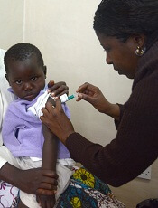
Credit: Caitlin Kleiboer
New research suggests that combining the use of malaria vaccines and insecticide-treated bed nets may actually increase the incidence of malaria.
The researchers used a mathematical model of malaria transmission to examine potential interactions between vaccines and bed nets.
They found that using insecticide-treated bed nets along with pre-erythrocytic vaccines (PEVs) or blood-stage vaccines (BSVs) increased the number of malaria cases.
However, using bed nets in conjunction with transmission-blocking vaccines (TBVs) resulted in fewer cases of malaria and increased the probability of eliminating the disease.
“The joint use of bed nets and vaccines will not always lead to consistent increases in the efficacy of malaria control,” said study author Mercedes Pascual, PhD, of the University of Michigan in Ann Arbor.
“Specifically, our study suggests that the combined use of some malaria vaccines with bed nets can lead to increased morbidity and mortality in older age classes.”
Dr Pascual and her colleagues described this research in Proceedings of the National Academy of Sciences.
The team noted that the malaria vaccine candidates currently under development fall into 3 categories, each focusing on a different stage of the malaria life cycle.
PEVs aim to reduce the chances that a person will be infected when bitten by a disease-carrying mosquito. BSVs don’t block infection but try to reduce the level of disease severity and the number of fatalities.
And TBVs don’t protect vaccinated individuals against infection or illness, but they prevent mosquitoes from spreading the disease to others after biting a vaccinated person.
Dr Pascual and her colleagues found that using bed nets in communities treated with BSVs can increase levels of morbidity—to levels even higher than those expected in the absence of nets. Furthermore, BSVs can’t promote malaria elimination on their own.
PEVs can promote malaria elimination, but the researchers found regions of decreased morbidity when PEV vaccination levels were low and increased morbidity when PEV vaccination levels were high.
This suggests that higher levels of PEV coverage and bed net use could result in malaria elimination, but it would involve crossing a peak of enhanced morbidity.
Finally, the researchers found that using bed nets in communities treated with TBVs always leads to significant decreases in morbidity and increases the probability of malaria elimination.
“Ironically, the vaccines that work best with bed nets are the ones that do not protect the vaccinated host—the bed net does that—but instead block transmission of malaria in mosquitoes that have found an opportunity to bite vaccinated hosts,” said study author Yael Artzy-Randrup, PhD, of the University of Amsterdam in The Netherlands.
Unraveling the interactions between bed nets and vaccines is especially challenging due to the complex and transient nature of malaria immunity, the researchers noted.
A child’s first malaria infection can result in severe, sometimes fatal, illness. If the child survives, he or she will gain partial immunity that reduces the risk of severe illness in the future.
Additional bites from infected mosquitoes can help the child retain that immunity, which would otherwise wane after 1 to 2 years. But the combination of bed nets and certain vaccines can undermine that natural immunity.
“This complexity is at the heart of why it has been so hard to develop any sort of malaria vaccine,” said study author Andrew Dobson, DPhil, of Princeton University in New Jersey. ![]()
Label changes report new side effects for hematology drugs

Credit: CDC
Several hematology drugs approved by the US Food and Drug Administration (FDA) have recently undergone label changes to reflect newly reported adverse events.
Changes have been made to the labels for the JAK1/2 inhibitor ruxolitinib (Jakafi), the anti-CD20 monoclonal antibody obinutuzumab (Gazyva), the factor Xa inhibitor rivaroxaban (Xarelto), and the hematopoietic stem cell mobilizer plerixafor (Mozobil).
Plerixafor
Plerixafor is FDA-approved for use in combination with granulocyte-colony stimulating factor to mobilize hematopoietic stem cells to the peripheral blood for collection and subsequent autologous transplantation in patients with non-Hodgkin lymphoma and multiple myeloma.
The product’s label was changed to include a new entry under the “Adverse Reactions” heading. Postmarketing experience suggested the drug may cause abnormal dreams and nightmares.
Rivaroxaban
Rivaroxaban is a factor Xa inhibitor that’s FDA-approved to reduce the risk of stroke and systemic embolism in patients with nonvalvular atrial fibrillation, for the treatment of deep vein thrombosis (DVT) and pulmonary embolism (PE), to reduce the risk of recurrent DVT and PE, and to prevent DVT, which may lead to PE, in patients undergoing knee or hip replacement surgery.
Postmarketing experience has led to two changes to the “Adverse Reactions” section of rivaroxaban’s label. Thrombocytopenia has been added as an adverse reaction, and the term “cytolytic hepatitis” has been replaced with “hepatitis (including hepatocellular injury).”
Obinutuzumab
Obinutuzumab is a CD20-directed cytolytic antibody that is FDA-approved in combination with chlorambucil to treat patients with previously untreated chronic lymphocytic leukemia.
The “Warnings and Precautions” section of obinutuzumab’s label has been changed to reflect that fatal infections have been reported in patients who received the drug.
The label has also been changed to coincide with changes in trial data. The label now states that obinutuzumab caused grade 3 or 4 neutropenia in 33% of patients and grade 3 or 4 thrombocytopenia in 10% of patients.
Ruxolitinib
Ruxolitinib is a JAK1/JAK2 inhibitor that’s FDA-approved to treat patients with polycythemia vera (PV) who cannot tolerate or don’t respond to hydroxyurea, as well as patients with intermediate or high-risk myelofibrosis.
Ruxolitinib’s label now includes a warning that symptoms of myeloproliferative neoplasms may return about a week after discontinuing treatment. The label also advises healthcare professionals to discourage patients form interrupting or discontinuing ruxolitinib without consulting their physician.
In addition, a warning about the risk of non-melanoma skin cancer associated with ruxolitinib, as well as advice for informing patients of this risk, have been added to ruxolitinib’s label.
The label has undergone significant changes in sections 6.1, “Clinical Trials Experience in Myelofibrosis” and 6.2 “Clinical Trial Experience in Polycythemia Vera.” It now includes additional information on the risk of thrombocytopenia, anemia, and neutropenia.
Under the “Special Populations” heading, recommendations were added to reduce the drug’s dose in patients with PV and moderate or severe renal impairment, as well as PV patients with hepatic impairment. ![]()
System can detect Candida better, faster than blood culture
Credit: Jeremy L. Grisham
A diagnostic system can detect sepsis pathogens with high sensitivity and specificity in 3 to 5 hours, eliminating the need for blood culture, according to a study published in Clinical Infectious Diseases.
The system consists of the T2Candida Panel and the T2Dx Instrument, and it provides direct detection of 5 yeast pathogens—Candida albicans, Candida tropicalis, Candida parapsilosis, Candida glabrata, and Candida krusei—in whole blood samples.
T2Candida and T2Dx were cleared for use by the US Food and Drug Administration in September. They are the first diagnostic products powered by T2MR, a magnetic resonance-based diagnostic technology platform that does not require blood culture and sample purification or preparation.
“The ability to determine the presence or absence of Candida within hours—compared to days [with blood culture]—is paradigm-changing for patients at risk for these infections,” said study author Eleftherios Mylonakis, MD, PhD, of Rhode Island Hospital and The Miriam Hospital in Providence.
“It will allow us to move from a ‘best-guess’ approach in treating high-risk patients, such as cancer and transplant patients and patients in the intensive care unit, to a more informed approach where we can quickly direct the best course of therapy, potentially improving patient outcomes and saving lives.”
Study findings
For this multicenter study, Dr Mylonakis and his colleagues collected blood specimens from 1801 hospitalized patients between the ages of 18 and 95 who had a blood culture ordered as part of routine care.
T2Candida and T2Dx demonstrated an overall specificity of 99.4% per assay and 98.1% per patient. The system yielded a specificity of 98.9% for C albicans/C tropicalis, 99.3% for C parapsilosis, and 99.9% for C krusei/C glabrata.
The system had an overall sensitivity of 91.1% per assay and 91.0% per patient. It yielded a sensitivity of 92.3% for C albicans/C tropicalis, 94.2% for C parapsilosis, and 88.1% for C krusei/C glabrata.
The mean time to a positive result for T2Candida and T2Dx was 4.4 hours, compared to 129 hours for blood culture and species identification. The mean time to negative result for T2Candida and T2Dx was 4.2 hours, compared to at least 120 hours for blood culture.
In one case described in the paper, T2Candida and T2Dx detected a Candida infection that blood culture missed in 12 successive tests.
Seven days after the T2Candida result was obtained, physicians performed an invasive procedure to obtain a tissue culture, which proved that the T2Candida result accurately identified a case of intra-abdominal candidiasis.
“Blood culture, the current standard of care for the diagnosis of Candida infections, is known to have poor sensitivity overall and has 38% sensitivity in proven and probable cases of invasive candidiasis,” said study author Cornelius J. Clancy, MD, of the University of Pittsburgh in Pennsylvania.
“In our case, the T2Candida Panel has shown that it can rapidly identify intra-abdominal candidiasis where 12 serial blood culture results were negative. In many patients at risk for candidiasis, the collection of tissue samples for diagnosis is not possible due to their underlying medical conditions. Achieving the level of sensitivity demonstrated in this case, without requiring an intra-abdominal sample, has the potential to positively impact the practice of medicine for these patients.” ![]()
Study offers explanation for gender gaps in academia

Credit: Rhoda Baer
New research offers an explanation for the lack of women in certain academic fields.
It isn’t that women don’t want to work long hours or can’t compete in highly selective fields, and it isn’t that they are less analytical than men, the researchers reported.
Instead, it appears that women are underrepresented in academic fields whose practitioners put a lot of emphasis on the importance of being brilliant—a quality some people assume women lack.
Sarah-Jane Leslie, PhD, of Princeton University in New Jersey, and her colleagues reported these findings in Science.
The researchers focused on a broad swath of academic disciplines, including those in the sciences, the humanities, social sciences, and math.
They focused on the culture of different fields, reasoning that stereotypes of women’s inferior intellectual abilities might help explain why women are underrepresented in fields—such as physics or philosophy—that idolize geniuses.
The team surveyed more than 1800 graduate students, post-doctoral researchers, and faculty members in 30 academic disciplines and, among other things, asked them what qualities were required for success in their fields.
Across the board, in the sciences, technology, engineering, and math (STEM) fields, as well as in the humanities and social sciences, women were found to be underrepresented in those disciplines whose practitioners put a premium on brilliance.
“We’re not saying brilliance—or valuing brilliance—is a bad thing,” said study author Andrei Cimpian, PhD, of the University of Illinois at Urbana-Champaign.
“And we’re not saying women are not brilliant or that being brilliant isn’t helpful to one’s academic career. Our data don’t address that. What they suggest is that conveying to your students a belief that brilliance is required for success may have a differential effect on males and females that are looking to pursue careers in your field.”
The team also tested 3 other hypotheses that might help explain women’s underrepresentation in some fields: that women avoid careers that require them to work long hours, that women are less able than men to get into highly selective fields, and that women are outnumbered by men in fields that require analytical, systematical reasoning.
“We found that none of these 3 alternative hypotheses was able to predict women’s representation across the academic spectrum,” Dr Leslie said. “A strong emphasis on brilliance among practitioners of particular fields was the best predictor of women’s underrepresentation in those fields.”
The researchers are still investigating whether women are actively avoiding fields that focus on cultivating brilliant individuals, or if practitioners in those fields are discriminating against women based on their beliefs about women’s aptitudes. A combination of the two is certainly plausible, according to Dr Cimpian.
“There is no convincing evidence in the literature that men and women differ intellectually in ways that would be relevant to their success across the entire range of fields we surveyed,” Dr Cimpian said. “So it is most likely that female underrepresentation is not the result of actual differences in intellectual ability but rather the result of perceived or presumed differences between women and men.” ![]()
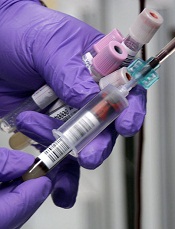
Antibiotics may enhance mosquitoes’ ability to transmit malaria

Credit: James Gathany
Mosquitoes’ ability to transmit malaria may be affected by antibiotics in the blood of those they bite, according to research published in Nature Communications.
Feeding on blood that contained the antibiotics penicillin and streptomycin (PS) hindered bacterial growth in mosquitoes’ guts.
The mosquitoes also became more susceptible to malaria infection, exhibited improved fertility, and lived longer than mosquitoes whose blood meals did not contain PS.
Study investigators said these findings do not suggest people should avoid taking antibiotics. But the study does indicate that more research is needed to explore how different combinations of antibiotics affect mosquitoes’ ability to spread malaria.
“Antibiotics are a valuable weapon in the fight against malaria and other diseases,” said study author Mathilde Gendrin, PhD, of Imperial College London in the UK.
“Our study suggests that the presence of antibiotics in people’s blood may have hidden effects on the guts of mosquitoes when mosquitoes ingest that blood, and that these effects could alter the mosquitoes’ ability to transmit malaria.”
Dr Gendrin and her colleagues chose to evaluate the effects of PS because these antibiotics are not used to combat malaria and don’t directly affect malaria parasites.
The team began their research by supplementing blood with therapeutic concentrations of PS and feeding the blood to Anopheles gambiae mosquitoes.
The investigators monitored the bacterial load in the mosquito gut over 3 blood feeds offered every 3 days. And they found that the proliferation of bacteria typically seen at 24 hours after a blood meal was reduced by 70% when mosquitoes received blood containing PS.
Next, the researchers conducted 3 experiments to determine whether receiving blood containing PS would influence the mosquitoes’ susceptibility to malaria infection.
In the first experiment, the team fed mosquitoes blood from rodents infected with Plasmodium berghei before or after the animals received PS.
The investigators assessed infection by counting the proportion of mosquitoes carrying oocysts (prevalence) and the number of oocysts per mosquito (intensity). In the presence of PS, infection prevalence increased by 21% (P=5.10-7), and the median intensity doubled (P=0.041).
In the second experiment, the researchers assessed the effect of PS on infection with human parasites. They fed mosquitoes blood containing Plasmodium falciparum gametocytes (cultured in vitro) and supplemented the blood with PS or buffer.
Again, the team observed higher infection prevalence (P=0.0033) and intensity (P=0.00026) in the presence of PS.
In the third experiment, the investigators fed mosquitoes blood freshly drawn from children carrying P falciparum gametocytes. The blood was supplemented with PS or buffer.
Once more, the intensity of infection significantly increased in the presence of PS (P=0.02). The 6.3% increase in infection prevalence was not statistically significant (P=0.4), but the researchers said this was likely due to the reduced statistical power of the experiment.
The team’s final experiments assessed the effects of PS on mosquitoes’ fertility and survival.
Exposure to PS led to a 32% higher proportion of egg-laying females (P=0.0038) and a 53% higher average number of eggs per female (P=0.016), although the proportion of eggs hatching into larvae was not affected.
Mosquito survival increased significantly after the insects ingested PS-treated human blood. The P value was 0.017 for the first blood meal and 0.0016 for all 3 blood meals.
“We only looked at two antibiotics in our study, so this is early stage research, and we don’t know what it means for other antibiotics,” Dr Gendrin noted.
“It’s possible other antibiotics might have no effect, or that they might make it harder for mosquitoes to transmit malaria. We would like to see much more research carried out to further understand our findings and to explore how other antibiotics might affect bacteria in the guts of mosquitoes.”
“Ultimately, we hope that understanding any hidden effects of different antibiotics would mean we can combat the spread of malaria more effectively,” added study author George Christophides, PhD, also of Imperial College London.
“For example, if particular antibiotics make mosquitoes more able to transmit malaria, the use of these could be combined with supply of bed nets in order to reduce mosquito bites. Additionally, if a doctor prescribed such antibiotics to a malaria patient, they could combine this with a drug such as atovaquone that can both treat malaria and block its transmission.” ![]()
Imaging reveals how HSPCs interact with niche

(green) in a zebrafish
Boston Children’s Hospital
Using a zebrafish model and enhanced imaging, a group of researchers discovered how hematopoietic stem and progenitor cells (HSPCs) interact with their niche.
Subsequent imaging in mice showed that HSPCs behaved the same way in mammals, which suggests similar results might be observed in humans.
In fact, the researchers are already using the results of this study to inform research on hematopoietic stem cell transplants.
“The same process occurs during a bone marrow transplant as occurs in the body naturally,” said senior study investigator Leonard Zon, MD, of Boston Children’s Hospital in Massachusetts.
“Our direct visualization gives us a series of steps to target, and, in theory, we can look for drugs that affect every step of that process.”
He and his colleagues described this research in a paper published in Cell, as well as in two animations on YouTube, one that’s general and one more technical.
“Stem cell and bone marrow transplants are still very much a black box,” said study author Owen Tamplin, PhD, also of Boston Children’s Hospital.
“Cells are introduced into a patient, and, later on, we can measure recovery of their blood system, but what happens in between can’t be seen. Now, we have a system where we can actually watch that middle step.”
The researchers already knew that HSPCs bud off from cells in the aorta, then circulate in the body until they find a niche where they’re prepped for creating blood.
With the current study, the team observed how this niche forms, using time-lapse imaging of naturally transparent zebrafish embryos and a genetic modification that tagged the HSPCs green.
On arrival in its niche (in the tail in zebrafish), the newborn HSPC attaches itself to the blood vessel wall. There, chemical signals prompt it to squeeze itself through the wall and into a space just outside the blood vessel. Other cells begin to interact with the HSPC, and nearby endothelial cells wrap themselves around it.
“We think that is the beginning of making a stem cell happy in its niche, like a mother cuddling a baby,” Dr Zon said.
As the HSPC is being “cuddled,” it’s brought into contact with a nearby stromal cell that helps keep it attached.
The “cuddling” was reconstructed from confocal and electron microscopy images of the zebrafish taken during this stage. Through a series of image slices, the researchers were able to reassemble the whole 3D structure—HSPC, cuddling endothelial cells, and stromal cells.
“Nobody’s ever visualized live how a stem cell interacts with its niche,” Dr Zon said. “This is the first time we get a very high-resolution view of the process.”
Next, the cuddled HSPC begins dividing. One daughter cell leaves the niche, while the other stays. Eventually, all the HSPCs leave and begin colonizing their future site of blood production. (In zebrafish, this is in the kidney, which is similar to mammalian bone marrow.)
Additional imaging in mice revealed evidence that HSPCs go through much the same process in mammals, which makes it likely in humans too.
These detailed observations are already informing the Zon lab’s attempt to improve stem cell transplants. By conducting a chemical screen in large numbers of zebrafish embryos, the researchers found that the compound lycorine promotes interaction between the HSPC and its niche, leading to greater numbers of HSPCs in the adult fish. ![]()
IPT doesn’t seem to benefit children with anemia

one of the trials took place
Credit: Gabrielle Tenenbaum
Intermittent preventive antimalarial treatment (IPT) does not provide much benefit for anemic children living in malaria-endemic regions, results of a review indicate.
Researchers reviewed 6 randomized, controlled trials and found that IPT, doses of antimalarial drugs given at regular intervals in case children had contracted malaria, did lead to an improvement in hemoglobin levels.
But this did not translate to a reduction in the incidence of anemia or the rate of death and hospitalization compared to children who received placebo.
“While we did note small benefits in hemoglobin levels when treating anemic children with IPT, there was no detectable effect on the number of deaths or hospital admissions,” said review author Mwaka Athuman, of Ifakara Health Institute in Dodoma, Tanzania, Africa.
“However, 3 of the trials were carried out in areas where malaria transmission was low, so any estimate of the protective effect of IPT would be expected to be modest. The summary of the evidence will assist people forming policy guidance as to whether IPT is worthwhile and provide a basis for researchers to consider whether additional studies are needed.”
The researchers reported these findings in the Cochrane Database of Systematic Reviews.
The team reviewed 6 trials that included a total of 3847 children with anemia. Three trials were conducted in areas of low malaria endemicity and 3 in areas of moderate-to-high endemicity.
In all trials, there was a group of children who received IPT and a control group receiving placebo. In some trials, children also received iron supplements, and this was taken into consideration when the researchers analyzed the data.
Data from 4 of the trials showed that IPT did increase the mean change in hemoglobin levels from baseline to follow-up at 12 weeks—on average, by 0.32 g/dL. The reviewers dubbed this moderate-quality evidence.
Results from the same 4 trials showed that the mean hemoglobin at 12 weeks’ follow-up was, on average, 0.35 g/dL higher in the IPT group than in the placebo group. This was considered low-quality evidence.
Regardless of improvements in hemoglobin, there was no significant difference in the number of children who had anemia at 12 weeks. The median risk of anemia across 4 trials was 579 per 1000 in the placebo group and 561 per 1000 in the IPT group. This was considered moderate-quality evidence.
Similarly, there was no significant difference in the rate of death and hospitalization at 6 months between children who received IPT and those who received placebo.
The median risk of both events combined was 34 per 1000 in the placebo group and 31 per 1000 in the IPT group. This was based on data from 3 trials and was considered moderate-quality evidence.
For all of these outcomes, there was no significant difference between children who received iron and those who did not, and there was no difference between children living in regions of low malaria endemicity and those living in regions of moderate-to-high malaria endemicity. ![]()
one of the trials took place
Credit: Gabrielle Tenenbaum
Intermittent preventive antimalarial treatment (IPT) does not provide much benefit for anemic children living in malaria-endemic regions, results of a review indicate.
Researchers reviewed 6 randomized, controlled trials and found that IPT, doses of antimalarial drugs given at regular intervals in case children had contracted malaria, did lead to an improvement in hemoglobin levels.
But this did not translate to a reduction in the incidence of anemia or the rate of death and hospitalization compared to children who received placebo.
“While we did note small benefits in hemoglobin levels when treating anemic children with IPT, there was no detectable effect on the number of deaths or hospital admissions,” said review author Mwaka Athuman, of Ifakara Health Institute in Dodoma, Tanzania, Africa.
“However, 3 of the trials were carried out in areas where malaria transmission was low, so any estimate of the protective effect of IPT would be expected to be modest. The summary of the evidence will assist people forming policy guidance as to whether IPT is worthwhile and provide a basis for researchers to consider whether additional studies are needed.”
The researchers reported these findings in the Cochrane Database of Systematic Reviews.
The team reviewed 6 trials that included a total of 3847 children with anemia. Three trials were conducted in areas of low malaria endemicity and 3 in areas of moderate-to-high endemicity.
In all trials, there was a group of children who received IPT and a control group receiving placebo. In some trials, children also received iron supplements, and this was taken into consideration when the researchers analyzed the data.
Data from 4 of the trials showed that IPT did increase the mean change in hemoglobin levels from baseline to follow-up at 12 weeks—on average, by 0.32 g/dL. The reviewers dubbed this moderate-quality evidence.
Results from the same 4 trials showed that the mean hemoglobin at 12 weeks’ follow-up was, on average, 0.35 g/dL higher in the IPT group than in the placebo group. This was considered low-quality evidence.
Regardless of improvements in hemoglobin, there was no significant difference in the number of children who had anemia at 12 weeks. The median risk of anemia across 4 trials was 579 per 1000 in the placebo group and 561 per 1000 in the IPT group. This was considered moderate-quality evidence.
Similarly, there was no significant difference in the rate of death and hospitalization at 6 months between children who received IPT and those who received placebo.
The median risk of both events combined was 34 per 1000 in the placebo group and 31 per 1000 in the IPT group. This was based on data from 3 trials and was considered moderate-quality evidence.
For all of these outcomes, there was no significant difference between children who received iron and those who did not, and there was no difference between children living in regions of low malaria endemicity and those living in regions of moderate-to-high malaria endemicity.
Investigation of simulated saline continues
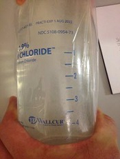
Credit: FDA
The US Food and Drug Administration (FDA) and the Centers for Disease Control and Prevention (CDC) are still investigating multiple instances of Wallcur’s simulated intravenous (IV) saline products being administered to patients.
So far, more than 40 patients have received infusions of Wallcur’s simulated IV saline solution, Practi-0.9% sodium chloride solution, which is intended for training purposes only. The product is not sterile and should not be injected in humans or animals.
There have been many adverse events associated with the infusions, including fever, chills, tremors, and headache. Some patients were hospitalized, and there has been 1 death, although it’s not clear if this death is directly related to the product.
Adverse events have been reported in 7 states: Florida, Georgia, Idaho, Louisiana, North Carolina, New York, and Colorado.
The FDA, in partnership with the CDC, has collected samples of Wallcur Practi 0.9% sodium chloride solution from clinics and distributors. These products are being tested to determine if they caused the adverse events observed in patients.
In addition, Wallcur has initiated a voluntary recall of Practi-0.9% sodium chloride IV solutions.
Most medical facilities that received the product said they were unaware that the IV solution bags were simulation products. However, at least one clinic recognized the Wallcur product was a simulation product upon receipt and returned it to the distributor.
The FDA said it is working with distributors who sold the simulated IV products and clinics that purchased and administered the products from Wallcur to determine how these products entered the supply chain and were administered to patients.
While Sodium Chloride 0.9% Injection (normal saline) has been in short supply, the FDA has been working with manufacturers to end the shortage.
The FDA has allowed the temporary distribution of additional IV normal saline from alternate sources: Fresenius Kabi USA, Baxter Healthcare Corp., and B. Braun Medical Inc. Currently, normal saline is available from several manufacturers, as posted on the FDA’s website.
FDA recommendations
The FDA is encouraging healthcare providers to ensure IV solution simulation products are removed from office inventory to eliminate the possible injection of Wallcur simulated products into patients.
Providers should visually inspect all current IV saline solution bags to ensure none of the bags are labeled “Wallcur,” “Practi-products,” “For clinical simulation,” or “Not for use in human or animal patients.”
If you have products labeled with any of these words or suspect you may have received other products intended for training purposes, separate simulation products from existing inventory, and contact your distributor for directions on how to return these products.
If you have received Wallcur Practi-products by mistake, please contact the distributor, or Wallcur, LLC of San Diego for return instructions.
Consider reviewing your office procedures and make sure there are procedures in place to visually inspect all future shipments of normal saline products to ensure they are for clinical use.
If you suspect that any Wallcur training IV products may have been administered to a patient, whether or not the incident has resulted in an adverse event:
- Evaluate all potentially exposed patients with new or ongoing symptoms
- Use appropriate treatment
- Report suspected cases to the state health department
- Report any adverse events following the use of these products to the FDA’s MedWatch program online or at 1-800-332-1088.
Patients who believe they received an injection of Wallcur simulated IV solution should contact their healthcare provider.
Patients who received simulated IV saline experienced fever, chills, muscle aches, and headaches almost immediately upon injection, and some required hospitalization. In most reported cases, these signs and symptoms were immediately recognized, and patients received appropriate medical attention.
Wholesalers, distributors, and suppliers of IV saline products should inspect their inventory to ensure they are not distributing simulated products as clinical-use products.
If you suspect you may have distributed Wallcur simulated IV solution to clients by mistake, immediately attempt to recall the products and warn clients of the potential risks. You should also contact Wallcur and your distributor and file a report with the FDA’s MedWatch program.
Platelet transfusions may increase risk of death
![]()
Platelet transfusions may increase the risk of death in patients with platelet consumptive disorders, results of a large study suggest.
Investigators observed a 2-fold increase in the risk of death among patients with thrombotic thrombocytopenic purpura (TTP) who received platelet transfusions and a 5-fold increase among transfused patients with heparin-induced thrombocytopenia (HIT).
There was no increased risk among patients with immune thrombocytopenia (ITP), however.
Aaron Tobian, MD, PhD, of the Johns Hopkins University School of Medicine in Baltimore, Maryland, and his colleagues reported these results in Blood. They were previously presented at the 2014 AABB Annual Meeting.
“Because these conditions are so rare, they’re difficult to study,” Dr Tobian noted. “There was some suggestion that transfusion may be harmful in these conditions, but it really was not known until now.”
“Our study is the first one to show that platelet transfusions are frequently administered to patients with ITP, HIT, and TTP, and that they’re associated with higher odds of arterial blood clots and mortality in TTP and HIT.”
For this study, Dr Tobian and his colleagues analyzed data from the Nationwide Inpatient Sample, a federal database that contains billing records for about 20% of all patients treated and discharged at about 1000 US community hospitals in 47 states.
The database contains information on about 8 million inpatient hospitalizations per year nationwide. The study covered the years 2007 to 2011.
During that time, there were 79,980 hospital admissions for ITP, 10,624 for TTP, and 6332 for HIT. Platelet transfusions were reported in 10.1% of hospitalizations for TTP, 7.1% for HIT, and 25.8% for ITP.
“Our analysis found no significantly increased risks from platelet transfusions in ITP,” said study author Ruchika Goel, MD, also of Johns Hopkins.
“But in TTP, a platelet transfusion increased the odds of a potentially lethal arterial blood clot more than 5-fold and doubled the odds of a heart attack.”
Specifically, in an age- and gender-adjusted analysis, the odds ratio (OR) for arterial thrombosis was 5.8, and the OR for acute myocardial infarction (AMI) was 2.0 in TTP patients who received platelet transfusions. The OR for stroke was 1.6, and the OR for venous thrombosis was 1.1.
Similarly, HIT patients had an increased risk of arterial thrombosis (OR=3.4) and AMI (OR=1.9) if they received a platelet transfusion. But they did not have an increased risk of venous thrombosis (OR=0.8) or stroke (OR=0.5).
Platelet transfusions among ITP patients were not significantly associated with venous thrombosis (OR=1.3), arterial thrombosis (OR=0.3), AMI (OR=1.3), or stroke (OR=1.3) after adjustment for age and gender.
The all-cause, in-hospital mortality rates were 8.8% for TTP patients, 3.4% for HIT patients, and 1.4% for ITP patients.
Patients with TTP and HIT had a significantly increased risk of all-cause mortality if they received platelet transfusions, with age- and gender-adjusted ORs of 2.0 and 5.2, respectively. But platelet transfusions were not significantly associated with mortality in ITP patients, with an OR of 1.1.
The investigators said they were surprised at the prevalence of platelet transfusions in this patient population, in spite of some practitioners’ concerns about the risks.
But Dr Tobian noted that, in some cases, doctors may not know a patient has a platelet disorder until they see the potentially deadly reaction to the transfusion. And in other cases, the transfusion may be used as a last resort.
He and his colleagues believe that, for patients with HIT and TTP, platelet transfusions should be reserved “only for severe, life-threatening bleeding refractory to other therapies or major surgery.”
Real-world data with rivaroxaban similar to trial data
Credit: CDC
A post-marketing study in patients with non-valvular atrial fibrillation (NVAF) suggests that rates and patterns of major bleeding associated with the use of rivaroxaban in routine clinical practice are generally consistent with those observed in a previous phase 3 trial.
The new study, which included more than 27,000 patients, showed a low rate of major bleeding. And gastrointestinal bleeding was more common than intracranial hemorrhage.
While the results are not intended for a direct comparison, the researchers said these data are generally consistent with data from the ROCKET-AF trial.
The results, published in Clinical Cardiology, are the initial findings from an ongoing, 5-year, observational study of patients using rivaroxaban daily over the course of their lives.
“These findings reaffirm the safety profile of Xarelto [rivaroxaban],” said study author W. Frank Peacock, MD, of the Baylor College of Medicine in Houston, Texas.
“We anticipate future findings from this 5-year observational study will continue to provide real-world information about the use of Xarelto in routine clinical practice.”
Dr Peacock and his colleagues analyzed data from January 1, 2013, to March 31, 2014, evaluating major bleeding in 27,467 NVAF patients treated with rivaroxaban in a real-world, clinical setting.
There were a total of 496 major bleeding events in 478 patients. So the incidence of major bleeding was 2.86 per 100 person-years, which was generally consistent with the rate reported in the ROCKET-AF trial.
Most bleeds were gastrointestinal (88.5%), followed by intracranial (7.5%). Fourteen patients each experienced 2 major bleeding events, and 2 patients each experienced 3 events.
Major bleeding was more likely in females and older patients. The incidence of major bleeding was 2.68 for males and 2.99 for females. The mean age of patients with major bleeding was 78.4, compared to 75.7 for those without major bleeding.
Comorbidities tended to be more prevalent among patients who experienced major bleeding. The most common were hypertension (95.6% vs 75.8%), coronary heart disease (64.2% vs 36.7%), heart failure (48.5% vs 23.7%), and renal disease (38.7% vs 16.7%).
Patients with major bleeding were less likely to be on medications such as statins, proton pump inhibitors, and amiodarone—29.1% vs 36.6%.
Fourteen patients died while in the hospital for major bleeding, for a fatal bleeding rate of 0.08 per 100 person-years. None of these patients had multiple bleeding events.
Fatal bleeding events included intracranial hemorrhage with intracerebral bleeding (n=7), gastrointestinal hemorrhage not otherwise specified (n=3), blood in the stool (n=2), subdural hemorrhage (n=1), and intracranial hemorrhage not otherwise specified (n=1).
Patients’ mean age at death was 82.4 years, and the mean time from hospitalization for the bleeding event to death was 3.9 days.
This ongoing study is funded by Janssen Scientific Affairs, LLC, and Bayer Healthcare, the companies developing rivaroxaban.