User login
Gene variations tied to drug-related hearing loss

Photo by Peter Barta
New research has revealed inherited genetic variations associated with rapid hearing loss in young cancer patients who receive cisplatin.
The drug is used to treat a range of cancers and is known to pose a risk of severe hearing loss, but the risk factors involved are not completely understood.
Now, researchers have found that variations in the gene ACYP2 are associated with an increased risk of cisplatin-related hearing loss.
Jun J. Yang, PhD, of St Jude Children’s Research Hospital in Memphis, Tennessee, and his colleagues reported this discovery in Nature Genetics.
The researchers checked the DNA of 238 young patients with brain tumors for more than 1.7 million common genetic variations. The team found that variations in ACYP2 were associated with as much as a 4-fold greater risk of cisplatin-related hearing loss.
The screening is among the first to survey the genetic landscape for clues to help explain why the risk of cisplatin-related hearing loss varies so widely among patients.
“This is an important first step in being able to pinpoint patients who are at higher risk of developing cisplatin toxicity and to learn how to better manage that risk,” said study author Clinton Stewart, PharmD, also of St Jude.
The researchers confirmed the association between the high-risk ACYP2 variants and cisplatin-related hearing loss in a separate group of 68 brain tumor patients. The association was independent of other risk factors for cisplatin-related hearing loss, including patient age and receipt of radiation therapy.
Twenty-four of the 306 patients in this study had at least one copy of the high-risk ACYP2 variant. All 24 patients had measurable hearing loss that occurred as early as weeks after beginning cisplatin therapy.
Overall, however, the ACYP2 variant explained a relatively small proportion of hearing damage. Just 12.4% of the 194 patients in this study with cisplatin-related hearing loss carried the ACYP2 variant.
“This suggests that other genes also contribute to the risk of hearing loss and are yet to be identified,” Dr Yang said. “Further research is needed to understand how the ACYP2 variations modify the risk . . . of cisplatin toxicity.”
Such studies could potentially lead to new medications to protect high-risk patients from cisplatin-related toxicity or help identify candidates for intensive monitoring of their hearing, Dr Stewart said. Early intervention could then be offered if problems are identified.
This study included patients enrolled in 1 of 3 trials designed by St Jude investigators for newly diagnosed pediatric brain tumors. The protocols involved similar treatment, including surgery to remove as much of the tumor as possible, followed by radiation, which was modified based on patient age and other risk factors.
The patients were scheduled to receive 4 rounds of cisplatin therapy. Patients’ hearing was tested before treatment began, after radiation therapy, after each round of chemotherapy, and then at regular standardized intervals. Analysis of the resulting data led to identification of ACYP2 and other variants.
“Our primary goal is to cure children with brain tumors, but we also have a duty to help patients survive with a high quality of life,” said Giles Robinson, MD, also of St Jude.
“Hearing loss can have a significant impact on a child’s quality of life, language development, and academic performance. There is no easy fix, but the more we know about the risk factors, the better we will understand how to use cisplatin.” ![]()
Signs may predict death in cancer patients

Researchers have identified 8 highly specific physical and cognitive signs that seem to be associated with imminent death in cancer patients.
The findings, published in Cancer, could offer clinicians the ability to better communicate with patients and their families.
The research might also help guide the medical team and caregivers when it comes to complex decision making, such as discontinuing tests and therapy, plans for hospital discharge, and hospice referral.
Previous studies in end-of-life care have focused on physicians prognosticating better. However, research on how to tell if a patient has entered the final days of life has been minimal, according to David Hui, MD, of the University of Texas MD Anderson Cancer Center in Houston.
“In the past, studies trying to understand the signs associated with impending death were conducted in people who were recognized as dying, so there’s a potential bias built into this model,” Dr Hui said.
“With our study, we observed a list of signs in patients from the time they were admitted to the palliative care unit. They were observed systematically, twice a day, without knowing if the patient would die or be discharged.”
Dr Hui and his colleagues observed 357 cancer patients, 57% of whom ultimately died. The researchers observed 52 physical and cognitive signs—identified by Dr Hui and his colleagues in previous research—twice a day from the patient’s admission to discharge or death.
Of those 52 signs, the 8 most highly associated with impending death within 3 days were:
- Nonreactive pupils
- Decreased response to verbal stimuli
- Decreased response to visual stimuli
- Inability to close eyelids
- Drooping of the nasolabial fold
- Neck hyperextension
- Grunting of vocal cords
- Upper gastrointestinal bleeding.
“When cancer patients reach the last days of life, this is an extremely emotional time for families; their stress levels cannot be understated,” Dr Hui said.
“Knowing when death is imminent would provide more information so caregivers can plan appropriately. For clinicians, having this information could help reassure families that we are providing the best care possible.”
Dr Hui stressed that this research is not yet practice-changing, but is an important step in understanding these 8 signs and their relation to impending death. In addition, the findings are only representative of imminent cancer death and should not be generalized to other causes of death.
Follow-up studies in different settings are planned. Dr Hui and his colleagues plan to look at the reliability of the identified signs, as well as evaluate this research in other countries and in the hospice setting. ![]()
Donor telomere length linked to survival after HSCT
![]()
Photo by Chad McNeeley
Leukocyte telomere length is associated with survival in patients who undergo hematopoietic stem cell transplant (HSCT) to treat severe aplastic anemia, according to research published in JAMA.
But it’s the donor’s telomere length—not the recipient’s—that makes the difference, the study showed.
Patients who received an HSCT from an unrelated donor had better overall survival at 5 years if that donor’s leukocytes had longer telomeres.
Shahinaz M. Gadalla, MD, PhD, of the National Cancer Institute in Rockville, Maryland, and colleagues conducted this research.
The group compared recipient and donor leukocyte telomere length prior to transplant with outcomes after unrelated HSCT for 330 patients with severe aplastic anemia.
The patients and their donors had pre-HSCT blood samples and other clinical results available at the Center for International Blood and Marrow Transplant Research. Patients underwent HSCT between 1989 and 2007 in 84 centers and were followed until March 2013.
The researchers categorized leukocyte telomere length for both recipients and donors based on the leukocyte telomere length tertiles in the donors: long (third tertile) and short (first and second tertiles combined).
The team found that longer donor leukocyte telomere length was associated with a higher overall survival. The 5-year overall survival was 56% in the longer telomere group and 40% in the shorter telomere group (P=0.009).
After adjusting for donor age and clinical factors associated with survival following HSCT in severe aplastic anemia, the risk of post-HSCT all-cause mortality remained approximately 40% lower in patients receiving HSCT from donors with long vs short leukocyte telomeres (hazard ratio [HR]=0.61).
There was no association between donor leukocyte telomere length and engraftment at 28 days (HR=0.94). Likewise, there was no association between telomere length and acute (HR=0.77) or chronic graft-vs-host disease (HR=0.81).
And recipient telomere length was not associated with overall survival (HR=0.91).
The researchers said these results suggest that donor leukocyte telomere length may have a role in long-term post-transplant survival. Authors of a related editorial explored what this discovery could mean for transplant centers. ![]()
Approved TKI could treat drug-resistant CML, ALL

Photo courtesy of CDC
New research indicates that a tyrosine kinase inhibitor (TKI) approved to treat advanced renal cell carcinoma could prove useful in treating patients with drug-resistant chronic myeloid leukemia (CML) or acute lymphoblastic leukemia (ALL).
The study showed that the TKI, axitinib, can inhibit BCR-ABL1 (T315I), a mutation known to confer drug resistance in CML and ALL.
“Since axitinib is already used to treat cancer, its safety is known,” said Kimmo Porkka, MD, PhD, of the University of Helsinki in Finland.
“[A] formal exploration of its clinical utility in drug-resistant leukemia can now be done in a fast-track mode. Thus, the normally very long path from lab bench to bedside is now significantly shortened.”
Dr Porkka and his colleagues described the newfound activity of axitinib in Nature.
The researchers used a drug sensitivity and resistance testing method developed at the University of Helsinki’s Institute for Molecular Medicine Finland (FIMM) to examine how patient-derived leukemia cells responded to a large panel of drugs.
In this way, the group identified axitinib as a promising drug candidate for CML and ALL. Axitinib effectively eliminated drug-resistant leukemia cells.
The TKI inhibited BCR-ABL1 (T315I) at biochemical and cellular levels by binding to the active form of ABL1 (T315I) in a mutation-selective binding mode.
The researchers said this suggests the T315I mutation shifts the conformational equilibrium of the kinase in favor of an active (DFG-in) A-loop conformation, which has more optimal binding interactions with axitinib.
“If you think of the targeted protein as a lock into which the cancer drug fits in as a key, the resistant protein changes in such a way that we need a different key,” said study author Brion Murray, PhD, of Pfizer Worldwide Research & Development in San Diego, California.
“In the case of axitinib, it acts as two distinct keys—one for renal cell carcinoma and one for leukemia.”
The researchers also treated a CML patient with axitinib and observed a “rapid reduction” of T315I-positive cells in the patient’s bone marrow.
“Further research will determine whether these findings have the potential to significantly improve the standard of care for this select group of CML patients and patients with other related leukemias,” Dr Murray concluded. ![]()
FDA inspection findings rarely reported in literature

Photo by Esther Dyson
A new study suggests that, when the US Food and Drug Administration (FDA) identifies problems at clinical trial sites, those findings are seldom reported in peer-reviewed articles later written about the research.
Only 4% of the articles analyzed in this study disclosed that an FDA inspection revealed significant problems at a trial site.
Charles Seife, of the Carter Institute of Journalism at New York University in New York, New York, reported this finding in JAMA Internal Medicine.
Seife noted that the FDA classifies its inspections based on the severity of the violations, and the most severe is “official action indicated (OAI),” which means objectionable conditions or practices that warrant regulatory action.
During the 2013 fiscal year, about 2% of the 644 inspections the FDA conducted at trial sites were classified as OAI.
With this in mind, Seife and his students set out to identify published trials in which an FDA inspection revealed significant problems and determine whether there was mention of it in the medical literature.
The group identified 57 trials in which an FDA inspection uncovered the following problems:
- Falsification or submission of false information (22 trials, 39%)
- Problems with adverse events reporting (14 trials, 25%)
- Protocol violations (42 trials, 74%)
- Inadequate or inaccurate recordkeeping (35 trials, 61%)
- Failure to protect the safety of patients and/or issues with oversight or informed consent (30 trials, 53%)
- Violations that were not otherwise characterized (20 trials, 35%).
Only 3 of the 78 publications (4%) that resulted from these trials mentioned the objectionable conditions or practices.
“The FDA does not typically notify journals when a site participating in a published clinical trial receives an OAI inspection, nor does it generally make any announcement intended to alert the public about the research misconduct that it finds,” Seife wrote.
“The documents the agency discloses tend to be heavily redacted. As a result, it is usually very difficult, or even impossible, to determine which published clinical trials are implicated by the FDA’s allegations of research misconduct.”
In a related editorial, Robert Steinbrook, MD, of the Yale School of Medicine in New Haven, Connecticut, and Rita F. Redberg, MD, of the University of California, San Francisco, wrote that Seife’s study highlights an important area for improved reporting of clinical trials and enhanced transparency at the FDA.
“A central responsibility of medical journals is maintaining and improving trust in the medical literature,” they wrote. “Journals should expect that investigators and sponsors of clinical trials would promptly notify them of substantial findings from FDA and other regulatory agency inspections and modify their reports of clinical trials as needed, either before or after publication.” ![]()
Study supports iron supplementation after blood donation

Photo by Charles Haymond
Low-dose oral iron supplementation can reduce the time to hemoglobin recovery after blood donation, according to a study published in JAMA.
Researchers found that a daily dose of ferrous gluconate (37.5 mg of elemental iron) reduced blood donors’ time to 80% hemoglobin recovery, whether the donors were iron-depleted (with ferritin levels of 26 ng/mL or lower) or iron-replete (with ferritin levels higher than 26 ng/mL).
Joseph E. Kiss, MD, of the Institute for Transfusion Medicine in Pittsburgh, Pennsylvania, and his colleagues conducted this study at 4 regional blood centers in the US.
The researchers randomized 215 subjects (who had not donated whole blood or red blood cells within 4 months) to receive one tablet of ferrous gluconate daily or no iron for 24 weeks after donating a unit of whole blood (500 mL).
The study’s primary outcomes were time to recovery of 80% of the post-donation decrease in hemoglobin and recovery of baseline ferritin levels.
The researchers found that subjects who received iron supplementation achieved 80% hemoglobin recovery more quickly than subjects who did not receive iron, regardless of ferritin levels.
In the low-ferritin group, the mean time to 80% hemoglobin recovery was 32 days in subjects who received iron and 158 days in those who did not (P<0.001). In the higher-ferritin group, the mean time to 80% hemoglobin recovery was 31 days in subjects who received iron and 78 days in those who did not (P=0.02).
In the low-ferritin group, the median time to recovery of baseline ferritin levels was 21 days in subjects who received iron and more than 168 days in subjects who did not. In the higher-ferritin group, the median time to recovery of baseline ferritin levels was 107 days in subjects who received iron and more than 168 days in those who did not.
The median time to recovery of iron stores was 76 days in all subjects who received iron supplements and more than 168 days in those who did not (P<0.001). Sixty-seven percent of subjects who did not receive iron had failed to recover their iron stores by 168 days.
The researchers said these findings raise important considerations regarding the 8-week minimum waiting period between blood donations that is required in the US and Canada. This is a shorter period than those allowed in many other countries.
The study suggests that hemoglobin recovery differs according to a donor’s pre-donation ferritin level, but, even in iron-replete donors, the mean recovery was only 70% at 8 weeks.
Prolonging the minimum wait time between blood donations would allow for more thorough recovery, the researchers noted, but it could also compromise the blood supply. Furthermore, increasing the waiting period might not be adequate for donors who do not take iron supplements. ![]()
Computer model simulates blood development

Photo courtesy of
University of Cambridge
A new computer model that simulates blood cell development could aid the discovery of novel treatments for hematologic malignancies, according to researchers.
“With this new computer model, we can carry out simulated experiments in seconds that would take many weeks to perform in the laboratory, dramatically speeding up research into blood development and the genetic mutations that cause leukemia,” said Bertie Gottgens, PhD, of the University of Cambridge in the UK.
Dr Gottgens and his colleagues explained this research in Nature Biotechnology.
To start, the researchers measured the activity of 48 genes in 3934 hematopoietic progenitor cells. They used the resulting dataset to construct the computer model of blood cell development, using computational approaches originally developed at Microsoft Research for the synthesis of computer code.
Subsequent lab experiments validated the accuracy of the model.
The researchers noted that the model can be used to simulate the activity of key genes implicated in hematologic malignancies. For example, around 1 in 5 children who develop leukemia have a faulty version of the RUNX1 gene, as do a similar proportion of adults with acute myeloid leukemia.
The computer model shows how RUNX1 interacts with other genes to control blood cell development. The gene produces the RUNX1 protein, which, in healthy patients, activates a network of key genes. In leukemia patients, an altered form of the protein is thought to suppress the network.
If the researchers change the “rules” in the network model, they can simulate the formation of abnormal leukemia cells. By tweaking the leukemia model until the behavior of the network reverts back to normal, the team can identify pathways that, potentially, could be targeted with drugs.
“Because the computer simulations are very fast, we can quickly screen through lots of possibilities to pick the most promising ones as pathways for drug development,” Dr Gottgens said.
“The cost of developing a new drug is enormous, and much of this cost comes from new candidate drugs failing late in the drug development process. Our model could significantly reduce the risk of failure, with the potential to make drug discovery faster and cheaper.” ![]()
Photo courtesy of
University of Cambridge
A new computer model that simulates blood cell development could aid the discovery of novel treatments for hematologic malignancies, according to researchers.
“With this new computer model, we can carry out simulated experiments in seconds that would take many weeks to perform in the laboratory, dramatically speeding up research into blood development and the genetic mutations that cause leukemia,” said Bertie Gottgens, PhD, of the University of Cambridge in the UK.
Dr Gottgens and his colleagues explained this research in Nature Biotechnology.
To start, the researchers measured the activity of 48 genes in 3934 hematopoietic progenitor cells. They used the resulting dataset to construct the computer model of blood cell development, using computational approaches originally developed at Microsoft Research for the synthesis of computer code.
Subsequent lab experiments validated the accuracy of the model.
The researchers noted that the model can be used to simulate the activity of key genes implicated in hematologic malignancies. For example, around 1 in 5 children who develop leukemia have a faulty version of the RUNX1 gene, as do a similar proportion of adults with acute myeloid leukemia.
The computer model shows how RUNX1 interacts with other genes to control blood cell development. The gene produces the RUNX1 protein, which, in healthy patients, activates a network of key genes. In leukemia patients, an altered form of the protein is thought to suppress the network.
If the researchers change the “rules” in the network model, they can simulate the formation of abnormal leukemia cells. By tweaking the leukemia model until the behavior of the network reverts back to normal, the team can identify pathways that, potentially, could be targeted with drugs.
“Because the computer simulations are very fast, we can quickly screen through lots of possibilities to pick the most promising ones as pathways for drug development,” Dr Gottgens said.
“The cost of developing a new drug is enormous, and much of this cost comes from new candidate drugs failing late in the drug development process. Our model could significantly reduce the risk of failure, with the potential to make drug discovery faster and cheaper.”
Ro-CHOP: Toxicity increases with efficacy
SAN FRANCISCO—Adding romidepsin to CHOP can enhance the regimen’s efficacy against peripheral T-cell lymphoma (PTCL), but the combination can also induce severe toxicity, results of a phase 1b/2 study have shown.
In patients with previously untreated PTCL, romidepsin plus CHOP elicited an overall response rate of about 69%.
But all patients experienced adverse events, a median of 49 per patient. In addition, rates of hematologic toxicities were high, and 3 patients experienced acute cardiac toxicity.
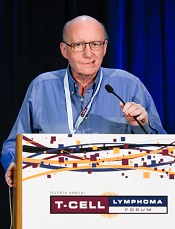
Bertrand Coiffier, MD, PhD, of CHU Lyon Sud in Pierre Benite, France, presented these findings at the 7th Annual T-cell Lymphoma Forum. Dr Coiffier and other researchers involved in this study receive funds from Celgene, the company developing romidepsin.
“CHOP is widely accepted,” Dr Coiffier noted. “It’s the most-used regimen for peripheral T-cell lymphoma, but it’s not the best one, and we certainly have regimens that do produce more [complete responses] and longer responses.”
He said researchers decided to test romidepsin in combination with CHOP because studies have suggested that romidepsin has very good efficacy in relapsed/refractory peripheral T-cell lymphoma, and the toxicities associated with romidepsin and CHOP alone have been managable.
So the researchers tested the combination in 37 patients with untreated PTCL, most of whom were male (n=20). The median age was 57, and 37.8% were older than 60. About 95% of patients had stage III/IV disease, and about 89% had an ECOG performance status less than 2.
Most patients had angioimmunoblastic T-cell lymphoma (n=17), followed by PTCL not otherwise specified (n=13), ALK- anaplastic large-cell lymphoma (n=3), enteropathy-associated T-cell lymphoma (n=1), hepatosplenic T-cell lymphoma (n=1), primary cutaneous CD4+ small/medium T-cell lymphoma (n=1), and “other” (n=1).
Early DLTs
The researchers used a standard “3+3” dose-escalation scheme, starting with a romidepsin dose of 10 mg/m2 given on days 1 and 8.
In the first 2 cycles, there were 3 dose-limiting toxicities (DLTs)—1 case of grade 3 syncope, 1 case of grade 3 general status alteration, and 1 case of grade 3 hematologic toxicity (neutropenia and thrombocytopenia) lasting longer than 7 days.
“So we looked at the definition of the criteria for DLT, and we thought that, this time, they were too severe,” Dr Coiffier said. “After a lot of discussion between all the investigators, we decided to modify the criteria for DLT regarding neutropenia or thrombocytopenia and to allow a little more toxicity before saying it’s a DLT.”
A DLT was initially defined as grade 3/4 non-hematologic toxicity, grade 3 hematologic toxicity lasting more than 7 days, or grade 4 hematologic toxicity lasting more than 3 days. The researchers modified the criteria so that hematologic toxicities would not be considered DLTs if they lasted less than 10 days for grade 3 or less than 7 days for grade 4.
When the team decreased the romidepsin dose to 8 mg/m2, they did not observe any DLTs according to the new criteria. The same was true when they raised the dose back up to 10 mg/m2.
There were, however, DLTs when the dose was increased to 12 mg/m2. In cohort 5, there was a case of grade 3 cardiac failure, and in cohort 6, there were 2 cases of grade 3 nausea.
Nevertheless, 12 mg/m2 became the phase 2 dose. In all, 25 patients received romidepsin at that dose.
Safety data
Twenty-six of 37 patients completed the 8 planned cycles of treatment. Five patients discontinued treatment due to progression and 6 due to toxicity (5 due to thrombocytopenia).
“One hundred percent of patients experienced at least one adverse event, but most of them were grade 1 or 2 [84%] and occurred during the first 2 cycles [38%],” Dr Coiffier said. “There were no deaths related to adverse events.”
Severe toxicities occurred during the expansion phase. There was a case of severe peripheral sensory neuropathy that led to treatment discontinuation, and there were 3 cases of acute cardiac toxicity. They all occurred after the first cycle, and none were fatal.
The rate of hematologic toxicity was high. Neutropenia occurred in all patients, thrombocytopenia in 94%, and anemia in 89%.
Grade 3/4 adverse events included neutropenia (85%), thrombocytopenia (35%), febrile neutropenia (19%), general status deterioration (13%), nausea/vomiting (10%), anemia (8%), hypophosphatemia (8%), fatigue (5%), mucositis (5%), decreased appetite (5%), hypocalcemia (3%), hyponatremia (3%), hypokalemia (3%), hypomagnesemia (3%), dysgeusia (3%), and peripheral sensory neuropathy (3%).
Response, survival, and next steps
About 51% of patients (18/35) achieved a complete response, and 17% (n=6) had a partial response. Twenty-six percent of patients (n=9) progressed.
The median follow-up was 30 months. The estimated 1-year progression-free survival was 57%, and the estimated 1-year overall survival was 82%.
“The [overall survival] curve is certainly much better than you would expect with just standard CHOP,” Dr Coiffier noted.
He added that this research has progressed to a phase 3 study comparing romidepsin and CHOP in combination to CHOP alone. There are 7 countries participating (France, Belgium, South Korea, Spain, Italy, Germany, and Portugal), and 100 patients have been enrolled thus far.
SAN FRANCISCO—Adding romidepsin to CHOP can enhance the regimen’s efficacy against peripheral T-cell lymphoma (PTCL), but the combination can also induce severe toxicity, results of a phase 1b/2 study have shown.
In patients with previously untreated PTCL, romidepsin plus CHOP elicited an overall response rate of about 69%.
But all patients experienced adverse events, a median of 49 per patient. In addition, rates of hematologic toxicities were high, and 3 patients experienced acute cardiac toxicity.
Bertrand Coiffier, MD, PhD, of CHU Lyon Sud in Pierre Benite, France, presented these findings at the 7th Annual T-cell Lymphoma Forum. Dr Coiffier and other researchers involved in this study receive funds from Celgene, the company developing romidepsin.
“CHOP is widely accepted,” Dr Coiffier noted. “It’s the most-used regimen for peripheral T-cell lymphoma, but it’s not the best one, and we certainly have regimens that do produce more [complete responses] and longer responses.”
He said researchers decided to test romidepsin in combination with CHOP because studies have suggested that romidepsin has very good efficacy in relapsed/refractory peripheral T-cell lymphoma, and the toxicities associated with romidepsin and CHOP alone have been managable.
So the researchers tested the combination in 37 patients with untreated PTCL, most of whom were male (n=20). The median age was 57, and 37.8% were older than 60. About 95% of patients had stage III/IV disease, and about 89% had an ECOG performance status less than 2.
Most patients had angioimmunoblastic T-cell lymphoma (n=17), followed by PTCL not otherwise specified (n=13), ALK- anaplastic large-cell lymphoma (n=3), enteropathy-associated T-cell lymphoma (n=1), hepatosplenic T-cell lymphoma (n=1), primary cutaneous CD4+ small/medium T-cell lymphoma (n=1), and “other” (n=1).
Early DLTs
The researchers used a standard “3+3” dose-escalation scheme, starting with a romidepsin dose of 10 mg/m2 given on days 1 and 8.
In the first 2 cycles, there were 3 dose-limiting toxicities (DLTs)—1 case of grade 3 syncope, 1 case of grade 3 general status alteration, and 1 case of grade 3 hematologic toxicity (neutropenia and thrombocytopenia) lasting longer than 7 days.
“So we looked at the definition of the criteria for DLT, and we thought that, this time, they were too severe,” Dr Coiffier said. “After a lot of discussion between all the investigators, we decided to modify the criteria for DLT regarding neutropenia or thrombocytopenia and to allow a little more toxicity before saying it’s a DLT.”
A DLT was initially defined as grade 3/4 non-hematologic toxicity, grade 3 hematologic toxicity lasting more than 7 days, or grade 4 hematologic toxicity lasting more than 3 days. The researchers modified the criteria so that hematologic toxicities would not be considered DLTs if they lasted less than 10 days for grade 3 or less than 7 days for grade 4.
When the team decreased the romidepsin dose to 8 mg/m2, they did not observe any DLTs according to the new criteria. The same was true when they raised the dose back up to 10 mg/m2.
There were, however, DLTs when the dose was increased to 12 mg/m2. In cohort 5, there was a case of grade 3 cardiac failure, and in cohort 6, there were 2 cases of grade 3 nausea.
Nevertheless, 12 mg/m2 became the phase 2 dose. In all, 25 patients received romidepsin at that dose.
Safety data
Twenty-six of 37 patients completed the 8 planned cycles of treatment. Five patients discontinued treatment due to progression and 6 due to toxicity (5 due to thrombocytopenia).
“One hundred percent of patients experienced at least one adverse event, but most of them were grade 1 or 2 [84%] and occurred during the first 2 cycles [38%],” Dr Coiffier said. “There were no deaths related to adverse events.”
Severe toxicities occurred during the expansion phase. There was a case of severe peripheral sensory neuropathy that led to treatment discontinuation, and there were 3 cases of acute cardiac toxicity. They all occurred after the first cycle, and none were fatal.
The rate of hematologic toxicity was high. Neutropenia occurred in all patients, thrombocytopenia in 94%, and anemia in 89%.
Grade 3/4 adverse events included neutropenia (85%), thrombocytopenia (35%), febrile neutropenia (19%), general status deterioration (13%), nausea/vomiting (10%), anemia (8%), hypophosphatemia (8%), fatigue (5%), mucositis (5%), decreased appetite (5%), hypocalcemia (3%), hyponatremia (3%), hypokalemia (3%), hypomagnesemia (3%), dysgeusia (3%), and peripheral sensory neuropathy (3%).
Response, survival, and next steps
About 51% of patients (18/35) achieved a complete response, and 17% (n=6) had a partial response. Twenty-six percent of patients (n=9) progressed.
The median follow-up was 30 months. The estimated 1-year progression-free survival was 57%, and the estimated 1-year overall survival was 82%.
“The [overall survival] curve is certainly much better than you would expect with just standard CHOP,” Dr Coiffier noted.
He added that this research has progressed to a phase 3 study comparing romidepsin and CHOP in combination to CHOP alone. There are 7 countries participating (France, Belgium, South Korea, Spain, Italy, Germany, and Portugal), and 100 patients have been enrolled thus far.
Anticoagulant now available in US pharmacies

Photo by Rhoda Baer
The oral factor Xa inhibitor edoxaban (Savaysa) is now available in US pharmacies, according to Daiichi Sankyo Company, Limited, the company developing the drug.
Savaysa is approved by the US Food and Drug Administration to reduce the risk of stroke and systemic embolism (SE) in patients with non-valvular atrial fibrillation (NVAF), as well as for the treatment of deep vein thrombosis and pulmonary embolism following 5 to 10 days of initial therapy with a parenteral anticoagulant.
According to Savaysa’s label, it should not be used in NVAF patients with creatinine clearance levels greater than 95 mL/min because, in that population, there is an increased risk of ischemic stroke with the drug compared to warfarin.
Daiichi Sankyo has developed resources for physicians and patients to help ensure patients can begin and/or remain on Savaysa per physician instructions.
The Savaysa Savings Plus program will include a reimbursement hotline to assist patients and prescribers who request help understanding a patient’s available coverage. Eligible patients who are prescribed Savaysa can enroll in a copay savings program and pay $4 per month through the Savaysa Savings Card.
Vouchers will also be available to provide patients and doctors with a way to try Savaysa at no cost to see if it is right for the patient.
In addition, the Savaysa Patient Assistance Program will offer assistance to qualified individuals, providing free product to eligible patients who are prescribed Savaysa, are uninsured, and are unable to identify alternative payment sources.
The approval of Savaysa in the US is based on data from the ENGAGE AF-TIMI 48 trial and the Hokusai-VTE trial.
FDA clears new blood-draw device

Photo courtesy of
Velano Vascular
The US Food and Drug Administration (FDA) has cleared for marketing a device that reduces the need for venipunctures for in-hospital blood draws.
Velano Vascular’s blood-draw device resembles a common syringe.
It allows peripheral intravenous catheters to be repurposed to draw blood from patients, thereby reducing the need for additional needle sticks among patients receiving medications and hydration via intravenous delivery.
The single-use device will soon be used for clinical evaluation in select hospitals, including the University of Pennsylvania in Philadelphia and University Hospitals Case Medical Center of Cleveland in Ohio.
“A fundamental benefit of this technology is reducing the ‘pin cushion effect,’ in which hospitalized patients are ‘stuck’ several times daily to obtain blood tests,” said Eric M. Stone, co-founder and chief executive officer of Velano Vascular, the company developing the blood-draw device.
“Oftentimes, the draw procedure is plagued by multiple failed attempts. The FDA’s clearance of this novel technology validates the existing clinical need and will allow us to expedite our efforts to bring this innovation to patients, healthcare providers, and hospitals around the world.”
According to research conducted by Velano Vascular, 1 of every 3 hospital patients is stuck 2 or more times daily for blood draws, with a significant subset of these patients receiving 3 or more blood draws, along with numerous needle sticks.
Twenty-eight percent of adult venipunctures and 44% of pediatric venipunctures require more than one stick to successfully draw blood.
“Traditional blood draws are one of the most common and most problematic healthcare procedures,” said Karen Daley, PhD, RN, past president of the American Nurses Association and a healthcare worker safety advocate.
“It is an antiquated technology that creates pain and anxiety for many patients, a significant safety risk for healthcare professionals, and a real inefficiency in our healthcare system. Velano Vascular has developed a common-sense solution to this pervasive, long-standing problem.”