User login
FDA approves drug for newly diagnosed MM
The US Food and Drug Administration (FDA) has expanded the existing indication for lenalidomide (Revlimid)—in combination with dexamethasone—to include patients with newly diagnosed multiple myeloma (MM).
The FDA previously approved lenalidomide in combination with dexamethasone to treat MM patients who had received at least 1 prior therapy.
Lenalidomide is also FDA-approved to treat mantle cell lymphoma patients who have failed 2 prior therapies, including bortezomib.
And the drug is approved to treat patients with transfusion-dependent anemia due to low- or intermediate-1-risk myelodysplastic syndromes associated with 5q deletion, with or without additional cytogenetic abnormalities.
“The approval of Revlimid as an option for use in all patients with multiple myeloma represents a new paradigm in the management of this disease,” said Kenneth Anderson, MD, of Dana-Farber/Brigham and Women’s Cancer Center in Boston, Massachusetts.
“We now have clinical evidence demonstrating that starting and keeping newly diagnosed multiple myeloma patients on Revlimid significantly improves progression-free survival.”
The FDA’s latest approval of lenalidomide was based on safety and efficacy results from phase 3 studies, particularly the FIRST trial.
The FIRST trial
In this phase 3 trial, researchers enrolled 1623 patients who were newly diagnosed with MM and not eligible for stem cell transplant.
Patients were randomized to receive lenalidomide and dexamethasone (Rd) in 28-day cycles until disease progression (n=535), 18 cycles of lenalidomide and dexamethasone (Rd18) for 72 weeks (n=541), or melphalan, prednisone, and thalidomide (MPT) for 72 weeks (n=547).
Response rates were significantly better with continuous Rd (75%) and Rd18 (73%) than with MPT (62%, P<0.001 for both comparisons). Complete response rates were 15%, 14%, and 9%, respectively.
The median progression-free survival was 25.5 months with continuous Rd, 20.7 months with Rd18, and 21.2 months with MPT.
This resulted in a 28% reduction in the risk of progression or death for patients treated with continuous Rd compared with those treated with MPT (hazard ratio[HR]=0.72, P<0.001) and a 30% reduction compared with Rd18 (HR=0.70, P<0.001).
The pre-planned interim analysis of overall survival showed a 22% reduction in the risk of death for continuous Rd vs MPT (HR=0.78, P=0.02), but the difference did not cross the pre-specified superiority boundary (P<0.0096).
Adverse events reported in 20% or more of patients in the continuous Rd, Rd18, or MPT arms included diarrhea (45.5%, 38.5%, 16.5%), anemia (43.8%, 35.7%, 42.3%), neutropenia (35.0%, 33.0%, 60.6%), fatigue (32.5%, 32.8%, 28.5%), back pain (32.0%, 26.9%, 21.4%), insomnia (27.6%, 23.5%, 9.8%), asthenia (28.2%, 22.8%, 22.9%), rash (26.1%, 28.0%, 19.4%), decreased appetite (23.1%, 21.3%, 13.3%), cough (22.7%, 17.4%, 12.6%), pyrexia (21.4%, 18.9%, 14.0%), muscle spasms (20.5%, 18.9%, 11.3%) and abdominal pain (20.5%, 14.4%, 11.1%).
The most frequently reported grade 3/4 events in the continuous Rd arm (until disease progression) were neutropenia (27.8%), anemia (18.2%), thrombocytopenia (8.3%), pneumonia (11.3%), asthenia (7.7%), fatigue (7.3%), back pain (7%), hypokalemia (6.6%), rash (7.3%), cataract (5.8%), dyspnea (5.6%), deep vein thrombosis (5.6%), and hyperglycemia (5.3%).
The incidence of invasive second primary malignancies was 3% in the continuous Rd arm, 6% in the Rd18 arm, and 5% in the MPT arm. The overall incidence of solid tumors was identical in the continuous Rd and MPT arms (3%) and 5% in the Rd18 arm. ![]()
Researchers map the human epigenome

to the Epigenome Roadmap
Image by Nik Spencer/Nature
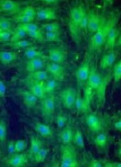
Scientists have created new maps of the human epigenome that may help unravel the complex links between DNA and disease.
Researchers supported by the Common Fund’s Epigenomics Program have mapped the epigenomes of more than 100 types of cells and tissues, providing new insight into which parts of the genome are used to make a particular type of cell.
The group published an article describing the epigenome maps in the journal Nature.
Twenty-three additional papers in Nature Publishing Group journals show how these maps can be used to study human biology.
The papers are available on Nature’s Epigenome Roadmap site.
For the Roadmap Epigenomics Project, scientists compared epigenomic signatures and established their differences across a variety of cell types. The resulting information may help us understand how changes to the genome and epigenome can lead to cancers and other conditions.
“These 111 reference epigenome maps are essentially a vocabulary book that helps us decipher each DNA segment in distinct cell and tissue types,” said Bing Ren, PhD, of the University of California, San Diego.
For cancer research, the new data will hasten a merging of genomic and epigenomic perspectives that was already underway, according to Joseph F. Costello, PhD, of the University of California, San Francisco.
“You’ve had cancer researchers studying the genome—the role of mutations, deletions, and so on—and others studying epigenomes,” Dr Costello said. “They’ve almost been working on parallel tracks, and they didn’t talk to each other all that much.”
“Over the past 5 or 6 years, there’s been a reframing of the discussion, because the most recurrent mutations in cancer affect epigenomic regulators. So the way mutations in the genome play out is through epigenomic mechanisms, and major pharmaceutical companies now view epigenomes as an important target.” ![]()
to the Epigenome Roadmap
Image by Nik Spencer/Nature
Scientists have created new maps of the human epigenome that may help unravel the complex links between DNA and disease.
Researchers supported by the Common Fund’s Epigenomics Program have mapped the epigenomes of more than 100 types of cells and tissues, providing new insight into which parts of the genome are used to make a particular type of cell.
The group published an article describing the epigenome maps in the journal Nature.
Twenty-three additional papers in Nature Publishing Group journals show how these maps can be used to study human biology.
The papers are available on Nature’s Epigenome Roadmap site.
For the Roadmap Epigenomics Project, scientists compared epigenomic signatures and established their differences across a variety of cell types. The resulting information may help us understand how changes to the genome and epigenome can lead to cancers and other conditions.
“These 111 reference epigenome maps are essentially a vocabulary book that helps us decipher each DNA segment in distinct cell and tissue types,” said Bing Ren, PhD, of the University of California, San Diego.
For cancer research, the new data will hasten a merging of genomic and epigenomic perspectives that was already underway, according to Joseph F. Costello, PhD, of the University of California, San Francisco.
“You’ve had cancer researchers studying the genome—the role of mutations, deletions, and so on—and others studying epigenomes,” Dr Costello said. “They’ve almost been working on parallel tracks, and they didn’t talk to each other all that much.”
“Over the past 5 or 6 years, there’s been a reframing of the discussion, because the most recurrent mutations in cancer affect epigenomic regulators. So the way mutations in the genome play out is through epigenomic mechanisms, and major pharmaceutical companies now view epigenomes as an important target.” ![]()
Product ‘solves engraftment problem’ with UCBT

Photo courtesy of NHS
SAN DIEGO—A product that promotes CD34 expansion has solved the problem of poor engraftment associated with umbilical cord blood transplant (UCBT), according to a speaker at the 2015 BMT Tandem Meetings.
John Wagner, MD, of the University of Minnesota in Minneapolis, supported this statement with early results from trials of HSC835, cord
blood units in which CD34 cells were expanded using the aryl hydrocarbon
receptor antagonist StemRegenin 1.
Dr Wagner and his colleagues conducted phase 1 and 2 trials of HSC835, which were supported by Novartis, the company developing the product.
Dr Wagner presented results of these trials at the BMT Tandem Meetings as abstract 29.*
The phase 1 trial included 18 patients who received double UCBT after myeloablative conditioning. They received an unmanipulated cord blood unit and an HSC835 unit.
In the phase 2 trial, 3 patients received a single HSC835 unit after myeloablative conditioning, and 3 received an HSC835 unit after non-myeloablative conditioning.
Manufacturing HSC835 took 15 days. Following expansion, the median CD34 cell dose increased 346-fold, and the median total nucleated cell (TNC) dose increased 848-fold.
Twenty-eight HSC835 products were produced, but 4 of them were not infused. Two products were contaminated, 1 was not infused due to patient relapse, and 1 product failed to expand. Dr Wagner noted that the failed unit started out at 50% viability, whereas the other units started at about 90% viability.
Phase 1
Eighteen patients were treated in the phase 1 trial. Eleven had acute lymphoblastic leukemia, 5 had acute myeloid leukemia, and 2 had myelodysplastic syndromes. The median age was 28 (range, 12 to 53).
The median TNC dose was 2.5 x 10⁷/kg for the unmanipulated cord blood unit and 5.1 x 10⁷/kg for the HSC835 unit. The median CD34 count was 0.4 x 10⁶/kg and 17.4 x 10⁶/kg, respectively. And the median CD3 count was 8.5 x 10⁶/kg and 2.5 x 10⁶/kg, respectively.
Most units were a 5/6 HLA match—50% for the unmanipulated unit and 67% for the HSC835 unit. Forty-four percent and 22% of the units, respectively, were 4/6 matches. And 6% and 11%, respectively, were 6/6 matches.
Neutrophil recovery was 100%, and the median time to recovery was 14.5 days.
Dr Wagner compared this to results in 121 matched historical controls who received UCBT with 2 unmanipulated units. Of those patients, 86% achieved neutrophil engraftment, and the median time to engraftment was 25 days (P<0.001).
In the current study, 6 patients had complete chimerism with HSC835, 6 had complete chimerism with the unmanipulated unit, and 6 had dual chimerism. In the dual-chimerism patients, both units were present, but all the T cells were derived from the unmanipulated unit.
“When we look at those patients who had engraftment of HSC835, whether it be this dual chimerism or complete chimerism, you see there is a very rapid recovery of 10.5 days, as compared to the historical control [recovery time] of 25 days,” Dr Wagner said. “And those that engrafted with the unmanipulated unit fall right where you’d expect them; that is, with the historical controls [23 days].”
Dr Wagner noted that the CD34 dose correlated with the pace of neutrophil recovery in patients who engrafted with the HSC835 unit, and patients with dual chimerism had the fastest neutrophil recovery.
Furthermore, the HSC835 unit predominated more than expected (P=0.05), winning out over the cord blood unit with a higher CD3 dose a disproportionate amount of time. The unit with a higher CD3 dose typically predominates two-thirds of the time in double UCBT.
“[These results] gave us enough information to say that [HSC835] could be a stand-alone product,” Dr Wagner said.
Phase 2
For phase 2, the researchers used a single HSC835 unit. They chose the lesser of 2 cord blood units (keeping the better unit as a backup), expanded it, and infused the resulting HSC835 unit into 3 patients who received myeloablative conditioning and 3 patients who did not.
In patients who received myeloablative conditioning, chimerism was complete at days 8, 12, and 14. Among the patients who received non-myeloablative conditioning, 1 had mixed chimerism at day 6. The other 2 had complete chimerism at days 5 and 7.
“So in conclusion, we believe that HSC835 is safe and effective in speeding neutrophil recovery after cord blood transplant,” Dr Wagner said. “These are very promising early results that compel us now to explore the single expanded product.”
Dr Wagner added that other considerations for HSC835 are that it may reduce the unit selection threshold, improve HLA match, enable re-cryopreservation (for transplant delays, backup, or multiple dosing), and perhaps allow for reduced-intensity conditioning with “mega-dose” grafts. ![]()
*Information in the abstract differs from that presented at the meeting.
Expanded MSCs can treat severe aGVHD
SAN DIEGO—Infusions of expanded mesenchymal stem cells (MSCs) can treat severe, steroid-resistant, acute graft-vs-host disease (aGVHD) in pediatric patients, according to a study presented at the 2015 BMT Tandem Meetings.
The MSC product, known as remestemcel-L, induced responses in all grades of aGVHD and all organ systems.
Response at day 28 was associated with improved survival at day 100, and clinically significant toxicities were minimal, according to investigators.
“The enrolled patients represent a very challenging population with severe graft-vs-host disease that was non-responsive to treatments, including steroids and, for many of these children, multiple immunosuppressive agents, so we believe these results are very promising,” said Joanne Kurtzberg, MD, of Duke University Medical Center in Durham, North Carolina.
Dr Kurtzberg and her colleagues reported the results in a poster presentation (abstract 492*). Three investigators involved in this research are employed by Mesoblast, Inc., the company developing remestemcel-L.
The study included 160 patients who had a median age of 10 years (range, 0.3 to 17.5 years). Eighty-four percent of patients had received an unrelated donor transplant, and 46% received a mismatched transplant.
At baseline, 19% of patients had grade B aGVHD, 28% had grade C, and 53% had grade D. Eighty-nine percent of patients had gastrointestinal involvement, 50% had skin involvement, and 29% had liver involvement. Forty-one percent of patients had 2 organs involved, and 15% had 3.
The median duration of aGVHD before study enrollment was 28 days (range, 1 to 237), and patients had failed a median of 3 immunosuppressive agents.
Treatment and outcomes
Patients received 8 bi-weekly, intravenous infusions of 2 × 10⁶ MSCs/kg for 4 weeks. They could receive additional weekly infusions if deemed eligible at day 28.
The patients received a median of 11 infusions (range, 1-20) and were exposed to the treatment for a median of 43.5 days.
Fifty-three percent of patients had at least 1 serious adverse event. Eight patients (5%) had serious events that investigators thought might be treatment-related. These included neutropenia, infusion-related reaction, pulmonary hemorrhage, respiratory distress, tachycardia, respiratory failure, and hypertension.
Three events (6%) that were considered possibly treatment-related (pulmonary hemorrhage, respiratory distress, and respiratory failure) ultimately resulted in death. Fifty-four patients (34%) died in all.
At day 28, the overall response rate (ORR) was 64%. The ORR was 74% for grade B aGVHD, 66% for grade C, and 59% for grade D. The ORR was 62% for gastrointestinal, 77% for skin, and 53% for liver aGVHD.
Response correlated with a significant improvement in survival at day 100. Eighty-one percent of patients who responded at day 28 were still alive at day 100, compared to 21% of non-responders (P<0.0001).
The investigators said this study provides support for remestemcel-L to treat aGVHD in children. A single-arm, phase 3 trial of pediatric patients with aGVHD is underway. ![]()
*Information in the abstract differs from that presented at the meeting.
Anticoagulant outperforms LMWH in NSTEMI

Image by Andre E.X. Brown
The factor Xa inhibitor fondaparinux may confer a lower risk of death and bleeding after heart attack than low-molecular-weight heparin (LMWH).
In a large study, patients who received fondaparinux after non-ST-segment elevation myocardial infarction (NSTEMI) had a lower risk of major bleeding and death, both in the hospital and after discharge, compared to patients who received LMWH.
However, both arms had similar rates of subsequent heart attack or stroke.
Karolina Szummer, MD, PhD, of the Karolinska Institutet in Stockholm, Sweden, and her colleagues disclosed these results in JAMA.
The researchers analyzed data from a Swedish registry that included 40,616 patients with NSTEMI. The patients received in-hospital treatment with fondaparinux (n=14,791; 36.4%) or LMWH (25,825; 63.6%) between September 2006 and June 2010, with follow-up through December 2010.
Patients in the fondaparinux arm were, on average, 2 years younger (72 years vs 74 years) than patients in the LMWH arm. Fondaparinux-treated patients also had fewer previous heart attacks (28.2% vs 32.2%), and fewer had been diagnosed with congestive heart failure (14.5% vs 18.7%), but more had undergone percutaneous coronary intervention (46.4% vs 38.9%).
The rate of prior bleeding events and previous hemorrhagic stroke was similar between the arms. Prior bleeding was reported in 6.1% of patients in both arms, and hemorrhagic stroke was reported in 1.4% of patients in the fondaparinux arm and 1.3% in the LMWH arm.
Following treatment, the absolute rate of severe in-hospital bleeding events was lower in the fondaparinux arm than in the LMWH arm—1.1% vs 1.8% (odds ratio [OR]=0.54).
The rate of severe bleeding while in the hospital or causing readmission was lower in the fondaparinux arm, both at 30 days—1.4% vs 2.1% (OR=0.56)—and at 180 days—1.9% vs 2.8% (OR=0.60).
In-hospital mortality was lower in the fondaparinux arm than the LMWH arm—2.7% and 4.0%, respectively (OR=0.75). The same pattern was observed for mortality at 30 days (OR=0.82) and 180 days (OR=0.76).
However, the rate of recurrent heart attack was similar in both arms. At 30 days, it was 9.0% in the fondaparinux arm and 9.5% in the LMWH arm (OR=0.94). And at 180 days, rates were 14.2% and 15.8%, respectively (OR=0.97).
Likewise, the rate of stroke did not differ significantly between the arms. At 30 days, it was 0.5% in the fondaparinux arm and 0.6% in the LMWH arm (OR=1.11). And at 180 days, rates were 1.7% and 2.0%, respectively (OR=0.98).
The results were similar in patients with varying degrees of kidney function and in the subset of patients who had undergone early percutaneous coronary intervention.
The researchers said these results provide an estimate of the treatment effect in a selected patient population. However, the effects may differ in clinical practice and should therefore be investigated in observational cohorts and in continuous registries. ![]()
FDA issues documents on drug compounding

Photo by Rhoda Baer
The US Food and Drug Administration (FDA) has issued 5 draft documents related to drug compounding and repackaging that aim to help entities comply with public health provisions.
The agency said these draft documents are applicable to pharmacies, federal facilities, outsourcing facilities, and physicians.
The new category of outsourcing facilities was created under the Drug Quality and Security Act (DQSA), which was enacted by Congress in November 2013.
It was enacted in response to a deadly fungal meningitis outbreak that was linked to contaminated sterile compounded drug products.
Drugs compounded in an outsourcing facility that meet certain conditions may be entitled to exemptions from certain provisions of the Federal Food, Drug, and Cosmetic Act (FD&C Act), including the new drug approval requirements and the requirement to label drug products with adequate directions for use.
Outsourcing facilities are subject to current good manufacturing practice requirements and inspections by the FDA according to a risk-based schedule.
Drugs produced by compounders that are not registered as outsourcing facilities must meet certain other conditions described in the FD&C Act, or they will be subject to all of the requirements applicable to drugs produced by conventional drug manufacturers.
“The draft guidance documents provide information to pharmacies, outsourcing facilities, healthcare entities, and others about these FDA-proposed policies, which are critical to protecting the public health,” said Janet Woodcock, MD, director of the FDA’s Center for Drug Evaluation and Research.
Descriptions of these documents follow.
This draft guidance provides an entity considering whether to register with the FDA as an outsourcing facility with information about the regulatory impact of registering.
For example, it explains that a facility engaged in only certain activities, including repackaging human drugs and compounding non-sterile drugs, should not register as an outsourcing facility because its drug products will not qualify for the exemptions provided in section 503B, including the exemption from the new drug approval requirements.
This draft guidance describes the conditions under which the FDA does not intend to take action for certain violations of the law when state-licensed pharmacies, federal facilities, or outsourcing facilities repackage certain drug products.
Repackaged drug products are generally not exempt from any of the provisions of the FD&C Act related to the production of drugs, and the compounding provisions of the FD&C Act do not address repackaging. Therefore, the FDA is issuing guidance to describe how it intends to address repackaging when done in a state-licensed pharmacy, federal facility, or outsourcing facility.
This draft guidance describes the conditions under which the FDA does not intend to take action for violations of certain sections of the Public Health Service Act (PHS Act) and the FD&C Act when state-licensed pharmacies, federal facilities, or outsourcing facilities mix, dilute, or repackage specific biological products without an approved BLA, or when such facilities or physicians prepare prescription sets of allergenic extracts without an approved BLA.
The draft guidance notes that a biological product that is mixed, diluted, or repackaged outside the scope of an approved BLA is an unlicensed biological product under section 351 of the PHS Act and may not be legally marketed without an approved BLA.
Additionally, the compounding provisions of the FD&C Act do not address biological products subject to licensure under section 351 of the PHS Act. Therefore, the FDA is issuing the guidance to describe how it intends to address these practices.
Entities registered as outsourcing facilities are required to report adverse events to the FDA. This draft guidance explains adverse event reporting for such facilities.
The draft memorandum of understanding (MOU) under section 503A of the FD&C Act describes the responsibilities of a state that chooses to sign the MOU in investigating and responding to complaints related to compounded human drug products distributed outside the state, and in addressing the interstate distribution of “inordinate amounts” of compounded human drug products.
These documents are the latest in a series of policy documents related to FDA oversight of drugs produced by state-licensed pharmacies, federal facilities, and outsourcing facilities.
The draft guidance documents are available for public comment for 90 days. The public has 120 days to comment on the draft MOU between the states and the FDA. ![]()
Day 90 CR not a good endpoint for auto-HSCT, doc says
Photo by Rhoda Baer
SAN DIGEO—A phase 3 study comparing conditioning regimens in multiple myeloma patients undergoing autologous transplant has yielded counterintuitive results.
Patients who received busulfan plus melphalan (bu-mel) had a lower complete response (CR) rate at 90 days and a higher rate of grade 3-4 non-hematologic
toxicity, compared to patients who received melphalan (mel) alone.
However, patients in the bu-mel arm enjoyed significantly longer progression-free survival (PFS).
These results suggest the rate of CR at 90 days is not a good endpoint for trials of autologous hematopoietic stem cell transplant (auto-HSCT), said Muzaffar H.
Qazilbash, MD, of The University of Texas MD Anderson Cancer Center in Houston.
Dr Qazilbash presented these and other findings at the 2015 BMT Tandem Meetings as abstract 83.*
“High-dose melphalan at 200 mg/m2 is considered the standard of care for autologous stem cell transplantation for multiple myeloma,” he said. “However, most patients have disease recurrence or progression after an autologous transplant, even with the use of post-transplant maintenance.”
A recent retrospective study suggested that bu-mel might be associated with a longer PFS compared to mel alone. So Dr Qazilbash and his colleagues decided to
investigate that possibility in a randomized, phase 3 trial.
The trial included 92 patients with symptomatic multiple myeloma who had received at least 2 cycles of induction therapy. They were all within 12 months of the start of induction, and all had adequate cardiac, pulmonary, renal, and liver function.
Forty-nine patients received bu-mel—a 130 mg/m2 daily infusion of busulfan for 4 days, followed by 2 daily doses of melphalan at 70 mg/m2. All patients in this arm also received phenytoin for seizure prophylaxis. The 43 patients in the mel-only arm received a single dose of melphalan at 200 mg/m2.
Baseline characteristics were similar between the 2 arms. The patients’ median ages were 58 in the bu-mel arm and 59 in the mel arm. At the time of transplant, 18% of patients in the bu-mel arm were in CR or near CR, as were 28% of patients in the mel arm.
Adverse events
At 100 days post-transplant, the transplant-related mortality was 0% in both arms.
“There was a significantly higher rate of grade 3-4 non-hematologic adverse events in the busulfan-plus-melphalan arm—78% vs 47% [P=0.002],” Dr Qazilbash noted. “And these adverse events were mostly infectious events.”
Grade 3 infection occurred in 71% of bu-mel-treated patients and 39% of mel-treated patients (P=0.003). Grade 2 gastrointestinal toxicities occurred in 69% and 48%, respectively (P=0.05).
There were no significant differences between the arms with regard to other adverse events. And there were no second primary malignancies in either arm.
Response
At 90 days, the CR rate was 16% in the bu-mel arm and 35% in the mel arm (P=0.05). The rates of CR plus very good partial response were 63% and 81%, respectively (P=0.06).
“The trial was stopped early, based on a prescribed rule in the trial design, because of a significantly higher CR rate in the control arm,” Dr Qazilbash said.
Still, he noted that, at 180 days, the CR rate was 26% in the bu-mel arm and 37% in the mel arm (P=0.36). And the 180-day rates of CR plus very good
partial response were 69% and 86%, respectively (P=0.65).
Maintenance
A majority of patients received maintenance therapy post-transplant—84% of patients in the bu-mel arm and 88% in the mel arm.
The median interval between auto-HSCT and maintenance was 4.6 months and 4.5 months, respectively. And the median duration of maintenance was 16 months and 14.2 months, respectively.
Maintenance largely consisted of lenalidomide. Sixty-five percent of patients in the bu-mel arm and 63% in the mel arm received the drug. Fewer patients received bortezomib—8% and 16%, respectively—or lenalidomide plus ixazomib—10% and 9%, respectively.
Survival
At a median follow-up of 19.5 months, bu-mel was associated with significantly longer PFS than mel alone (P=0.047). And a multivariate analysis showed that bu-mel was an independent predictor of PFS.
Dr Qazilbash noted that there was no significant difference in overall survival between the bu-mel and mel arms (P=0.97), but the follow-up is less than 2 years, so this is expected.
“Bu-mel was associated with a significantly lower day 90 CR rate, a significantly higher rate of grade 3-4 non-hematologic toxicity, no significant difference in transplant-related mortality . . . , and a significantly longer PFS,” Dr Qazilbash summarized.
He therefore concluded that, based on this study, day 90 CR is not an adequate endpoint for auto-HSCT trials. This trial was amended to change the primary
endpoint from day 90 CR to PFS, and accrual has been increased to 205 patients.
Dr Qazilbash and others involved in this study received research funding from Otsuka, makers of busulfan. Dr Qazilbash also reported relationships (advisory
board, honoraria) with Celgene, Millennium, and Onyx. ![]()
*Information in the abstract differs from that presented at the meeting.
Photo by Rhoda Baer
SAN DIGEO—A phase 3 study comparing conditioning regimens in multiple myeloma patients undergoing autologous transplant has yielded counterintuitive results.
Patients who received busulfan plus melphalan (bu-mel) had a lower complete response (CR) rate at 90 days and a higher rate of grade 3-4 non-hematologic
toxicity, compared to patients who received melphalan (mel) alone.
However, patients in the bu-mel arm enjoyed significantly longer progression-free survival (PFS).
These results suggest the rate of CR at 90 days is not a good endpoint for trials of autologous hematopoietic stem cell transplant (auto-HSCT), said Muzaffar H.
Qazilbash, MD, of The University of Texas MD Anderson Cancer Center in Houston.
Dr Qazilbash presented these and other findings at the 2015 BMT Tandem Meetings as abstract 83.*
“High-dose melphalan at 200 mg/m2 is considered the standard of care for autologous stem cell transplantation for multiple myeloma,” he said. “However, most patients have disease recurrence or progression after an autologous transplant, even with the use of post-transplant maintenance.”
A recent retrospective study suggested that bu-mel might be associated with a longer PFS compared to mel alone. So Dr Qazilbash and his colleagues decided to
investigate that possibility in a randomized, phase 3 trial.
The trial included 92 patients with symptomatic multiple myeloma who had received at least 2 cycles of induction therapy. They were all within 12 months of the start of induction, and all had adequate cardiac, pulmonary, renal, and liver function.
Forty-nine patients received bu-mel—a 130 mg/m2 daily infusion of busulfan for 4 days, followed by 2 daily doses of melphalan at 70 mg/m2. All patients in this arm also received phenytoin for seizure prophylaxis. The 43 patients in the mel-only arm received a single dose of melphalan at 200 mg/m2.
Baseline characteristics were similar between the 2 arms. The patients’ median ages were 58 in the bu-mel arm and 59 in the mel arm. At the time of transplant, 18% of patients in the bu-mel arm were in CR or near CR, as were 28% of patients in the mel arm.
Adverse events
At 100 days post-transplant, the transplant-related mortality was 0% in both arms.
“There was a significantly higher rate of grade 3-4 non-hematologic adverse events in the busulfan-plus-melphalan arm—78% vs 47% [P=0.002],” Dr Qazilbash noted. “And these adverse events were mostly infectious events.”
Grade 3 infection occurred in 71% of bu-mel-treated patients and 39% of mel-treated patients (P=0.003). Grade 2 gastrointestinal toxicities occurred in 69% and 48%, respectively (P=0.05).
There were no significant differences between the arms with regard to other adverse events. And there were no second primary malignancies in either arm.
Response
At 90 days, the CR rate was 16% in the bu-mel arm and 35% in the mel arm (P=0.05). The rates of CR plus very good partial response were 63% and 81%, respectively (P=0.06).
“The trial was stopped early, based on a prescribed rule in the trial design, because of a significantly higher CR rate in the control arm,” Dr Qazilbash said.
Still, he noted that, at 180 days, the CR rate was 26% in the bu-mel arm and 37% in the mel arm (P=0.36). And the 180-day rates of CR plus very good
partial response were 69% and 86%, respectively (P=0.65).
Maintenance
A majority of patients received maintenance therapy post-transplant—84% of patients in the bu-mel arm and 88% in the mel arm.
The median interval between auto-HSCT and maintenance was 4.6 months and 4.5 months, respectively. And the median duration of maintenance was 16 months and 14.2 months, respectively.
Maintenance largely consisted of lenalidomide. Sixty-five percent of patients in the bu-mel arm and 63% in the mel arm received the drug. Fewer patients received bortezomib—8% and 16%, respectively—or lenalidomide plus ixazomib—10% and 9%, respectively.
Survival
At a median follow-up of 19.5 months, bu-mel was associated with significantly longer PFS than mel alone (P=0.047). And a multivariate analysis showed that bu-mel was an independent predictor of PFS.
Dr Qazilbash noted that there was no significant difference in overall survival between the bu-mel and mel arms (P=0.97), but the follow-up is less than 2 years, so this is expected.
“Bu-mel was associated with a significantly lower day 90 CR rate, a significantly higher rate of grade 3-4 non-hematologic toxicity, no significant difference in transplant-related mortality . . . , and a significantly longer PFS,” Dr Qazilbash summarized.
He therefore concluded that, based on this study, day 90 CR is not an adequate endpoint for auto-HSCT trials. This trial was amended to change the primary
endpoint from day 90 CR to PFS, and accrual has been increased to 205 patients.
Dr Qazilbash and others involved in this study received research funding from Otsuka, makers of busulfan. Dr Qazilbash also reported relationships (advisory
board, honoraria) with Celgene, Millennium, and Onyx.
*Information in the abstract differs from that presented at the meeting.
Antibodies can fight lymphoma

Researchers say they have developed a method that simulates Epstein-Barr virus (EBV) infection, and the immune cells that are activated as a result can kill non-Hodgkin lymphoma cells efficiently.
The team made use of antibodies that exhibit a piece of viral protein. The antibodies contain binding sites that target molecules on the surface of lymphoma cells.
The researchers used genetic engineering methods to fuse protein pieces of EBV to the “rear” end of the antibody protein.
As exposure to EBV is common, many people already have memory T cells that can mount a rapid immune response upon a new encounter with this pathogen.
The antibodies attach to the cancerous B cells and are subsequently engulfed into the cell interior. There, the antibody protein is degraded, and the individual fragments are presented by molecules on the surface of the cancer cells.
As a result, the viral protein is also exhibited on the cell surface, thus making it look like an EBV infection to the immune system.
The researchers found that T cells effectively killed the “infected” lymphoma cells in vitro. In blood cells from individuals who had been infected with EBV in the past, the antigen-armed antibodies successfully activated memory T cells.
“This is a clear indication that our antigen-armed antibodies can also induce an immune response against lymphoma cells in a living organism,” said study author Henri-Jacques Delecluse, MD, PhD, of the German Cancer Research Center (Deutsches Krebsforschungszentrum, DKFZ) in Heidelberg.
To activate the immune system in as many people as possible, Dr Delecluse and his colleagues also inserted larger pieces of EBV proteins into their antibodies.
Depending on a person’s genetic makeup, the cells could then cut out various smaller protein segments and present them on their surface.
“A problem with antibody-based cancer therapies is that the tumor cells make the surface molecule targeted by the antibody disappear from their surface,” Dr Delecluse said. “To prevent this situation, we used a mixture of antibodies that target 4 different B-cell surface molecules.”
For more details, see the researchers’ article in Blood.
Neural stem cells may fight chemo brain

Photo by Janet Stevens
Human neural stem cell treatments are showing promise for reversing learning and memory deficits after chemotherapy, according to an article in Cancer Research.
Investigators found that transplanting stem cells in rats a week after they completed a series of chemotherapy sessions restored a range of cognitive functions, as
measured a month later via behavioral testing.
In contrast, animals that did not receive stem cells showed significant learning and memory impairment.
“Our findings provide the first solid evidence that transplantation of human neural stem cells can be used to reverse chemotherapeutic-induced damage of healthy tissue in the brain,” said study author Charles Limoli, PhD, of the University of California, Irvine.
For this study, Dr Limoli and his colleagues transplanted adult neural stem cells into the brains of rats that had received cyclophosphamide.
The cells migrated throughout the hippocampus, where they survived and differentiated into multiple neural cell types. Additionally, these cells triggered the secretion of neurotrophic growth factors that helped rebuild wounded neurons.
The investigators also found that engrafted cells protected the host neurons, thereby preventing the loss or promoting the repair of damaged neurons and their finer structural elements, referred to as dendritic spines.
“This research suggests that stem cell therapies may one day be implemented in the clinic to provide relief to patients suffering from cognitive impairments incurred as a result of their cancer treatments,” Dr Limoli said. “While much work remains, a clinical trial analyzing the safety of such approaches may be possible within a few years.”
Physical activity lowers risk of VTE, other vascular events

Middle-aged women who are physically active a few times a week have a lower risk of vascular events than inactive women, according to research published in Circulation.
Women who engaged in physical activity 2 to 3 times per week were about 20% less likely to develop heart disease, stroke, or venous thromboembolism (VTE) than women who reported little or no activity.
However, more frequent activity was associated with an increased risk of vascular events.
“Inactive middle-aged women should try to do some activity regularly,” said study author Miranda Armstrong, PhD, of the University of Oxford in the UK.
“However, to prevent heart disease, stroke, and blood clots, women don’t need to do very frequent activity, as this seems to provide little additional benefit above that of moderately frequent activity.”
Dr Armstrong and her colleagues analyzed 1.1 million women in the UK with no history of cancer, heart disease, stroke, VTE, or diabetes who joined the Million Women Study from 1996 to 2001. Their average age at study entry was 56.
The women reported their level of physical activity—hours spent walking, cycling, gardening, and doing housework—at the beginning of the study and 3 years later. The researchers then examined hospital admissions and deaths in relation to participants’ responses.
During an average follow-up of 9 years, 49,113 women had a first coronary heart disease event, 17,822 had a first cerebrovascular event, and 14,550 had a first VTE.
Women who reportedly engaged in moderate physical activity had a significantly lower risk of each event compared to inactive women (P<0.001 for all).
On the other hand, the risk of cerebrovascular disease and VTE were significantly higher in women who engaged in physical activity daily, when compared to women who reported physical activity 2 to 3 times a week (P<0.001).
For coronary heart disease, women who reported daily activity had a significantly higher risk only if that activity was strenuous (P=0.002)—not for any physical activity (P=0.8).