User login
Team uses 3D printing to create drug carrier

Photo by Aaron Logan
Researchers have used a 3D printer to create a carrier that allows for local and sustained delivery of the immunosuppressive drug cyclosporine A (CsA) after cell transplantation.
The carrier is a combination of microspheres and hydrogel. In murine experiments, it delivered a local, sustained load of CsA in an amount that eliminated the need for additional drugs to treat immune rejection.
The researchers described these results in Cell Transplantation.
“Our objective was to show the feasibility of using a subcutaneous, 3D-printed drug delivery system to achieve local and sustained CsA release and to investigate the local immunosuppressive effects of the CsA after cell transplantation,” said study author Dong-Woo Cho, PhD, of the Pohang University of Science and Technology in Korea.
“The improved load-bearing capacity of the combined microsphere and hydrogel system, and its ability to maintain its integrity and shape during the implantation period, helped to deliver a sustained CsA release, preventing the acceleration of the secretion of cytokines related to immune rejection.”
The researchers noted that CsA improves the success rate of transplants, but systemic administration requires high doses that can have severe side effects. The benefit of a carrier is that it provides local drug delivery.
Other research groups have attempted CsA delivery via either microspheres or hydrogels, but most encountered serious problems, such as embolisms or organ damage due to migration of the microspheres from the injection site.
In addition, weak mechanical properties in some delivery systems caused premature dissolution and placed limitations on drug load quantity.
However, Dr Cho’s group said their carrier’s improved structure and load-bearing capacity allowed for sustained release of CsA at the desired site.
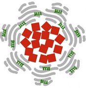
Their carrier is a hybrid of a CsA-poly (lactic-co-glycolic) acid microsphere-loaded hydrogel and a polymeric framework, which ensures the carrier can endure external force under physiological conditions.
In in vitro experiments with the carrier, the researchers observed decreased expression of cytokines, which are secreted by spleen cells activated by Concanavalin A and are related to immune rejection.
The team also implanted in mice drug carriers seeded with xenogeneic cells, and they observed significant suppression of T-cell-mediated rejection for 4 weeks.
The researchers believe this study could help overcome existing cell transplantation limitations caused by systemic immunosuppression. They said their carrier could be a promising solution for treating a range of diseases that require cell-based therapy. ![]()
Photo by Aaron Logan
Researchers have used a 3D printer to create a carrier that allows for local and sustained delivery of the immunosuppressive drug cyclosporine A (CsA) after cell transplantation.
The carrier is a combination of microspheres and hydrogel. In murine experiments, it delivered a local, sustained load of CsA in an amount that eliminated the need for additional drugs to treat immune rejection.
The researchers described these results in Cell Transplantation.
“Our objective was to show the feasibility of using a subcutaneous, 3D-printed drug delivery system to achieve local and sustained CsA release and to investigate the local immunosuppressive effects of the CsA after cell transplantation,” said study author Dong-Woo Cho, PhD, of the Pohang University of Science and Technology in Korea.
“The improved load-bearing capacity of the combined microsphere and hydrogel system, and its ability to maintain its integrity and shape during the implantation period, helped to deliver a sustained CsA release, preventing the acceleration of the secretion of cytokines related to immune rejection.”
The researchers noted that CsA improves the success rate of transplants, but systemic administration requires high doses that can have severe side effects. The benefit of a carrier is that it provides local drug delivery.
Other research groups have attempted CsA delivery via either microspheres or hydrogels, but most encountered serious problems, such as embolisms or organ damage due to migration of the microspheres from the injection site.
In addition, weak mechanical properties in some delivery systems caused premature dissolution and placed limitations on drug load quantity.
However, Dr Cho’s group said their carrier’s improved structure and load-bearing capacity allowed for sustained release of CsA at the desired site.
Their carrier is a hybrid of a CsA-poly (lactic-co-glycolic) acid microsphere-loaded hydrogel and a polymeric framework, which ensures the carrier can endure external force under physiological conditions.
In in vitro experiments with the carrier, the researchers observed decreased expression of cytokines, which are secreted by spleen cells activated by Concanavalin A and are related to immune rejection.
The team also implanted in mice drug carriers seeded with xenogeneic cells, and they observed significant suppression of T-cell-mediated rejection for 4 weeks.
The researchers believe this study could help overcome existing cell transplantation limitations caused by systemic immunosuppression. They said their carrier could be a promising solution for treating a range of diseases that require cell-based therapy. ![]()
Skiing accident claims life of leukemia expert

Photo courtesy of RPCI
Meir Wetzler, MD, Chief of the Leukemia Section at the Roswell Park Cancer Institute (RPCI) in Buffalo, New York, has died at the age of 60.
Dr Wetzler passed away on February 23, in a Denver, Colorado, hospital a little more than 2 weeks after a skiing accident.
He was nationally prominent in his field and served on the Chronic Myelogenous Leukemia (CML) Treatment Committee of the National Comprehensive Cancer Network, helping set the standard of care for CML patients.
Originally from Israel, Dr Wetzler earned his medical degree at Hebrew University’s Hadassah Medical School in Jerusalem and did his residency in internal medicine at Kaplan Hospital in Rehovot before coming to the US.
From 1988 to 1992, he served 2 fellowships—in clinical immunology/biologic therapy and medical oncology—at MD Anderson Cancer Center in Houston, Texas. He joined the Leukemia Division of RPCI in 1994.
Dr Wetzler’s colleagues said he worked tirelessly with cooperative groups and pharmaceutical companies to attract new trials to RPCI for the benefit of his patients.
“He gave a piece of himself in everything he did, from his research to his care for patients to his interactions with his team of colleagues,” said Kara Eaton-Weaver, RPCI’s Executive Director of the Patient and Family Experience. “Meir was a transformational leader who built a culture of empathy, compassion, integrity, and innovation.”
“He was like a father,” said Linda Lutgen-Dunckley, a pathology resource technician at RPCI. “Everybody was part of a team, and nobody was less important than he was. He felt everybody played their part on the team.”
Dr Wetzler is survived by his wife, Chana, and their 4 children: Mor, Shira, Adam, and Modi.
The Dr Meir Wetzler Memorial Fund for Leukemia Research has been established to benefit leukemia research. A portion of the donations will be used to plant a tree in his memory in RPCI’s Kaminski Park & Gardens. To donate directly, visit giving.roswellpark.org/wetzler.
To send a personal message to Dr Wetzler’s family, direct it to Jamie Genovese at Roswell Park Cancer Institute, Department of Medicine, Elm and Carlton Streets, Buffalo, NY 14263. Messages can also be dropped off at RPCI’s Leukemia Center. ![]()
Haplo-BMT feasible in high-risk malignancies

Photo by Chad McNeeley
SAN DIEGO—Haploidentical bone marrow transplant (haplo-BMT) is a feasible option for patients with high-risk hematologic malignancies who don’t have timely access to an HLA-matched donor, according to a speaker at the 2015 BMT Tandem Meetings.
Haplo-BMT using myeloablative conditioning, T-cell replete grafts, and post-transplant cyclophosphamide elicited “excellent” rates of engraftment, graft-vs-host disease (GVHD), and transplant-related mortality, said Heather Symons, MD, of The Johns Hopkins University School of Medicine in Baltimore, Maryland.
Dr Symons presented these results at the meeting as abstract 6*, which was chosen as one of the meeting’s “Best Abstracts.” The research was funded by Otsuka Pharmaceuticals.
Dr Symons and her colleagues conducted a phase 2 study of 96 patients with high-risk hematologic malignancies and a median age of 42 (range, 1-65). Males made up 58% of the population.
Diagnoses included acute and chronic leukemias, lymphomas, multiple myeloma, and myelodysplastic syndromes. Some patients were in complete remission, and some were in chemo-sensitive partial remission. There was also a mix of minimal residual disease positivity and negativity.
“Given the heterogeneity of these patients, we further classified our patients by disease risk index,” Dr Symons said. “We used the revised disease risk index as published by Armand in 2013. The disease risk index, or DRI, assigned patients into overall survival risk groups based on disease type and status.”
So 6 patients had a low DRI, 61 had an intermediate DRI, and 29 had a high DRI.
For most patients (n=73), conditioning consisted of intravenous busulfan (pharmacokinetically adjusted) on days –6 to –3 and cyclophosphamide (50 mg/kg/day) on days –2 and –1. But 23 patients (those with acute lymphocytic leukemia or lymphoblastic lymphoma) received cyclophosphamide (50 mg/kg/day) on days –5 and –4 and total body irradiation (200 cGy twice daily) on days –3 to -1.
All patients received T-cell-replete bone marrow from haploidentical, related donors. The median number of HLA mismatches was 4.
Post-transplant immunosuppression consisted of cyclophosphamide (50 mg/kg/day) on days 3 and 4, followed by mycophenolate mofetil for 30 days and tacrolimus for 6 months.
‘Excellent’ outcomes
The median follow-up was 18 months (range, 3-59). The median time to neutrophil engraftment was 24 days, and the median time to platelet engraftment was 29 days. Ninety-one percent of patients had donor chimerism greater than 95% at day 60.
The cumulative incidence of acute GVHD was 17% for grades 2-4 and 7% for grades 3-4. The cumulative incidence of chronic GHVD was 15%. For moderate-to-severe chronic GVHD, it was 5%.
Twenty-four percent of patients had CMV reactivation, and 22% had hemorrhagic cystitis.
The rate of relapse was 36% at 1 year and 44% at 3 years. The transplant-related mortality rate was 6% at 100 days and 11% at 1 year.
Ten patients died—2 from GVHD, 1 of cardiomyopathy, 2 of veno-occlusive disease, 1 of drug-induced liver injury, 2 due to infection, and 2 of unknown causes.
Overall survival was 72% at 1 year, 57% at 2 years, and 51% at 3 years. Event-free survival was 56% at 1 year, 51% at 2 years, and 47% at 3 years.
A multivariate analysis revealed that overall survival decreased with increasing age and increasing DRI.
Compared to patients younger than 20, the hazard ratio (HR) was 6.3 for patients ages 20 to 50 (P=0.02) and 4.7 for patients older than 50 (P=0.04). Compared to patients with a low or intermediate DRI, the HR was 2.2 for those with a high DRI (P=0.03).
The analysis also indicated that increasing age and donor CMV-positivity conferred worse event-free survival.
Compared to patients younger than 20, the HR was 3.6 for patients ages 20 to 50 (P=0.04) and 4.0 for patients older than 50 (P=0.03). Compared to patients who had a CMV-negative donor, the HR was 2.2 for patients who had a CMV-positive donor (P=0.01).
“In conclusion, myeloablative haploidentical bone marrow transplantation with post-transplantation cyclophosphamide for high-risk hematologic malignancies has excellent rates of engraftment, graft-vs-host disease, and transplant-related mortality, with results that are similar to those described in myeloablative HLA-matched bone marrow transplantation,” Dr Symons said.
“Overall, this seems a feasible option for high-risk patients who lack timely access to an HLA-matched donor and warrants continued study. We are soon to start enrolling patients on a Pediatric Blood & Marrow Transplant Consortium trial using myeloablative conditioning, haploidentical donors, and post-transplantation cyclophosphamide.” ![]()
*Information in the abstract differs from that presented at the meeting.
Protein appears key to success with clopidogrel
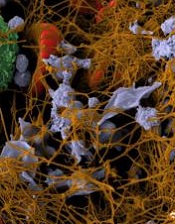
Image by Andre E.X. Brown
The platelet protein RASA3 is critical to treatment success with the antiplatelet drug clopidogrel (Plavix), according to research published in The Journal of Clinical Investigation.
The study shows that RASA3 is part of a cellular pathway that is crucial for platelet activity during thrombus formation.
Researchers believe this newfound information could aid the development of new antiplatelet compounds and antidotes to clopidogrel.
“We believe these findings could lead to improved strategies for treatment following a heart attack and a better understanding of why people respond differently to antiplatelet drugs such as aspirin and Plavix,” said study author Wolfgang Bergmeier, PhD, of the University of North Carolina at Chapel Hill.
Since the 1970s, scientists have known that clopidogrel has an anticlotting effect on platelets. In 2001, they found the compound’s target—the adenosine diphosphate receptor P2Y12. But they didn’t know how this receptor communicates with other proteins in the cell pathways important for platelet activation.
Researchers have since learned that P2Y12 communicates with RAP1, a small GTPase that cycles between an inactive GDP-bound form and an active GTP-bound form. RAP1 is tightly regulated by GEFs, which stimulate GTP loading, and GAPs, which catalyze GTP hydrolysis.
Dr Bergmeier’s group and others previously showed that RAP1 regulates platelet adhesion and thrombosis.
Now, his team has found evidence suggesting the GAP RASA3 is the “missing link” between P2Y12 and RAP1 in platelets. And agents that inhibit P2Y12, like clopidogrel, prevent thrombosis mainly through their effect on RASA3/RAP1 signaling.
The researchers used deep sequencing techniques to show that RASA3 was the only highly expressed GAP gene for RAP1 in platelets. And they hypothesized that a malfunctioning RASA3 protein would lead to the activation and clearance of platelets.
Experiments showed that mice with a RASA3 mutation had 3% to 5% of the typical platelet count. The rest of their platelets were being activated and cleared from circulation.
However, when the researchers disabled the major GEF proteins, platelet counts rose to normal levels. This suggests a tightly controlled balance between GEF and GAP proteins, especially RASA3, is vital for platelet activity.
“These experiments show that this RAP1 GEF-GAP pathway is crucial for platelets to jump into action to plug a hole in the endothelium,” Dr Bergmeier said. “And now we know that RASA3 is a critical negative regulator, a ‘brake’ on the process.”
“We have good reason to believe that the RAP1 ‘switch,’ controlled by the same GEF and GAP proteins, also regulates the active state of human platelets. We expect this research will provide critical information for improving antiplatelet therapies, possibly including approaches that eliminate some of the patient-to-patient variability and the increased bleeding risk associated with current antiplatelet drugs.” ![]()
NSAIDs may increase bleeding, cardiovascular events

Photo courtesy of CDC
A large, retrospective study has shown that nonsteroidal anti-inflammatory drugs (NSAIDs) can increase the risk of bleeding and cardiovascular events in patients who are receiving antithrombotic therapy after myocardial infarction (MI).
The risks were increased regardless of the type of antithrombotic treatment patients received, the types of NSAIDs they were prescribed, or the duration of NSAID use.
Researchers reported these findings in JAMA.
Anne-Marie Schjerning Olsen, MD, PhD, of Copenhagen University Hospital Gentofte in Hellerup, Denmark, and her colleagues conducted the research. They used nationwide administrative registries in Denmark (2002-2011) to analyze patients 30 years of age or older who were admitted with first-time MI and were alive 30 days after hospital discharge.
The team looked at subsequent treatment with aspirin, clopidogrel, or other oral anticoagulants and their combinations, as well as ongoing, concomitant, prescription NSAID use. They assessed the risk of bleeding requiring hospitalization and a composite cardiovascular outcome (cardiovascular death, nonfatal recurrent MI, and stroke).
The study included 61,971 patients with a mean age of 68 years. Thirty-four percent of these patients filled at least 1 NSAID prescription.
At a median follow-up of 3.5 years, 18,105 patients (29.2%) had died. There were 5288 bleeding events (8.5%) and 18,568 cardiovascular events (30.0%).
A multivariate analysis showed that NSAID use increased the risk of bleeding (hazard ratio, 2.02) and cardiovascular events (hazard ratio, 1.40). The increased risk of these events was present regardless of the type of antithrombotic treatment, the types of NSAIDs used, or the duration of NSAID use.
Dr Schjerning Olsen and her colleagues said additional research is needed to confirm these findings, but physicians should exercise caution when prescribing NSAIDs to patients with a recent MI.
Authors of a related editorial pointed out that this study only included prescription NSAID use. In countries where NSAIDs are available over the counter, physicians may be unaware that patients are taking these drugs. ![]()
Variant may predict risk of toxicity in ALL

Kristine Crews, Barthelemy
Diouf, and William Evans
Photo by Seth Dixon
A gene variant may help predict vincristine-related toxicity in children with acute lymphoblastic leukemia (ALL).
A study of more than 300 children with ALL showed that patients with an inherited variant in the gene CEP72 were more likely to develop peripheral neuropathy after receiving the chemotherapy drug vincristine.
If these results can be replicated in additional populations, they may provide a basis for safer dosing of the drug, according to investigators.
William Evans, PharmD, of St Jude Children’s Research Hospital in Memphis, Tennessee, and his colleagues conducted this research and shared the results in JAMA.
The investigators analyzed patients in 2 prospective clinical trials for childhood ALL that included treatment with 36 to 39 doses of vincristine. The team performed genetic analyses and assessed vincristine-induced peripheral neuropathy in 321 patients in whom DNA data were available.
This included 222 patients with a median age of 6 years who were enrolled in a St Jude Children’s Research Hospital study from 1994 to 1998, as well as 99 patients with a median age of 11.4 years who were enrolled in a Children’s Oncology Group (COG) study from 2007 to 2010.
Grade 2 to 4 vincristine-induced peripheral neuropathy occurred in 28.8% of patients (64/222) in the St Jude cohort and in 22.2% (22/99) in the COG cohort.
The investigators found that a single nucleotide polymorphism (SNP) in the promoter region of the CEP72 gene, which encodes a centrosomal protein involved in microtubule formation, was significantly associated with vincristine-induced neuropathy (P=6.3×10⁻⁹).
This SNP had a minor allele frequency of 37%, and 16% of patients (50/321) were homozygous for the risk allele (TT at rs924607).
Among patients with the high-risk CEP72 genotype, 56% (28/50) had at least 1 episode of grade 2 to 4 neuropathy. This was significantly higher than in patients with the CEP72 CC or CT genotypes. About 21% of those patients (58/271) had at least 1 episode of grade 2 to 4 neuropathy (P=2.4×10⁻⁶).
In addition, the severity of neuropathy was greater in patients who were homozygous for the TT genotype than in patients with the CC or CT genotype—2.4-fold greater by Poisson regression (P<0.0001) and 2.7-fold greater based on the mean grade of neuropathy (P=0.004).
Finally, in lab experiments, the investigators found that reducing CEP72 expression in human neurons and leukemia cells increased their sensitivity to vincristine.
In a related editorial, Howard L. McLeod, PharmD, of the Moffitt Cancer Center in Tampa, Florida, noted that this study has several strengths: genome-wide discovery in patients from well-conducted clinical trials, replication in a multicenter cohort, statistical robustness, and laboratory correlative findings that contribute to biologic plausibility.
However, he also pointed out that vincristine remains a component of the most widely accepted treatment regimens for childhood ALL.
“It is not clear that vincristine can be removed from the treatment options for a child with CEP72 variants, although this study suggests that the resulting increase in leukemia cellular sensitivity makes vincristine dose reductions possible without compromising antileukemic effect,” he wrote.
“However, there is value in the association of CEP72 with vincristine-induced peripheral neuropathy (VIPN). The ability to objectively ascribe a degree of heightened VIPN risk will allow for greater transparency in discussions of risk and benefits of therapy with patients and their family members.”
“This also may lead to developmental therapeutic approaches to modulate CEP72 function as either primary prevention or treatment of chronic VIPN. This study also represents an initial robust effort to generate predictors for adverse drug reactions in cancer care.” ![]()
Nanoparticles destroy blood clots faster
iron oxide (red), albumin
(gray), and tPA (green)
Image courtesy of
Paolo Decuzzi lab
Results of preclinical experiments suggest that nanoparticles made of iron oxide and coated with tissue plasminogen activator (tPA) and albumin can directly target blood clots and destroy them faster than free tPA injected into the bloodstream.
Researchers found these nanoparticles could destroy blood clots 100 times faster than free tPA. And when the nanoparticles were heated using alternating magnetic fields, they destroyed clots 1000 times faster than free tPA.
Paolo Decuzzi, PhD, of Fondazione Istituto Italiano di Tecnologia in Genoa, Italy, and his colleagues described these experiments in Advanced Functional Materials.
The researchers created iron oxide nanoparticles and coated them with tPA. Typically, a small volume of concentrated tPA is injected into a patient’s blood upstream of a confirmed or suspected clot. From there, some of the tPA reaches the clot, but much of it may travel past or around the clot, potentially ending up anywhere in the circulatory system.
“We have designed the nanoparticles so that they trap themselves at the site of the clot, which means they can quickly deliver a burst of the commonly used, clot-busting drug tPA where it is most needed,” Dr Decuzzi said.
He and his colleagues used iron oxide as the nanoparticles’ core so the particles can be used for magnetic resonance imaging, remote guidance with external magnetic fields, and for further accelerating clot dissolution with localized magnetic heating.
“We think it is possible to use a static magnetic field first to help guide the nanoparticles to the clot, then alternate the orientation of the field to increase the nanoparticles’ efficiency in dissolving clots,” Dr Decuzzi said.
The team also coated the nanoparticles in albumin, which provides a sort of camouflage. It gives the nanoparticles time to reach a clot before the immune system recognizes the particles as invaders and attacks them.
“The nanoparticle protects the drug from the body’s defenses, giving the tPA time to work,” said Alan Lumsden, MD, of Houston Methodist Hospital in Texas.
“But it also allows us to use less tPA, which could make hemorrhage less likely. We are excited to see if the technique works as phenomenally well for our patients as what we saw in these experiments.”
The researchers tested the nanoparticles using human tissue cultures to see where the tPA landed and how long it took to destroy fibrin-rich clots. They also introduced blood clots in mice, injected the nanoparticles into the bloodstream, and used optical microscopy to follow the dissolution of the clots.
The nanoparticles destroyed clots about 100 times faster than free tPA.
Although free tPA is usually injected at room temperature, a number of studies have suggested the drug is most effective at higher temperatures (40° C or about 104° F). The same seems to be true for tPA delivered via the iron oxide nanoparticles.
By exposing the nanoparticles to external, alternating magnetic fields, the researchers created friction and heat. Warmer tPA (42° C or about 108° F) was released faster and increased the rate of clot dissolution 10-fold (to 1000 times greater than free tPA).
Dr Decuzzi said the next steps with this research will be testing the nanoparticles’ safety and effectiveness in other animal models, with the ultimate goal of clinical trials. He said his group will continue to examine the feasibility of using magnetic fields to guide and heat the nanoparticles.
“We are optimistic because the [US Food and Drug Administration] has already approved the use of iron oxide as a contrast agent in MRIs,” Dr Decuzzi said. “And we do not anticipate needing to use as much of the iron oxide at concentrations higher than what’s already been approved. The other chemical aspects of the nanoparticles are natural substances you already find in the bloodstream.” ![]()
iron oxide (red), albumin
(gray), and tPA (green)
Image courtesy of
Paolo Decuzzi lab
Results of preclinical experiments suggest that nanoparticles made of iron oxide and coated with tissue plasminogen activator (tPA) and albumin can directly target blood clots and destroy them faster than free tPA injected into the bloodstream.
Researchers found these nanoparticles could destroy blood clots 100 times faster than free tPA. And when the nanoparticles were heated using alternating magnetic fields, they destroyed clots 1000 times faster than free tPA.
Paolo Decuzzi, PhD, of Fondazione Istituto Italiano di Tecnologia in Genoa, Italy, and his colleagues described these experiments in Advanced Functional Materials.
The researchers created iron oxide nanoparticles and coated them with tPA. Typically, a small volume of concentrated tPA is injected into a patient’s blood upstream of a confirmed or suspected clot. From there, some of the tPA reaches the clot, but much of it may travel past or around the clot, potentially ending up anywhere in the circulatory system.
“We have designed the nanoparticles so that they trap themselves at the site of the clot, which means they can quickly deliver a burst of the commonly used, clot-busting drug tPA where it is most needed,” Dr Decuzzi said.
He and his colleagues used iron oxide as the nanoparticles’ core so the particles can be used for magnetic resonance imaging, remote guidance with external magnetic fields, and for further accelerating clot dissolution with localized magnetic heating.
“We think it is possible to use a static magnetic field first to help guide the nanoparticles to the clot, then alternate the orientation of the field to increase the nanoparticles’ efficiency in dissolving clots,” Dr Decuzzi said.
The team also coated the nanoparticles in albumin, which provides a sort of camouflage. It gives the nanoparticles time to reach a clot before the immune system recognizes the particles as invaders and attacks them.
“The nanoparticle protects the drug from the body’s defenses, giving the tPA time to work,” said Alan Lumsden, MD, of Houston Methodist Hospital in Texas.
“But it also allows us to use less tPA, which could make hemorrhage less likely. We are excited to see if the technique works as phenomenally well for our patients as what we saw in these experiments.”
The researchers tested the nanoparticles using human tissue cultures to see where the tPA landed and how long it took to destroy fibrin-rich clots. They also introduced blood clots in mice, injected the nanoparticles into the bloodstream, and used optical microscopy to follow the dissolution of the clots.
The nanoparticles destroyed clots about 100 times faster than free tPA.
Although free tPA is usually injected at room temperature, a number of studies have suggested the drug is most effective at higher temperatures (40° C or about 104° F). The same seems to be true for tPA delivered via the iron oxide nanoparticles.
By exposing the nanoparticles to external, alternating magnetic fields, the researchers created friction and heat. Warmer tPA (42° C or about 108° F) was released faster and increased the rate of clot dissolution 10-fold (to 1000 times greater than free tPA).
Dr Decuzzi said the next steps with this research will be testing the nanoparticles’ safety and effectiveness in other animal models, with the ultimate goal of clinical trials. He said his group will continue to examine the feasibility of using magnetic fields to guide and heat the nanoparticles.
“We are optimistic because the [US Food and Drug Administration] has already approved the use of iron oxide as a contrast agent in MRIs,” Dr Decuzzi said. “And we do not anticipate needing to use as much of the iron oxide at concentrations higher than what’s already been approved. The other chemical aspects of the nanoparticles are natural substances you already find in the bloodstream.”
Older age doesn’t decrease HRQOL among PBSC donors
Photo courtesy of Canterbury
District Health Board
SAN DIEGO—New research indicates that older stem cell donors have somewhat poorer overall health before they donate, but their health-related quality of life (HRQOL) post-donation is similar to that of younger donors.
In fact, the older donors included in this study actually fared better than their younger counterparts in some respects.
Galen E. Switzer, PhD, of the University of Pittsburgh in Pennsylvania, presented these results at the 2015 BMT Tandem Meetings (abstract 27*).
“[Older donors] may be at greater physical and psychological risk because of their age and comorbid conditions,” Dr Switzer noted. “So it’s critical for us to understand the health-related quality of life experiences of these donors.”
With that in mind, he and his colleagues evaluated 163 subjects who donated peripheral blood stem cells (PBSCs) to relatives in need of a transplant. The team compared donors over the age of 60 (n=104, median age 66 years) to those aged 18 to 60 (n=59, median age 41 years).
The investigators collected data via structured telephone interviews 2 weeks before PBSC donation and at 4 weeks and 1 year post-donation.
A comparison of sociodemographic factors revealed that older PBSC donors were significantly less likely to be employed (P<0.001) but more likely be white (P=0.009), be married (P=0.044), and have children (P<0.001).
Pre- and post-donation HRQOL
Pre-donation, older donors had significantly poorer physical health (P=0.001) and better mental health (P=0.036) than younger donors. But there was no significant difference between the age groups with regard to the incidence of depression or anxiety.
Similarly, there were no significant differences with regard to ambivalence, satisfaction, or medical concerns about donation. However, older donors were more likely to consult their physician about donation (P=0.049), and they had fewer work/family concerns (P=0.049) than younger donors.
At 4 weeks post-donation, there were no significant differences between the age groups with regard to general physical health, mental health, or any of 12 donation-related symptoms. However, younger donors were significantly more likely to report that donation was painful (P=0.025).
Older donors were significantly less likely to report work/family concerns, such as missing work, family worry, or worry about what others would think (P=0.001). They were less likely to have other donation-related concerns as well, such as worrying about who would pay for the procedure (P=0.034). And they were less likely to say they would feel responsible if the transplant did not have a favorable outcome (P=0.022).
At 1 year post-donation, there were no significant differences between the age groups with regard to overall physical and mental health, depression, ambivalence, satisfaction, 11 of 12 donation side effects, physical difficulty, psychological difficulty, or “other concerns.”
However, older donors reported significantly less anxiety, fewer medical concerns, and fewer work/family concerns (P<0.05 for all). They were also less likely to feel responsible for transplant outcomes and less likely to have problems sleeping, which was 1 of the 12 donation side effects (P<0.05 for both).
“So the overall conclusion, I think, is really reassuring,” Dr Switzer said. “Despite having somewhat poorer overall general physical health at pre-donation, older donors experience similar—and, in some domains, better—donation-related health-related quality of life than younger donors. So they seem to be doing at least as well and, in some domains, better than their younger counterparts.”
*Information in the abstract differs from that presented at the meeting.
Treatment likely doesn’t increase risk of cancer

Pimecrolimus, a topical cream used to treat eczema in children, does not appear to confer an increased risk of hematologic and other cancers, according to researchers.
In 2001 and 2002, respectively, the US Food and Drug Administration and the European Medicines Agency approved pimecrolimus to treat eczema in children 2 years of age and older.
The product was approved with a black box warning describing a potential risk of malignancy.
To investigate this risk, and as part of the post-marketing commitments for the approval of pimecrolimus, researchers initiated the Pediatric Eczema Elective Registry (PEER) study in 2004.
David J. Margolis, MD, PhD, of the University of Pennsylvania in Philadelphia, and his colleagues analyzed data from this study, comparing the incidence of malignancy among PEER subjects to expected cancer rates from the Surveillance, Epidemiology and End Results program.
The team reported their findings in JAMA Dermatology.
The PEER study enrolled 7457 children (26,792 person-years) who used an average of 793 g of pimecrolimus while on study.
As of May 2014, 5 malignancies were reported in these children: 2 leukemias, 2 lymphomas, and 1 osteosarcoma.
No skin cancers were reported, and none of the findings regarding cancer incidence were statistically significant.
The researchers therefore said it’s unlikely that topical pimecrolimus, as it was used in the PEER study, is associated with an increased risk of malignancy.
This research and the PEER study were both funded by Valeant Pharmaceuticals International, makers of pimecrolimus.
In a related editorial in JAMA Dermatology, Jon M. Hanifin, MD, of Oregon Health and Science University in Portland, said he hopes this research will improve the management of eczema by countering any concerns about pimecrolimus use that were raised by the treatment’s black box warning.
“The positive and optimistic report of pimecrolimus post-marketing surveillance by Margolis et al should help reduce the physician and pharmacist concerns that have restricted the use of these effective topical alternatives to corticosteroids,” Dr Hanifin wrote. “The interim results should help bring relief to a larger segment of the many young individuals with [eczema].”
FDA approves first HDAC inhibitor for MM

The US Food and Drug Administration (FDA) has granted accelerated approval for panobinostat (Farydak) to treat patients with multiple myeloma (MM).
Panobinostat is the first histone deacetylase (HDAC) inhibitor approved to treat MM.
The drug can now be used in combination with bortezomib and dexamethasone to treat patients who have received at least 2 prior standard therapies, including bortezomib and an immunomodulatory agent (IMiD).
Panobinostat was approved with a boxed warning alerting patients and healthcare professionals that severe diarrhea and severe and fatal cardiac events, arrhythmias, and electrocardiogram changes have occurred in patients receiving the drug.
Panobinostat was approved with a Risk Evaluation and Mitigation Strategy as well, which consists of a communication plan to inform healthcare professionals of these risks and how to minimize them.
Data supporting approval
In November 2014, the FDA’s Oncologic Drugs Advisory Committee advised the agency that, based on the data reviewed, the benefits of panobinostat did not outweigh its risks for patients with relapsed MM.
After the meeting, Novartis, the company developing the HDAC inhibitor, submitted additional information supporting the use of panobinostat for a different indication: MM patients who have received at least 2 prior standard therapies, including bortezomib and an IMiD.
The FDA’s accelerated approval of panobinostat is based on that data—efficacy and safety results in a subgroup analysis of 193 patients enrolled in the phase 3 PANORAMA-1 trial. These patients had received prior treatment with both bortezomib and an IMiD.
In these patients, treatment with panobinostat, bortezomib, and dexamethasone resulted in superior progression-free survival, when compared to treatment with bortezomib, dexamethasone, and placebo—10.6 months and 5.8 months, respectively (hazard ratio=0.52).
The most common adverse events (incidence ≥ 20%) in clinical studies of panobinostat have been diarrhea, fatigue, nausea, peripheral edema, decreased appetite, pyrexia, and vomiting.
The most common non-hematologic laboratory abnormalities (incidence ≥ 40%) were hypophosphatemia, hypokalemia, hyponatremia, and increased creatinine. The most common hematologic laboratory abnormalities (incidence ≥ 60%) were thrombocytopenia, lymphopenia, leukopenia, neutropenia, and anemia.
Panobinostat can cause fatal and serious toxicities, including severe diarrhea and cardiac toxicities.
The most frequent (≥ 5%) treatment-emergent serious adverse events for patients treated with the HDAC inhibitor were pneumonia (18%), diarrhea (11%), thrombocytopenia (7%), fatigue (6%), and sepsis (6%). Additional serious adverse events included hemorrhage, myelosuppression, infections, hepatotoxicity, and embryo-fetal toxicity.
Panobinostat development
The FDA previously granted panobinostat priority review and orphan product designation. Priority review provides an expedited review of drugs that are intended to treat a serious disease or condition and may provide a significant improvement over available therapy. Orphan product designation is given to drugs intended to treat rare diseases.
Now, the FDA has granted panobinostat accelerated approval, which allows for conditional approval of a drug based on clinical data showing the drug has an effect on a surrogate endpoint reasonably likely to predict clinical benefit to patients.
Continued approval of panobinostat may be contingent upon verification of a clinical benefit in confirmatory trials conducted by Novartis. An improvement in overall survival or disease-related symptoms has not yet been established for the HDAC inhibitor.
For more details on panobinostat, see the full prescribing information.