User login
Immunotoxin could treat B-cell malignancies, team says
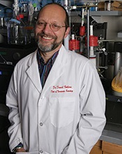
Photo courtesy of the
University of Minnesota
A bispecific ligand-directed diphtheria toxin known as DT2219 shows promise for treating patients with relapsed/refractory B-cell malignancies, according to researchers.
DT2219 produced responses in 2 of 25 patients analyzed in a phase 1 study. The maximum tolerated dose of DT2219 was not reached, although 2 patients experienced dose-limiting toxicities.
“In this phase 1 trial, we found a safe dose of the drug that has biological activity,” said Daniel Vallera, PhD, of the University of Minnesota in Minneapolis.
“We are planning a phase 2 trial with this drug. It will focus on giving more cycles of treatment, which we believe will dramatically enhance the response rates.”
Dr Vallera and his colleagues detailed the phase 1 results in Clinical Cancer Research.
To develop DT2219, the researchers chose 2 antibody fragments that each selectively bind to CD19 and CD22. They used genetic engineering to attach these two antibodies to the bacterial diphtheria toxin.
When the antibody fragments bind to the two targets on the cancer cell, the entire drug enters the cell, and the toxin kills the cell.
To test DT2219, the researchers enrolled 25 patients with chemo-refractory pre-B acute lymphoblastic leukemia (n=10), chronic lymphocytic leukemia (n=5), or non-Hodgkin lymphoma (n=10). All tumors had CD19 and/or CD22 proteins.
Patients had received a median of 3 prior therapies (range, 2 to 5), and 8 patients had undergone an unsuccessful stem cell transplant (5 autologous and 3 allogeneic).
Patients received DT2219 intravenously over 2 hours every other day for 4 total doses. The dose was escalated from 0.5 μg/kg/day to 80 μg/kg/day in 9 dose cohorts until a dose-limiting toxicity occurred.
All but 1 patient received a single course of DT2219. That patient received a second, 4-dose course after attaining a partial response.
Outcomes
The 12 patients who received doses ranging from 0.5 mg/kg/day to 20 mg/kg/day had minimal or no adverse events (AEs). But all 13 patients who received 4 doses of DT2219 at 40 mg/kg or greater every other day experienced treatment-related AEs.
Grade 1-2 treatment-related AEs included capillary leak syndrome (n=7), ALT/AST elevation (n=4), fatigue (n=3), fever (n=3), hypokalemia (n=2), hypoalbuminemia (n=1), hearing loss (n=1), hypocalcemia (n=1), anemia (n=1), and vomiting (n=1).
Grade 3-4 treatment-related AEs were thrombocytopenia (n=2), neutropenia (n=1), neutropenic fever (n=1), capillary leak syndrome (n=1), hypokalemia (n=1), and leg weakness (n=1). The grade 3 leg weakness and grade 3 capillary leak syndrome were dose-limiting toxicities.
The maximum tolerated dose was not reached, but clinical responses occurred between doses of 40 to 80 µg/kg.
All 25 patients were evaluable for response, but only 9 patients in the highest dose cohorts had measurable drug levels.
Two patients had durable, objective responses. One patient had chronic lymphocytic leukemia, and the other had diffuse large B-cell lymphoma. The latter patient’s response was a complete remission that occurred after 2 treatment cycles.
“We were surprised that the drug was effective enough to entirely eliminate the cancer in one of our patients,” Dr Vallera said. “Further, we expected the patients to make antibodies against the bacterial toxin and, thus, reject our drug. Surprisingly, this did not occur in the majority of our patients [70%].”
“We need to study more patients to understand why they did not produce neutralizing antibodies. However, we also have been working to create a less immunogenic form of the toxin for the next-generation drug.”
“Another important fact about our drug is that it was home-grown, meaning there was no commercial partner, which is rare. The drug was funded mostly with private donations, including individuals that have lost loved ones to cancer.” ![]()
Risks and benefits of long-term ticagrelor

Photo courtesy of AstraZeneca
SAN DIEGO—New research suggests that adding the antiplatelet drug ticagrelor to aspirin as long-term therapy after a heart attack can significantly reduce the risk of cardiovascular death, heart attack, or stroke, but it can also increase the risk of major bleeding.
Marc S. Sabatine, MD, of Brigham and Women’s Hospital in Boston, Massachusetts, presented these results at the American College of Cardiology’s 64th Annual Scientific Session (abstract 400-18). The findings were published in NEJM as well.
The research was sponsored by AstraZeneca, the company developing ticagrelor.
The trial, known as PEGASUS-TIMI 54, included 21,162 patients who had experienced a heart attack in the previous 1 to 3 years. Each had another factor, such as age or diabetes, that put them at risk for a second heart attack.
The patients were randomized to receive aspirin plus twice-daily doses of ticagrelor at 90 mg, ticagrelor at 60 mg, or placebo.
The twice-daily 90 mg dose of ticagrelor is already approved for patients with acute coronary syndrome. The researchers included a lower dose in this study to investigate whether platelet inhibition needed 2 years after a heart attack might be different from what is needed 2 hours after a heart attack.
The team found that both ticagrelor doses reduced the rate of the primary endpoint, which was a composite of cardiovascular death, heart attack, and stroke. At 3 years, the rate was 7.85% in the 90 mg group, 7.77% in the 60 mg group, and 9.04% in the placebo group (P=0.008 for 90 mg vs placebo and P=0.004 for 60 mg vs placebo).
“The benefit we saw was remarkably consistent across the individual components of the endpoint and in all the major subgroups of patients,” Dr Sabatine said. “Moreover, we followed patients for an average of just under 3 years, and our event curves continue to spread out over time, suggesting that the benefit continues to accrue over time.”
“Efficacy was virtually identical with both ticagrelor doses,” he added. “Risk of bleeding and dyspnea tended to be, as predicted, a bit more with the 90 mg than the 60 mg dose, but the trial wasn’t designed to compare those two dose levels.”
The rates of TIMI major bleeding were 2.60% in the 90 mg group, 2.30% in the 60 mg group, and 1.06% in the placebo group (P<0.001 for each ticagrelor dose vs placebo).
The rates of dyspnea were 18.93% in the 90 mg group, 15.84% in 60 mg group, and 6.38% in the placebo group (P<0.001 for each ticagrelor dose vs placebo).
The rates of dyspnea leading to treatment discontinuation were 6.5% in the 90 mg group, 4.55% in the 60 mg group, and 0.79% in the placebo group (P<0.001 for each ticagrelor dose vs placebo).
Although the differences in dyspnea and TIMI major bleeding between the 2 ticagrelor dose groups were not statistically significant, Dr Sabatine and his colleagues said the 60 mg dose may offer a more attractive benefit-risk profile.
“Now that we have the evidence, when faced with a patient who has had a heart attack, based on these data, I would continue treatment with ticagrelor as long as the patient tolerated it,” Dr Sabatine concluded. ![]()
Research paves way for new SCID treatment
Image courtesy of the Salk
Institute of Biological Studies
Researchers say they have devised a way to correct a genetic defect in cells from a patient with X-linked severe combined immunodeficiency (SCID-X1).
The group generated induced pluripotent stem cells (iPSCs) using a sample from an infant with SCID-X1, then used the gene-editing technique TALEN to correct the defect in those iPSCs.
And this allowed the iPSCs to generate mature natural killer (NK) cells and T-cell precursors.
The researchers said this is the first evidence of genomic correction of iPSCs derived from a patient with SCID-X1 that resulted in the regeneration of mature lymphoid cells in vitro.
And the work, which is published in Cell Stem Cell, holds promise for the development of new treatments for this disease.
“This work demonstrates a new method that could lead to a more effective and less invasive treatment for this devastating disease,” said study author Inder Verma, PhD, of The Salk Institute of Biological Studies in La Jolla, California.
“It also has the potential to lay the foundation to cure other deadly and rare blood disorders.”
Dr Verma and his colleagues began this research by securing a sample of bone marrow from a deceased infant with SCID-X1. The subject harbored a novel splice-site mutation (c.468+3A > C) of the IL-2Rγ gene, which results in a lack of functional NK and T cells.
The researchers used the subject’s sample to create iPSC lines and used TALEN to correct the mutation in those iPSCs.
“We use TALEN-based genome editing to change just one nucleotide in one gene to correct the deficiency,” said study author Tushar Menon, PhD, also of the Salk Institute. “The technique is literally that precise.”
The team then compared the corrected iPSCs to uncorrected SCID-X1 iPSCs and control iPSCs generated from cord-blood-derived endothelial cells and dermal fibroblasts.
Like control cells, the corrected iPSCs were able to differentiate into mature NK cells that expressed both inhibitory and activating receptors (KIR/CD158b and CD16, respectively).
The corrected iPSCs could also differentiate into T-cell precursors but not mature T cells. The researchers are currently working on producing mature T cells.
“Ultimately, we hope these efforts will help lead to the ‘holy grail’ in the field: the ability to create stem cells from iPSCs capable of generating all types of blood and immune cells,” Dr Verma said.
He and his colleagues noted, however, that further improvements of their protocol will be needed to obtain sufficient cells for clinical use. ![]()
New approach to gene therapy for hemophilia

that can develop hemophilia B
Gene therapy that produces a potent clotting factor holds promise for treating hemophilia B, researchers have reported in Blood.
The group administered adeno-associated viral-8 (AAV-8) vectors encoding the clotting factor FIX-Padua to mice and dogs with hemophilia B.
The treatment appeared to be safe and effective, did not prompt the formation of inhibitory antibodies, and even eradicated pre-existing inhibitors in one of the dogs.
“Our findings may provide a new approach to gene therapy for hemophilia and perhaps other genetic diseases that have similar complications from inhibiting antibodies,” said study author Valder R. Arruda, MD, PhD, of The Children’s Hospital of Philadelphia in Pennsylvania.
For years, researchers have investigated gene therapy strategies that deliver DNA sequences carrying genetic code to produce clotting factor in patients. But this approach has been hindered by the body’s immune response against vectors.
Those responses, which defeated initial benefits seen in experimental human gene therapy, were dose-dependent. So Dr Arruda and his colleagues decided to test gene therapy that used lower doses of AAV-8 vector to produce FIX-Padua.
Dr Arruda was part of a team that discovered FIX-Padua in a young Italian man with thrombophilia. A mutation in the factor IX gene produced the clotting factor, which was named after the patient’s city of residence.
FIX-Padua is hyperfunctional, clotting blood 8 to 12 times more strongly than wild-type factor IX. Therefore, in the current study, the researchers needed to strike a balance—to relieve severe hemophilia in dogs by using a dose strong enough to allow clotting but not enough to cause thrombosis or stimulate immune reactions.
“Our ultimate goal is to translate this approach to humans by adapting this variant protein found in one patient to benefit other patients with the opposite disease,” Dr Arruda said
He and his colleagues tested the safety of canine FIX-Padua (AAV-cFIX-Padua) in 3 dogs, all with naturally occurring types of hemophilia B.
Two of the dogs had never been exposed to clotting factor and had never developed antibodies. Injections of AAV-cFIX-Padua changed their hemophilia from severe to mild. They had no bleeding episodes for up to 2 years and did not develop inhibitory antibodies or thrombosis.
The third dog, Wiley, already had inhibitory antibodies before receiving AAV-cFIX-Padua. Like his peers, Wiley responded to the treatment, and that response persisted for 3 years. AAV-cFIX-Padua also eradicated his inhibitors, which marks the first time this occurred in an animal model with pre-existing antibodies.
Another set of experiments in mice suggested the gene therapy is safe and effective. Mice that received AAV encoding human FIX-Padua (AAV-hFIX-Padua) did not develop antibodies.
And the researchers found that AAV-hFIX-Padua was comparable to wild-type human FIX with regard to long-term expression and toxicity.
Dr Arruda noted that larger studies are needed in dogs with pre-existing inhibitors to confirm these encouraging early results.
In the meantime, at least one clinical trial is making use of FIX-Padua in adult patients with hemophilia B—at the University of North Carolina at Chapel Hill, under Paul Monahan, MD. Leaders of a separate trial being planned at Spark Therapeutics in Philadelphia, under Katherine A. High, MD, are contemplating using FIX-Padua as well. ![]()
Group uses gene editing to fight lymphoma
Aubrey, Gemma Kelly,
and Marco Herold
Photo courtesy of the
Walter and Eliza Hall Institute
The gene-editing technique CRISPR/Cas9 can be used to target and kill lymphoma cells with high accuracy, according to preclinical research published in Cell Reports.
Using a lentiviral CRISPR/Cas9 platform, researchers were able to kill human Burkitt lymphoma cells by locating and deleting MCL-1, a gene known to be essential for cancer cell survival.
These results suggest the technology could be used as a direct treatment for diseases arising from genetic errors.
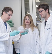
“Our study showed that the CRISPR technology can directly kill cancer cells by targeting factors that are essential for their survival and growth,” said study author Brandon Aubrey, a PhD candidate at the Walter and Eliza Hall Institute of Medical Research in Parkville, Victoria, Australia.
Aubrey and his colleagues said they engineered a lentiviral vector platform that allows for efficient cell transduction and subsequent inducible expression of small guide RNAs with concomitant constitutive expression of Cas9.
After finding they could use this system to knock out the pro-apoptotic BH3-only protein BIM in human and mouse cell lines, the team wanted to determine if it could target genes that are essential for sustained cell growth.
So they used the technique to delete MCL-1 in human Burkitt lymphoma cells. And they observed a “very high frequency” of cell killing.
The researchers also used their system to produce hematopoietic-cell-restricted TRP53-knockout mice. Along with mutations that caused loss of the TRP53 protein, the team found they had generated novel mutant TRP53 proteins that could promote lymphoma development.
“[W]e showed, for the first time, that it is possible for CRISPR technology to be used in cancer therapy,” said Marco Herold, PhD, of the Walter and Eliza Hall Institute.
“In addition to its very exciting potential for disease treatment, we have shown that it has the potential to identify novel mutations in cancer-causing genes and genes that ‘suppress’ cancer development, which will help us to identify how they initiate or accelerate the development of cancer.” ![]()
Aubrey, Gemma Kelly,
and Marco Herold
Photo courtesy of the
Walter and Eliza Hall Institute
The gene-editing technique CRISPR/Cas9 can be used to target and kill lymphoma cells with high accuracy, according to preclinical research published in Cell Reports.
Using a lentiviral CRISPR/Cas9 platform, researchers were able to kill human Burkitt lymphoma cells by locating and deleting MCL-1, a gene known to be essential for cancer cell survival.
These results suggest the technology could be used as a direct treatment for diseases arising from genetic errors.
“Our study showed that the CRISPR technology can directly kill cancer cells by targeting factors that are essential for their survival and growth,” said study author Brandon Aubrey, a PhD candidate at the Walter and Eliza Hall Institute of Medical Research in Parkville, Victoria, Australia.
Aubrey and his colleagues said they engineered a lentiviral vector platform that allows for efficient cell transduction and subsequent inducible expression of small guide RNAs with concomitant constitutive expression of Cas9.
After finding they could use this system to knock out the pro-apoptotic BH3-only protein BIM in human and mouse cell lines, the team wanted to determine if it could target genes that are essential for sustained cell growth.
So they used the technique to delete MCL-1 in human Burkitt lymphoma cells. And they observed a “very high frequency” of cell killing.
The researchers also used their system to produce hematopoietic-cell-restricted TRP53-knockout mice. Along with mutations that caused loss of the TRP53 protein, the team found they had generated novel mutant TRP53 proteins that could promote lymphoma development.
“[W]e showed, for the first time, that it is possible for CRISPR technology to be used in cancer therapy,” said Marco Herold, PhD, of the Walter and Eliza Hall Institute.
“In addition to its very exciting potential for disease treatment, we have shown that it has the potential to identify novel mutations in cancer-causing genes and genes that ‘suppress’ cancer development, which will help us to identify how they initiate or accelerate the development of cancer.” ![]()
Genotyping can help predict bleeding risk with warfarin

An analysis of data from the ENGAGE AF-TIMI 48 trial has shown that patients with a genetic sensitivity to warfarin had higher rates of bleeding during the first several months of treatment and benefitted from treatment with a different anticoagulant.
The research, published in The Lancet, suggests that using genetic analyses to identify patients who are most at risk of bleeding with warfarin could offer safety benefits, particularly in the first 90 days of treatment.
“We were able to look at patients from around the world who were being treated with warfarin and found that certain genetic variants make a difference for an individual’s risk for bleeding,” said study author Jessica L. Mega, MD, of Brigham and Women’s Hospital in Boston, Massachusetts.
“For these patients who are sensitive or highly sensitive responders based on genetics, we observed a higher risk of bleeding in the first several months with warfarin, and consequently, a big reduction in bleeding when treated with the drug edoxaban instead of warfarin.”
The FDA label for warfarin notes that genetic variants in 2 genes—CYP2C9 and VKORC1—can assist in determining the right warfarin dosage. But a conclusive link between variation in these genes and bleeding has been debated.
By leveraging data from the ENGAGE AF-TIMI 48 trial—in which patients with atrial fibrillation received warfarin or 2 different doses of edoxaban—investigators were able to observe connections between genetic differences and patient outcomes.
A subgroup of patients was genotyped for variants in CYP2C9 and VKORC1, and the results were used to identify normal responders, sensitive responders, and highly sensitive responders to warfarin.
Dr Mega and her colleagues looked at data from 14,348 patients. Of the 4833 patients taking warfarin, 61.7% were normal responders, 35.4% were sensitive responders, and 9.2% were highly sensitive responders.
In the first 90 days of treatment, normal responders were over-anticoagulated a median of 2.2% of the time, compared to 8.4% of the time for sensitive responders, and 18.3% of the time for highly sensitive responders (Ptrend<0.0001).
Both sensitive and highly sensitive responders also had an increased risk of bleeding in the first 90 days when compared to normal responders. The hazard ratios were 1.31 for sensitive responders (P=0.0179) and 2.66 for highly sensitive responders (P<0.0001).
As a result, during the first 90 days, edoxaban was more effective than warfarin at reducing bleeding in sensitive and highly sensitive responders.
Compared with warfarin, both the higher and lower doses of edoxaban reduced bleeding more in sensitive and highly sensitive responders than in normal responders (Pinteraction=0.0066 and 0.0036 for the higher and lower doses, respectively).
“These findings demonstrate the power of genetics in personalizing medicine and tailoring specific therapies for our patients,” said Marc S. Sabatine, MD, also of Brigham and Women’s Hospital.
The ENGAGE AF-TIMI 48 trial was supported by research grants from Daiichi Sankyo, makers of edoxaban. ![]()
iPSCs reveal new insight into Fanconi anemia

Image by James Thompson
Induced pluripotent stem cells (iPSCs) may help elucidate the pathogenesis of bone marrow failure (BMF) in Fanconi anemia (FA), researchers say.
They generated iPSCs from FA patients and found evidence suggesting that hematopoietic consequences originate at the earliest hematopoietic stage.
Specifically, hemoangiogenic progenitor cells (HAPCs) from FA-iPSCs produced significantly fewer hematopoietic and endothelial cells than controls.
“Although various consequences in hematopoietic stem cells have been attributed to FA-BMF, its cause is still unknown,” said study author Megumu K. Saito, MD, PhD, of Kyoto University in Japan.
“To address the issue, our team established iPSCs from 2 FA patients who have the FANCA gene mutation that is typical in FA. We were then able to obtain fetal-type immature blood cells [KDR+ CD34+ HAPCs] from these iPSCs.”
The researchers assessed differentiation in the FA-iPSC-derived HAPCs (FA-HAPCs) and found they produced significantly fewer CD34+ CD45+ hematopoietic precursors—and later, myeloid and erythroid lineage hematopoietic cells—than control cells. Likewise, FA-HAPCs produced fewer CD31+ endothelial cells than controls.
Cell cycle distribution in FA-HAPCs was comparable to that of controls, and FA-HAPCs were not apoptotic. This, according to the researchers, suggests a defect in FA-HAPCs’ ability to differentiate into hematopoietic and endothelial cells.
Further study of FA-HAPCs revealed significant downregulation of transcription factors that are critical for hematopoietic differentiation. This suggests the FA pathway might be involved in maintaining the transcriptional network critical for determining the differentiation propensity of HAPCs, the researchers said.
They also identified 227 genes that were significantly upregulated and 396 genes that were significantly downregulated in FA-HAPCs. The downregulated genes included those associated with mesodermal differentiation, vascular formation, and hematopoiesis.
“These data indicate that the hematopoietic consequences in FA patients originate from the earliest hematopoietic stage and highlight the potential usefulness of iPSC technology for explaining how FA-BMF occurs,” Dr Saito said.
“Since conducting a comprehensive analysis of patient-derived affected stem cells is not feasible without iPSC technology, the technology provides an unprecedented opportunity to gain further insight into this disease.”
Dr Saito and colleagues described this research in STEM CELLS Translational Medicine. ![]()
Image by James Thompson
Induced pluripotent stem cells (iPSCs) may help elucidate the pathogenesis of bone marrow failure (BMF) in Fanconi anemia (FA), researchers say.
They generated iPSCs from FA patients and found evidence suggesting that hematopoietic consequences originate at the earliest hematopoietic stage.
Specifically, hemoangiogenic progenitor cells (HAPCs) from FA-iPSCs produced significantly fewer hematopoietic and endothelial cells than controls.
“Although various consequences in hematopoietic stem cells have been attributed to FA-BMF, its cause is still unknown,” said study author Megumu K. Saito, MD, PhD, of Kyoto University in Japan.
“To address the issue, our team established iPSCs from 2 FA patients who have the FANCA gene mutation that is typical in FA. We were then able to obtain fetal-type immature blood cells [KDR+ CD34+ HAPCs] from these iPSCs.”
The researchers assessed differentiation in the FA-iPSC-derived HAPCs (FA-HAPCs) and found they produced significantly fewer CD34+ CD45+ hematopoietic precursors—and later, myeloid and erythroid lineage hematopoietic cells—than control cells. Likewise, FA-HAPCs produced fewer CD31+ endothelial cells than controls.
Cell cycle distribution in FA-HAPCs was comparable to that of controls, and FA-HAPCs were not apoptotic. This, according to the researchers, suggests a defect in FA-HAPCs’ ability to differentiate into hematopoietic and endothelial cells.
Further study of FA-HAPCs revealed significant downregulation of transcription factors that are critical for hematopoietic differentiation. This suggests the FA pathway might be involved in maintaining the transcriptional network critical for determining the differentiation propensity of HAPCs, the researchers said.
They also identified 227 genes that were significantly upregulated and 396 genes that were significantly downregulated in FA-HAPCs. The downregulated genes included those associated with mesodermal differentiation, vascular formation, and hematopoiesis.
“These data indicate that the hematopoietic consequences in FA patients originate from the earliest hematopoietic stage and highlight the potential usefulness of iPSC technology for explaining how FA-BMF occurs,” Dr Saito said.
“Since conducting a comprehensive analysis of patient-derived affected stem cells is not feasible without iPSC technology, the technology provides an unprecedented opportunity to gain further insight into this disease.”
Dr Saito and colleagues described this research in STEM CELLS Translational Medicine.
News reports on stem cell research often unrealistic, team says

Media coverage of translational stem cell research might generate unrealistic expectations, according to a pair of researchers.
The team analyzed reports on stem cell research published in major daily newspapers in Canada, the US, and the UK between 2010 and 2013.
They found that most reports were highly optimistic about the future of stem cell therapies and indicated that therapies would be available for clinical use within 5 to 10 years or sooner.
The researchers said that, as spokespeople, scientists need to be mindful of harnessing public expectations.
“As the dominant voice in respect to timelines for stem cell therapies, the scientists quoted in these stories need to be more aware of the importance of communicating realistic timelines to the press,“ said Kalina Kamenova, PhD, of the University of Alberta in Edmonton, Canada.
Dr Kamenova conducted this research with Timothy Caulfield, also of the University of Alberta, and the pair disclosed their results in Science Translational Medicine.
The researchers examined 307 news reports covering translational research on stem cells, including human embryonic stem cells (21.5%), induced pluripotent stem cells (12.1%), cord blood stem cells (2.9%), other tissue-specific stem cells such as bone marrow or mesenchymal stem cells (23.8%), multiple types of stem cells (18.9%), and stem cells of an unspecified type (20.8%).
The team assessed perspectives on the future of stem cell therapies and found that 57.7% of news reports were optimistic, 10.4% were pessimistic, and 31.9% were neutral.
In addition, 69% of all news stories citing timelines predicted that stem cell therapies would be available within 5 to 10 years or sooner.
“The approval process for new treatments is long and complicated, and only a few of all drugs that enter preclinical testing are approved for human clinical trials,” Dr Kamenova pointed out. “It takes, on average, 12 years to get a new drug from the lab to the market and [an] additional 11 to 14 years of post-market surveillance.”
“Our findings showed that many scientists have often provided, either by implication or direct quotes, authoritative statements regarding unrealistic timelines for stem cell therapies,” Caulfield added.
“[M]edia hype can foster unrealistic public expectations about clinical translation and increased patient demand for unproven stem cell therapies. Care needs to be taken by the media and the research community so that advances in research and therapy are portrayed in a realistic manner.”
Study seems to support liberal transfusion strategy
![]()
Photo courtesy of UAB Hospital
Results of a large, randomized trial suggest a liberal transfusion strategy may benefit patients undergoing cardiac surgery.
Patients who received blood transfusions when their hemoglobin (Hb) levels were below 9 g/dL fared better than patients who only received transfusions once their Hb levels were below 7.5 g/dL.
The “low” Hb group had a slightly higher incidence of serious complications and a significantly higher rate of 90-day mortality than the “high” Hb group.
However, the researchers noted that the latter finding, while important, is difficult to interpret because the trial was not primarily designed to compare the difference in the number of deaths.
“Although only a hypothesis, the suggestion that it might be better rather than worse to transfuse patients who are only mildly anemic goes against the evidence about when to transfuse in non-cardiac surgery settings,” said Barnaby Reeves, DPhil, of the University of Bristol in the UK.
“Transfusing more rather than fewer patients would create a challenge for hospitals. With an aging population and possibly an increase in heart disease, obesity, and diabetes, it can only become more difficult in the future to maintain the national blood supply in the UK and in other developed countries around the world. Our findings emphasize the importance of interventions to reduce blood loss in the first place.”
Dr Reeves and his colleagues reported their findings in NEJM.
The team conducted their randomized, controlled trial to determine whether transfusing cardiac surgery patients at a lower Hb level would be safer or more cost-effective, as has been shown in other patient groups.
Individuals older than 16 who were undergoing non-emergency cardiac surgery were recruited to the trial at 17 UK hospitals. Patients with an Hb level of less than 9 g/dL after their operations were randomized to have a transfusion either when they became substantially anemic—with an Hb level of less than 7.5 g/dL—or right away, when they were mildly anemic—with an Hb level of less than 9 g/dL.
To compare the two transfusion strategies, the researchers assessed the incidence of serious infection, ischemic event, heart attack, infarction of the gut, and acute kidney injury in the first 3 months after the operation.
The team analyzed data for 2003 patients. Nearly all of the patients in the high Hb group received a transfusion (92.2%), compared to just over half of patients in the low Hb group (53.4%).
Slightly more patients in the low Hb group than the high Hb group had one or more of the aforementioned serious complications—35.1% and 33%, respectively (P=0.30). And significantly more patients had died at 90 days in the low Hb group than the high group—4.2% and 2.6%, respectively (P=0.045).
The researchers found no significant differences between the high and low Hb groups with respect to other information measured to assess recovery, but some of the other findings in the trial showed a trend in the same direction.
In addition, healthcare costs up to 3 months after surgery were similar in the high and low Hb groups.
“Even though the high group were given more blood, it was interesting that this did not lead to them costing more once the costs of treating complications were added to the analysis,” said Sarah Wordsworth, PhD, of the University of Oxford in the UK.
Based on the overall pattern of findings, the researchers proposed that a high or liberal transfusion threshold may be better after cardiac surgery. This challenges most prevailing guidelines and current health policy.
“Existing national and international transfusion guidelines recommend that blood transfusions only be given to patients who develop very low hemoglobin concentrations,” said Gavin Murphy, FRCS, of the University of Leicester in the UK.
“We have shown that this strategy may increase the number of deaths in cardiac surgery. This was the largest randomized trial ever conducted in the UK in a surgical or cardiac surgery population. It was the largest trial ever conducted that has considered indications for transfusion in cardiac surgery, and recruited over twice the number of patients recruited in all the previous trials put together. It . . . recruited patients from the majority of [National Health Service] cardiac surgery centers in the UK and therefore reflects current UK practice and is relevant to UK patients.”
ZFNs can correct sickle cell mutation

and a normal one
Image by Betty Pace
Gene editing via zinc-finger nucleases (ZFNs) shows early promise for treating sickle cell disease (SCD), according to preclinical research published in Blood.
Investigators used ZFNs to correct the SCD mutation in CD34+ hematopoietic stem and progenitor cells (HSPCs) derived from the bone marrow of SCD patients.
And these modified cells were able to produce normal red blood cells in vitro.
“This is a very exciting result,” said study author Donald Kohn, MD, of the University of California, Los Angeles.
“It suggests the future direction for treating genetic diseases will be by correcting the specific mutation in a patient’s genetic code.”
For this research, Dr Kohn and his colleagues took ZFNs designed to flank the SCD mutation and delivered them with a homologous donor template (either an integrase-defective lentiviral vector [IDLV] or a DNA oligonucleotide).
The investigators first tested this gene-editing system in CD34+ HSPCs derived from healthy donors and observed “high levels” of gene modification at the beta-globin locus. They also discovered off-target cleavage in the delta-globin gene but noted that this gene is “functionally dispensable.”
Additional experiments revealed that the modified CD34+ HSPCs could differentiate into erythroid, myeloid, and lymphoid cell types in vitro and in NSG mice.
So the investigators tested ZFN messenger RNA and the IDLV donor in CD34+ cells derived from the bone marrow of SCD patients. The team cultured cells in erythroid expansion medium and, after expansion but before enucleation, harvested the cells.
Genomic analysis revealed correction of the SCD mutation in 18.4±6.7% of the reads, and with that came the production of wild-type hemoglobin.
The investigators said these results show that ZFNs can provide site-specific gene correction and lay the groundwork for a potential therapy to treat SCD. The next steps will be to make the gene correction process more efficient and conduct additional research to determine if the method is effective and safe enough to move to clinical trials.
“This is a promising first step in showing that gene correction has the potential to help patients with sickle cell disease,” said Megan Hoban, a graduate student at UCLA. “The study data provide the foundational evidence that the method is viable.”
Another research group has reported similarly promising early results using induced pluripotent stem cells and CRISPR/Cas9 to correct the SCD mutation.