Article Type
Changed
Wed, 05/01/2019 - 07:56
Display Headline
Dabigatran-induced esophagitis

A 74-year-old man presented to the gastroenterology clinic with a 2-day history of retrosternal discomfort. His vital signs were normal, and laboratory testing showed a normal leukocyte count.

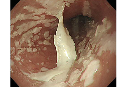
Esophagogastroduodenoscopy (EGD) revealed longitudinal sloughing mucosal casts in the middle and lower esophagus (Figure 1).

Endoscopy shows longitudinal sloughing mucosal casts in the middle and lower esophagus
Figure 1. Endoscopy shows longitudinal sloughing mucosal casts in the middle and lower esophagus.
Esophageal candidiasis would be suspected on the basis of endoscopic findings of white or yellowish plaque-like lesions that could not be washed away.1 However, in this patient, the lesions were exfoliative, a picture compatible with chemical or “pill esophagitis.” The patient had no risk factors for esophageal candidiasis such as human immunodeficiency virus infection, alcohol abuse, or diabetes mellitus. Further, no Candida species were detected in EGD samples sent for microscopic study.

The patient had been taking dabigatran 110 mg twice daily for 2 years because of nonvalvular atrial fibrillation. He was also taking amlodipine 2.5 mg/day for hypertension.

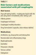
Risk factors and medications associated with pill esophagitis
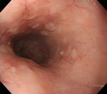
Risk factors and medications commonly associated with pill esophagitis are shown in Table 1. Further questioning of the patient revealed that he took his medications with very little water. Apixaban was prescribed as an alternative to dabigatran, in addition to the proton-pump inhibitor rabeprazole 10 mg/day. The symptoms disappeared within a few days. At follow-up 1 month later, EGD confirmed that the esophageal mucosa had completely healed (Figure 2).

DABIGATRAN-INDUCED ESOPHAGITIS

Complete healing of the esophageal mucosa 1 month later
Figure 2. Complete healing of the esophageal mucosa 1 month later.
Dabigatran, an oral direct thrombin inhibitor, is widely used for thromboprophylaxis. Unlike other direct oral anticoagulants, it contains tartaric acid as an excipient, which reduces variability in the drug’s absorption. However, if the tablet gets stuck in the esophagus, the acid leaching out can damage the esophageal mucosa.

Although no study has investigated the overall prevalence of dabigatran-induced esophagitis, a retrospective database review of 91 patients taking dabigatran and undergoing upper-gastrointestinal endoscopy reported that 19 (20.9%) had endoscopic signs of dabigatran-induced esophagitis.2

Typical symptoms are the acute onset of chest pain, epigastralgia, odynophagia, and dysphagia. But patients can also have no symptoms or only mild symptoms.2,3

Despite dabigatran’s anticoagulant activity, there have been few reports of bleeding, perhaps because  the lesions tend to be superficial on the surface of the esophageal mucosa.

Symptoms usually resolve within 1 week after stopping dabigatran and starting a proton pump inhibitor. To prevent mucosal injury, patients should be instructed to take dabigatran with sufficient water and to remain in an upright position for at least 30 minutes afterward.4

References
  1. Baehr PH, McDonald GB. Esophageal infections: risk factors, presentation, diagnosis, and treatment. Gastroenterology 1994; 106(2):509–532. pmid:7980741
  2. Toya Y, Nakamura S, Tomita K, et al. Dabigatran-induced esophagitis: the prevalence and endoscopic characteristics. J Gastroenterol Hepatol 2016; 31(3):610–614. doi:10.1111/jgh.13024
  3. Ueta E, Fujikawa T, Imagawa A. A case of a slightly symptomatic exfoliative oesophagitis. BMJ Case Rep 2015; pii:bcr2015211925. doi:10.1136/bcr-2015-211925
  4. Ootani A, Hayashi Y, Miyagi Y. Dabigatran-induced esophagitis. Clin Gastroenterol Hepatol 2014; 12(7):e55–e56. doi:10.1016/j.cgh.2013.09.010
Article PDF
Author and Disclosure Information

Yusaku Kajihara, MD, FACP
Department of Gastroenterology, Fuyoukai Murakami Hospital, Aomori, Japan

Address: Yusaku Kajihara, MD, FACP, Department of Gastroenterology, Fuyoukai Murakami Hospital, 3-3-14 Hamada, Aomori 030-0843, Japan; y_kaji2012@yahoo.co.jp

Issue
Cleveland Clinic Journal of Medicine - 86(5)
Publications
Topics
Page Number
303-304
Legacy Keywords
dabigatran, esophagitis, pill esophagitis, candidiasis, Candida, tartaric acid, endoscopy, Yusaku Kajihara
Sections
Author and Disclosure Information

Yusaku Kajihara, MD, FACP
Department of Gastroenterology, Fuyoukai Murakami Hospital, Aomori, Japan

Address: Yusaku Kajihara, MD, FACP, Department of Gastroenterology, Fuyoukai Murakami Hospital, 3-3-14 Hamada, Aomori 030-0843, Japan; y_kaji2012@yahoo.co.jp

Author and Disclosure Information

Yusaku Kajihara, MD, FACP
Department of Gastroenterology, Fuyoukai Murakami Hospital, Aomori, Japan

Address: Yusaku Kajihara, MD, FACP, Department of Gastroenterology, Fuyoukai Murakami Hospital, 3-3-14 Hamada, Aomori 030-0843, Japan; y_kaji2012@yahoo.co.jp

Article PDF
Article PDF
Related Articles

A 74-year-old man presented to the gastroenterology clinic with a 2-day history of retrosternal discomfort. His vital signs were normal, and laboratory testing showed a normal leukocyte count.

Esophagogastroduodenoscopy (EGD) revealed longitudinal sloughing mucosal casts in the middle and lower esophagus (Figure 1).

Endoscopy shows longitudinal sloughing mucosal casts in the middle and lower esophagus
Figure 1. Endoscopy shows longitudinal sloughing mucosal casts in the middle and lower esophagus.
Esophageal candidiasis would be suspected on the basis of endoscopic findings of white or yellowish plaque-like lesions that could not be washed away.1 However, in this patient, the lesions were exfoliative, a picture compatible with chemical or “pill esophagitis.” The patient had no risk factors for esophageal candidiasis such as human immunodeficiency virus infection, alcohol abuse, or diabetes mellitus. Further, no Candida species were detected in EGD samples sent for microscopic study.

The patient had been taking dabigatran 110 mg twice daily for 2 years because of nonvalvular atrial fibrillation. He was also taking amlodipine 2.5 mg/day for hypertension.

Risk factors and medications associated with pill esophagitis
Risk factors and medications commonly associated with pill esophagitis are shown in Table 1. Further questioning of the patient revealed that he took his medications with very little water. Apixaban was prescribed as an alternative to dabigatran, in addition to the proton-pump inhibitor rabeprazole 10 mg/day. The symptoms disappeared within a few days. At follow-up 1 month later, EGD confirmed that the esophageal mucosa had completely healed (Figure 2).

DABIGATRAN-INDUCED ESOPHAGITIS

Complete healing of the esophageal mucosa 1 month later
Figure 2. Complete healing of the esophageal mucosa 1 month later.
Dabigatran, an oral direct thrombin inhibitor, is widely used for thromboprophylaxis. Unlike other direct oral anticoagulants, it contains tartaric acid as an excipient, which reduces variability in the drug’s absorption. However, if the tablet gets stuck in the esophagus, the acid leaching out can damage the esophageal mucosa.

Although no study has investigated the overall prevalence of dabigatran-induced esophagitis, a retrospective database review of 91 patients taking dabigatran and undergoing upper-gastrointestinal endoscopy reported that 19 (20.9%) had endoscopic signs of dabigatran-induced esophagitis.2

Typical symptoms are the acute onset of chest pain, epigastralgia, odynophagia, and dysphagia. But patients can also have no symptoms or only mild symptoms.2,3

Despite dabigatran’s anticoagulant activity, there have been few reports of bleeding, perhaps because  the lesions tend to be superficial on the surface of the esophageal mucosa.

Symptoms usually resolve within 1 week after stopping dabigatran and starting a proton pump inhibitor. To prevent mucosal injury, patients should be instructed to take dabigatran with sufficient water and to remain in an upright position for at least 30 minutes afterward.4

References
  1. Baehr PH, McDonald GB. Esophageal infections: risk factors, presentation, diagnosis, and treatment. Gastroenterology 1994; 106(2):509–532. pmid:7980741
  2. Toya Y, Nakamura S, Tomita K, et al. Dabigatran-induced esophagitis: the prevalence and endoscopic characteristics. J Gastroenterol Hepatol 2016; 31(3):610–614. doi:10.1111/jgh.13024
  3. Ueta E, Fujikawa T, Imagawa A. A case of a slightly symptomatic exfoliative oesophagitis. BMJ Case Rep 2015; pii:bcr2015211925. doi:10.1136/bcr-2015-211925
  4. Ootani A, Hayashi Y, Miyagi Y. Dabigatran-induced esophagitis. Clin Gastroenterol Hepatol 2014; 12(7):e55–e56. doi:10.1016/j.cgh.2013.09.010
Issue
Cleveland Clinic Journal of Medicine - 86(5)
Issue
Cleveland Clinic Journal of Medicine - 86(5)
Page Number
303-304
Page Number
303-304
Publications
Publications
Topics
Article Type
Display Headline
Dabigatran-induced esophagitis
Display Headline
Dabigatran-induced esophagitis
Legacy Keywords
dabigatran, esophagitis, pill esophagitis, candidiasis, Candida, tartaric acid, endoscopy, Yusaku Kajihara
Legacy Keywords
dabigatran, esophagitis, pill esophagitis, candidiasis, Candida, tartaric acid, endoscopy, Yusaku Kajihara
Sections
Disallow All Ads
Content Gating
No Gating (article Unlocked/Free)
Alternative CME
Disqus Comments
Default
Gate On Date
Wed, 04/24/2019 - 13:15
Un-Gate On Date
Wed, 04/24/2019 - 13:15
Use ProPublica
CFC Schedule Remove Status
Wed, 04/24/2019 - 13:15
Hide sidebar & use full width
render the right sidebar.
Article PDF Media