User login
A 50-year-old woman presented to the clinic with an intensely pruritic rash that had come on suddenly and extended over the dorsal aspect of both arms. She said that she had not begun taking any new medicines, and had no recent exposures to any new chemicals. She did, however, note that she’d recently spent some time in the sun.
Her history included schizoaffective disorder, bipolar disorder, reactive airways disease, type 2 diabetes mellitus, and hypertension.
She was taking a number of medications including trihexyphenidyl, flu- phenazine, mirtazapine, ibuprofen, propranolol, acetaminophen, albuterol/ipratropium inhaled, and triamcinolone inhaled. She reported being allergic to lithium, erythromycin, and haloperidol.
Her skin exam showed erythematous plaques over the dorsum of both forearms (Figure 1). There were no lesions on her left forearm where she had been wearing a watch, and there were no lesions on her face or lower extremities. Her vital signs were normal.
We ordered a punch biopsy.
FIGURE 1
Plaque on forearms, except under watchband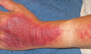
What is your diagnosis?
Diagnosis: Polymorphous light eruption
Biopsy findings
The biopsy showed extensive spongiosis and edema of the dermis with a deep lymphohistiocytic infiltrate, consistent with polymorphous light eruption—an idiopathic, delayed type hypersensitivity to UVA and UVB light.1-3
Characteristics
Polymorphous light eruption may affect up to 10% of the population, with a predilection for females.3,4 The prevalence increases in northern latitudes. All skin types may be affected, but those with Fitzpatrick skin types I or II are most frequently affected.4
Polymorphous light eruption may appear spontaneously at any age and may manifest as plaques (as was the case with our patient), papules, or rarely, vesicles (Figure 2). It tends to present early in the spring and summer, with the first significant UV exposure of the year. The rash typically develops 1 to 4 days after sun exposure, and with time, remits spontaneously. Occasionally, though, it will last as long as there is significant sun exposure.
FIGURE 2
Polymorphous light eruption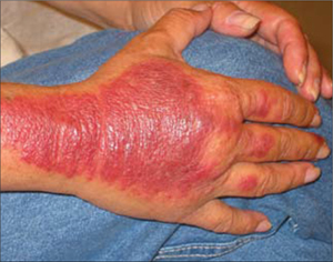
A condition that mimics a phototoxic drug reaction
The differential diagnosis for polymorphous light eruption includes phototoxic drug reaction, systemic lupus erythematosus, and porphyria cutanea tarda.
A phototoxic drug reaction is a non-immunologic reaction that manifests 2 to 6 hours after exposure to sunlight.1-3 It can be difficult to differentiate from polymorphous light eruption without a biopsy. A phototoxic drug reaction can be likened to sunburn, with a mild form causing slight erythema and a severe form causing vesicles or bullae.
Our patient certainly used medicines known to cause phototoxic reactions (ibuprofen, fluphenazine). However, she’d had no reactions with prior sun exposure. The significant dermal edema (hence the plaque) without any vesicles or bullae made a phototoxic drug reaction diagnosis less likely. The biopsy clarified the issue.
Systemic lupus erythematosus
SLE was also a possibility with our patient. Sunlight can precipitate a lupus rash, so the photodistribution made lupus plausible. Its abrupt onset and marked pruritus spoke against it, though, as did the negative serum antinuclear antibody test, which had been ordered.
Porphyria cutanea tarda
This disorder, which can also be precipitated by sunlight, was also a possibility with our patient. It tends to present with vesicles or bullae in sun-exposed areas, especially the dorsum of the hands.2 The bullae generally have no surrounding erythema, which made it an unlikely diagnosis for our patient. A diagnosis of porphyria cutanea tarda hinges on increased porphyrin levels, measured in a 24-hour urine collection.
Steroids, antihistamines, sunblock, and long sleeves
Treatment for polymorphous light eruption includes topical steroids and antihistamines.1-3 Patients can attempt to prevent future episodes by applying broad-spectrum (UVA and UVB coverage) sunblock and wearing long-sleeved garments when going out in the sun.
Desensitization with phototherapy is often necessary. UVA, UVB, and PUVA have all proven beneficial.5
Hydroxychloroquine. Recalcitrant cases may require hydroxychloroquine during the summer months.6
Nonadherence hinders our patient’s recovery
We started our patient on oral antihistamines and topical steroids and recommended that she avoid direct sunlight.
Our patient, however, didn’t avoid sun exposure, and when we saw her on follow-up, her pruritis had improved, but the lesions were essentially unchanged.
We subsequently lost our patient to follow-up.
1. James WD, Berger TG, Elston DA. Andrews’ Diseases of the Skin, Clinical Dermatology. 10th ed. Philadelphia, Pa: W.B. Saunders; 2006.
2. Habif TP. Clinical Dermatology: A Color Guide to Diagnosis and Therapy. 4th ed. Philadelphia, Pa: Mosby; 2004.
3. Fitzpatrick TB, Johnson RA, Wolff K, Suurmond D. Color Atlas and Synopsis of Clinical Dermatology, Common and Serious Diseases. 4th ed. New York, NY: McGraw Hill; 2001.
4. Fesq H, Ring J, Abeck D. Management of polymorphous light eruption. Clinical course, pathogenesis, diagnosis and intervention. Am J Clin Dermatol 2003;4:399-406.
5. Murphy GM, Logan RA, Lovell CR, Morris RW, Hawk JL, Magnus IA. Prophylactic PUVA and UVB therapy in polymorphic light eruption—a controlled trial. Br J Dermatol 1987;116:531-538.
6. Murphy GM, Hawk JLM, Magnus IA. Hydroxychloroquine in polymorphic light eruption: a controlled trial with drug and visual sensitivity monitoring. Br J Dermatol 1987;116:379-386.
A 50-year-old woman presented to the clinic with an intensely pruritic rash that had come on suddenly and extended over the dorsal aspect of both arms. She said that she had not begun taking any new medicines, and had no recent exposures to any new chemicals. She did, however, note that she’d recently spent some time in the sun.
Her history included schizoaffective disorder, bipolar disorder, reactive airways disease, type 2 diabetes mellitus, and hypertension.
She was taking a number of medications including trihexyphenidyl, flu- phenazine, mirtazapine, ibuprofen, propranolol, acetaminophen, albuterol/ipratropium inhaled, and triamcinolone inhaled. She reported being allergic to lithium, erythromycin, and haloperidol.
Her skin exam showed erythematous plaques over the dorsum of both forearms (Figure 1). There were no lesions on her left forearm where she had been wearing a watch, and there were no lesions on her face or lower extremities. Her vital signs were normal.
We ordered a punch biopsy.
FIGURE 1
Plaque on forearms, except under watchband
What is your diagnosis?
Diagnosis: Polymorphous light eruption
Biopsy findings
The biopsy showed extensive spongiosis and edema of the dermis with a deep lymphohistiocytic infiltrate, consistent with polymorphous light eruption—an idiopathic, delayed type hypersensitivity to UVA and UVB light.1-3
Characteristics
Polymorphous light eruption may affect up to 10% of the population, with a predilection for females.3,4 The prevalence increases in northern latitudes. All skin types may be affected, but those with Fitzpatrick skin types I or II are most frequently affected.4
Polymorphous light eruption may appear spontaneously at any age and may manifest as plaques (as was the case with our patient), papules, or rarely, vesicles (Figure 2). It tends to present early in the spring and summer, with the first significant UV exposure of the year. The rash typically develops 1 to 4 days after sun exposure, and with time, remits spontaneously. Occasionally, though, it will last as long as there is significant sun exposure.
FIGURE 2
Polymorphous light eruption
A condition that mimics a phototoxic drug reaction
The differential diagnosis for polymorphous light eruption includes phototoxic drug reaction, systemic lupus erythematosus, and porphyria cutanea tarda.
A phototoxic drug reaction is a non-immunologic reaction that manifests 2 to 6 hours after exposure to sunlight.1-3 It can be difficult to differentiate from polymorphous light eruption without a biopsy. A phototoxic drug reaction can be likened to sunburn, with a mild form causing slight erythema and a severe form causing vesicles or bullae.
Our patient certainly used medicines known to cause phototoxic reactions (ibuprofen, fluphenazine). However, she’d had no reactions with prior sun exposure. The significant dermal edema (hence the plaque) without any vesicles or bullae made a phototoxic drug reaction diagnosis less likely. The biopsy clarified the issue.
Systemic lupus erythematosus
SLE was also a possibility with our patient. Sunlight can precipitate a lupus rash, so the photodistribution made lupus plausible. Its abrupt onset and marked pruritus spoke against it, though, as did the negative serum antinuclear antibody test, which had been ordered.
Porphyria cutanea tarda
This disorder, which can also be precipitated by sunlight, was also a possibility with our patient. It tends to present with vesicles or bullae in sun-exposed areas, especially the dorsum of the hands.2 The bullae generally have no surrounding erythema, which made it an unlikely diagnosis for our patient. A diagnosis of porphyria cutanea tarda hinges on increased porphyrin levels, measured in a 24-hour urine collection.
Steroids, antihistamines, sunblock, and long sleeves
Treatment for polymorphous light eruption includes topical steroids and antihistamines.1-3 Patients can attempt to prevent future episodes by applying broad-spectrum (UVA and UVB coverage) sunblock and wearing long-sleeved garments when going out in the sun.
Desensitization with phototherapy is often necessary. UVA, UVB, and PUVA have all proven beneficial.5
Hydroxychloroquine. Recalcitrant cases may require hydroxychloroquine during the summer months.6
Nonadherence hinders our patient’s recovery
We started our patient on oral antihistamines and topical steroids and recommended that she avoid direct sunlight.
Our patient, however, didn’t avoid sun exposure, and when we saw her on follow-up, her pruritis had improved, but the lesions were essentially unchanged.
We subsequently lost our patient to follow-up.
1. James WD, Berger TG, Elston DA. Andrews’ Diseases of the Skin, Clinical Dermatology. 10th ed. Philadelphia, Pa: W.B. Saunders; 2006.
2. Habif TP. Clinical Dermatology: A Color Guide to Diagnosis and Therapy. 4th ed. Philadelphia, Pa: Mosby; 2004.
3. Fitzpatrick TB, Johnson RA, Wolff K, Suurmond D. Color Atlas and Synopsis of Clinical Dermatology, Common and Serious Diseases. 4th ed. New York, NY: McGraw Hill; 2001.
4. Fesq H, Ring J, Abeck D. Management of polymorphous light eruption. Clinical course, pathogenesis, diagnosis and intervention. Am J Clin Dermatol 2003;4:399-406.
5. Murphy GM, Logan RA, Lovell CR, Morris RW, Hawk JL, Magnus IA. Prophylactic PUVA and UVB therapy in polymorphic light eruption—a controlled trial. Br J Dermatol 1987;116:531-538.
6. Murphy GM, Hawk JLM, Magnus IA. Hydroxychloroquine in polymorphic light eruption: a controlled trial with drug and visual sensitivity monitoring. Br J Dermatol 1987;116:379-386.